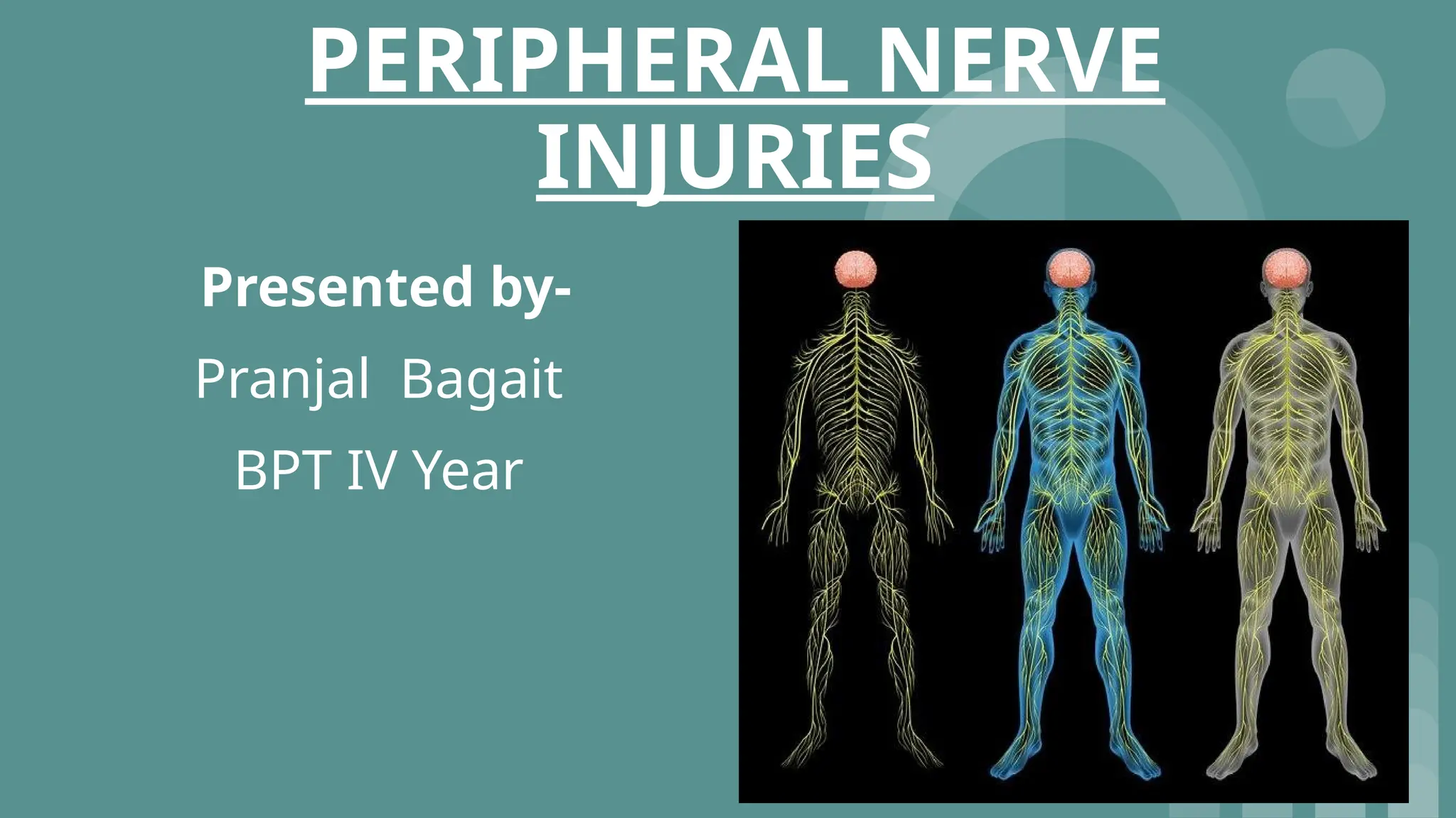
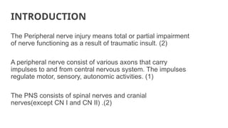
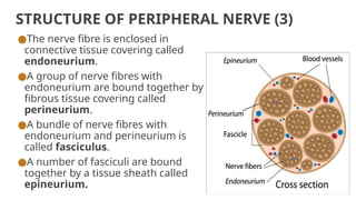
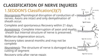
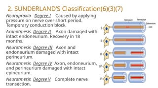
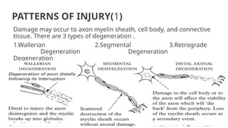
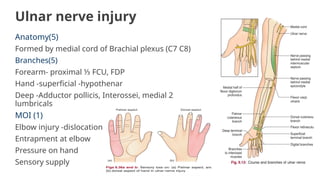
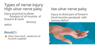
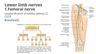
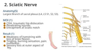
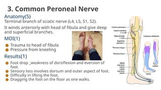
PERIPHERAL NERVE
INJURIES
Presented by-
Pranjal Bagait
BPT IV Year
CONTENTS
●Introduction
●Structure of peripheral nerve
●Classification
●Patterns of injury
●Mechanism of injury
●ULE Nerve Injuries
●LLE Nerve injuries
●Investigations
●Treatment
INTRODUCTION
The Peripheral nerve injury means total or partial impairment
of nerve functioning as a result of traumatic insult. (2)
A peripheral nerve consist of various axons that carry
impulses to and from central nervous system. The impulses
regulate motor, sensory, autonomic activities. (1)
The PNS consists of spinal nerves and cranial
nerves(except CN I and CN II) .(2)
STRUCTURE OF PERIPHERAL NERVE (3)
●The nerve fibre is enclosed in
connective tissue covering called
endoneurium.
●A group of nerve fibres with
endoneurium are bound together by
fibrous tissue covering called
perineurium.
●A bundle of nerve fibres with
endoneurium and perineurium is
called fasciculus.
●A number of fasciculi are bound
together by a tissue sheath called
epineurium.
CLASSIFICATION OF NERVE INJURIES
1.SEDDON’S Classification(3)(7)
Neuropraxia-Physiological disruption of conduction of
nerves. Axons are intact and only demyelination of
sheath occur.
Complete and spontaneous Recovery within 21 days.
Axonotmesis -Complete interruption of axon and myelin
sheath but internal structure of nerve is preserved.
Wallerian degeneration occurs.
Recovery takes several months and may not be
complete
Neurotmesis- The structure of nerve is damaged due to
cutting of segment
Recovery only after nerve repair.
2. SUNDERLAND’S Classification(6)(3)(7)
Neurapraxia Degree I Caused by applying
pressure on nerve over short period.
Temporary conduction block,
Axonotmesis Degree II Axon damaged with
intact endoneurium. Recovery in 18
months.
Neurotmesis Degree III Axon and
endoneurium damaged with intact
perineurium.
Neurotmesis Degree IV Axon, endoneurium,
and perineurium damaged with intact
epineurium.
Neurotmesis Degree V Complete nerve
transection.
PATTERNS OF INJURY(1)
Damage may occur to axon myelin sheath, cell body, and connective
tissue. There are 3 types of degeneration .
1.Wallerian 2.Segmental 3.Retrograde
Degeneration Degeneration
Degeneration
MECHANISM OF INJURY (3)
1. Direct injury -cut, laceration
2. Infections-Leprosy
3. Mechanical injury - traction, overstretching, compression
(CTS), friction, dislocations, avulsion
4. Cooling and freezing-frost bite
5. Thermal injuries
6. Electrical injuries-shock
7. Ischaemia -VIC
8. Radiation in cancer
9. RTA
10.External compression-orthosis, prosthesis, plaster cast
UPPER LIMB NERVE INJURIES
1. Brachial Plexus injury
● Erb's paralysis
● Klumpke's paralysis
● Axillary nerve
● Radial nerve
● Median nerve
● Ulnar nerve
LOWER LIMB NERVE INJURIES
1. Lumbo-sacral Plexus injury
● Femoral nerve
● Sciatic nerve
● Common peroneal nerve
BRACHIAL PLEXUS INJURY
Brachial plexus is network of nerves formed by anterior rami of lower 4
cervical nerves (C5 -C8) and 1 upper thoracic nerve (T1). It sends
signals to upper limb.(5)
Causes of injury(1)
● Birth trauma
● Fall on shoulder
● RTA
● Tumors
● Sports injuries
It consist mainly of 2 types of injury
● Erb's paralysis
● Klumpke’s paralysis
Erb’s paralysis(5)
Site of injury -Injury to upper trunk. (C5, C6)
Injury to erb’s point where 6 nerves meet,
*When damage to C5C6 is more proximal, nerve to
rhomboids and long thoracic nerve may be affected.
MOI- undue separation of head of humerus from
shoulder.
Muscles paralysed - Biceps , brachialis, deltoid,
supraspinatus, infraspinatus, shoulder adductors.
Deformity-Policeman’s tip hand or waiter’s tip hand.
Disability - Abduction & lateral rotation of arm at
shoulder.
-Flexion and supination of forearm.
-Biceps and supinator jerks lost.
- Sensory loss over lower deltoid.
Klumpke's paralysis(5)
Site of injury -Lower trunk of brachial plexus (partly C8 mainly
T1).
MOI - Forced abduction of arm at birth.
Trauma .
Muscles paralyzed - Intrinsic muscle of hand(T1).
Ulnar flexor of wrist and fingers (C8).
Deformity - claw hand (hyperextension of MCP joint & flexion of
IP joint)
Disability -loss of biceps & supinator jerk
Complete claw hand
Cutaneous anaesthesia
Horner syndrome
Axillary Nerve
Anatomy (5)
Derived from posterior cord of brachial plexus (C5C6).
Branches(5) -
Anterior -motor innervation to deltoid.
Posterior-motor innervation to teres minor.
Superior lateral cutaneous nerve - sensory innervation over inferior deltoid.
MOI (1)-
● Anterior & inferior shoulder dislocation
● FOOSH
● Contusion to deltoid
Result (1)
● Weakness of shoulder between 15-90 degrees .
● Sensory loss over outer aspect of shoulder .
Radial nerve injury
Anatomy(5)
Branch of posterior cord of Brachial plexus (C5-C8, T1)
Branches (5)
Before radial groove (post.cutaneous nerve of arm)-long
and medial head of triceps
After radial groove -lateral head of triceps, Anconeus,
Brachioradialis, ECRL
After crossing elbow before piercing supinator (deep
branch)-ECRB, Supinator
After piercing supinator(PIN) -extensor muscle of forearm
MOI (1)
● Humerus fracture
● Gunshot injury
● Crutch paralysis
● Stabbing
● Carcinomas
Types of injury(3)
High radial nerve palsy Low radial nerve palsy
Nerve injury in radial groove. All
muscles are paralysed except triceps
and anconeus brachioradialis,
Results (1)
● Weakness and wasting of muscles
supplies
● Deformity-wrist drop -finger
flexed (weak extensor)
● Sensory loss on dorsum of hand
and forearm
● Loss of tricep reflex
Nerve is injured around elbow
and the muscles supplied by
radial nerve in distal arm are
spare
ECRL, ECRB
Median Nerve injury
Anatomy(5)
Formed by joining lateral and medial cord of brachial
plexus (C7 C8)
Branches (5)
No branch in arm
Muscular branch -FCR, PL, FDS
AIN - proximal ⅓- All flexor muscle except FCU & medial
½ of FDP
Palmar cutaneous branch-Thenar muscle, 1 & 2
lumbricals
Articular branch & vascular branch
MOI(1)
● Dislocation in shoulder
● Compression in forearm (AIN Branch)
● Compression at wrist
Types of nerve injury
High median nerve palsy low median nerve palsy
Injury proximal to elbow,. All muscles are
paralysed of forearm and hand. There is sensory
deficit.
Result (1)
● Weakness of abduction & opposition of
thumb
● Weakness of pronation of forearm
● Ulnar deviation on wrist flexion
● Wasting of thenar muscle
● Sensory loss (mainly on index & middle
finger)
● Deformity - Ape hand deformity, hand of
benediction,
Common disease-
carpal tunnel syndrome
Injury in distal ⅓ of forearm causing
paralysis of hand muscles (sparring
forearm muscle). Sensory deficit.
Ulnar nerve injury
Anatomy(5)
Formed by medial cord of Brachial plexus (C7 C8)
Branches(5)
Forearm- proximal ⅓ FCU, FDP
Hand -superficial -hypothenar
Deep -Adductor pollicis, Interossei, medial 2
lumbricals
MOI (1)
Elbow injury -dislocation
Entrapment at elbow
Pressure on hand
Sensory supply
Types of nerve injury
High ulnar nerve palsy low ulnar nerve palsy
Injury proximal to elbow
Paralysis of all muscles of
forearm & hand
Sensory
deficit
Result(1)
● Ulnar claw hand - weakness of
muscles supplied
Injury to distal part of forearm
hand muscles paralysed with
sensory deficit
Lower limb nerves
1.Femoral nerve
Largest Branch of lumbar plexus L2,
L3,L4
Branches(5)
MOI(1)
● Fracture of upper femur
● CDH
● Hip surgery
Result(1)
● Weakness of hip flexion.
● Weakness of knee extension with wasting of thigh muscle.
● Sensory loss over anterior and medial aspect of thigh .
● Knee jerk lost .
● Difficulty in walking.
2. Sciatic Nerve
Anatomy(5)
Largest Branch of sacral plexus (L4, L5 S1, S2, S3).
MOI (1)
● CDH, traumatic hip dislocation
● Penetrating injuries
● Entrapment at sciatic notch
Result (1)
● Weakness of hamstring with
loss of knee flexion .
● Tingling, burning sensation, pain,
numbness .
● Sensory loss at outer aspect of
leg.
3. Common Peroneal Nerve
Anatomy(5)
Terminal branch of sciatic nerve (L4, L5, S1, S2).
It winds anteriorly with head of fibula and give deep
and superficial branches.
MOI(1)
● Trauma to head of fibula
● Pressure from kneeling
Results(1)
● Foot drop _weakness of dorsiflexion and eversion of
foot.
● Sensory loss involves dorsum and outer aspect of foot.
● Difficulty in lifting the foot.
● Dragging the foot on the floor as one walks.
Investigation
1.Electromyography -record electrical activity of muscle at
rest and during activity
2.NCV -suggest whether nerve is injured, whether it is
partial or complete
3.MRI
4.Special tests
Treatment(3)
1.Conservative
● Splintage of paralysed limb
● Joint mobility preservation
● Analgesic -NSAID -prednisolone
2. Operative
● Nerve repair
● Nerve suture
● Neurolysis
● Reconstructive surgery
References
1. Kenneth W. Lindsay
2. Suraj kumar
3. Maheshwari
4. K. Sembulingam
5. BD. Chaurasia -VOl. 1,VOL.2 Edition 7
6. https://pubmed.ncbi.nlm.nih.gov/
7. S. Sunder

PERIPHERAL NERVE INJURIES (presented by pranjal).pptx

  • 1.
  • 2.
    CONTENTS ●Introduction ●Structure of peripheralnerve ●Classification ●Patterns of injury ●Mechanism of injury ●ULE Nerve Injuries ●LLE Nerve injuries ●Investigations ●Treatment
  • 3.
    INTRODUCTION The Peripheral nerveinjury means total or partial impairment of nerve functioning as a result of traumatic insult. (2) A peripheral nerve consist of various axons that carry impulses to and from central nervous system. The impulses regulate motor, sensory, autonomic activities. (1) The PNS consists of spinal nerves and cranial nerves(except CN I and CN II) .(2)
  • 4.
    STRUCTURE OF PERIPHERALNERVE (3) ●The nerve fibre is enclosed in connective tissue covering called endoneurium. ●A group of nerve fibres with endoneurium are bound together by fibrous tissue covering called perineurium. ●A bundle of nerve fibres with endoneurium and perineurium is called fasciculus. ●A number of fasciculi are bound together by a tissue sheath called epineurium.
  • 5.
    CLASSIFICATION OF NERVEINJURIES 1.SEDDON’S Classification(3)(7) Neuropraxia-Physiological disruption of conduction of nerves. Axons are intact and only demyelination of sheath occur. Complete and spontaneous Recovery within 21 days. Axonotmesis -Complete interruption of axon and myelin sheath but internal structure of nerve is preserved. Wallerian degeneration occurs. Recovery takes several months and may not be complete Neurotmesis- The structure of nerve is damaged due to cutting of segment Recovery only after nerve repair.
  • 6.
    2. SUNDERLAND’S Classification(6)(3)(7) NeurapraxiaDegree I Caused by applying pressure on nerve over short period. Temporary conduction block, Axonotmesis Degree II Axon damaged with intact endoneurium. Recovery in 18 months. Neurotmesis Degree III Axon and endoneurium damaged with intact perineurium. Neurotmesis Degree IV Axon, endoneurium, and perineurium damaged with intact epineurium. Neurotmesis Degree V Complete nerve transection.
  • 7.
    PATTERNS OF INJURY(1) Damagemay occur to axon myelin sheath, cell body, and connective tissue. There are 3 types of degeneration . 1.Wallerian 2.Segmental 3.Retrograde Degeneration Degeneration Degeneration
  • 8.
    MECHANISM OF INJURY(3) 1. Direct injury -cut, laceration 2. Infections-Leprosy 3. Mechanical injury - traction, overstretching, compression (CTS), friction, dislocations, avulsion 4. Cooling and freezing-frost bite 5. Thermal injuries 6. Electrical injuries-shock 7. Ischaemia -VIC 8. Radiation in cancer 9. RTA 10.External compression-orthosis, prosthesis, plaster cast
  • 9.
    UPPER LIMB NERVEINJURIES 1. Brachial Plexus injury ● Erb's paralysis ● Klumpke's paralysis ● Axillary nerve ● Radial nerve ● Median nerve ● Ulnar nerve LOWER LIMB NERVE INJURIES 1. Lumbo-sacral Plexus injury ● Femoral nerve ● Sciatic nerve ● Common peroneal nerve
  • 10.
    BRACHIAL PLEXUS INJURY Brachialplexus is network of nerves formed by anterior rami of lower 4 cervical nerves (C5 -C8) and 1 upper thoracic nerve (T1). It sends signals to upper limb.(5) Causes of injury(1) ● Birth trauma ● Fall on shoulder ● RTA ● Tumors ● Sports injuries It consist mainly of 2 types of injury ● Erb's paralysis ● Klumpke’s paralysis
  • 11.
    Erb’s paralysis(5) Site ofinjury -Injury to upper trunk. (C5, C6) Injury to erb’s point where 6 nerves meet, *When damage to C5C6 is more proximal, nerve to rhomboids and long thoracic nerve may be affected. MOI- undue separation of head of humerus from shoulder. Muscles paralysed - Biceps , brachialis, deltoid, supraspinatus, infraspinatus, shoulder adductors. Deformity-Policeman’s tip hand or waiter’s tip hand. Disability - Abduction & lateral rotation of arm at shoulder. -Flexion and supination of forearm. -Biceps and supinator jerks lost. - Sensory loss over lower deltoid.
  • 12.
    Klumpke's paralysis(5) Site ofinjury -Lower trunk of brachial plexus (partly C8 mainly T1). MOI - Forced abduction of arm at birth. Trauma . Muscles paralyzed - Intrinsic muscle of hand(T1). Ulnar flexor of wrist and fingers (C8). Deformity - claw hand (hyperextension of MCP joint & flexion of IP joint) Disability -loss of biceps & supinator jerk Complete claw hand Cutaneous anaesthesia Horner syndrome
  • 13.
    Axillary Nerve Anatomy (5) Derivedfrom posterior cord of brachial plexus (C5C6). Branches(5) - Anterior -motor innervation to deltoid. Posterior-motor innervation to teres minor. Superior lateral cutaneous nerve - sensory innervation over inferior deltoid. MOI (1)- ● Anterior & inferior shoulder dislocation ● FOOSH ● Contusion to deltoid Result (1) ● Weakness of shoulder between 15-90 degrees . ● Sensory loss over outer aspect of shoulder .
  • 14.
    Radial nerve injury Anatomy(5) Branchof posterior cord of Brachial plexus (C5-C8, T1) Branches (5) Before radial groove (post.cutaneous nerve of arm)-long and medial head of triceps After radial groove -lateral head of triceps, Anconeus, Brachioradialis, ECRL After crossing elbow before piercing supinator (deep branch)-ECRB, Supinator After piercing supinator(PIN) -extensor muscle of forearm MOI (1) ● Humerus fracture ● Gunshot injury ● Crutch paralysis ● Stabbing ● Carcinomas
  • 15.
    Types of injury(3) Highradial nerve palsy Low radial nerve palsy Nerve injury in radial groove. All muscles are paralysed except triceps and anconeus brachioradialis, Results (1) ● Weakness and wasting of muscles supplies ● Deformity-wrist drop -finger flexed (weak extensor) ● Sensory loss on dorsum of hand and forearm ● Loss of tricep reflex Nerve is injured around elbow and the muscles supplied by radial nerve in distal arm are spare ECRL, ECRB
  • 16.
    Median Nerve injury Anatomy(5) Formedby joining lateral and medial cord of brachial plexus (C7 C8) Branches (5) No branch in arm Muscular branch -FCR, PL, FDS AIN - proximal ⅓- All flexor muscle except FCU & medial ½ of FDP Palmar cutaneous branch-Thenar muscle, 1 & 2 lumbricals Articular branch & vascular branch MOI(1) ● Dislocation in shoulder ● Compression in forearm (AIN Branch) ● Compression at wrist
  • 17.
    Types of nerveinjury High median nerve palsy low median nerve palsy Injury proximal to elbow,. All muscles are paralysed of forearm and hand. There is sensory deficit. Result (1) ● Weakness of abduction & opposition of thumb ● Weakness of pronation of forearm ● Ulnar deviation on wrist flexion ● Wasting of thenar muscle ● Sensory loss (mainly on index & middle finger) ● Deformity - Ape hand deformity, hand of benediction, Common disease- carpal tunnel syndrome Injury in distal ⅓ of forearm causing paralysis of hand muscles (sparring forearm muscle). Sensory deficit.
  • 18.
    Ulnar nerve injury Anatomy(5) Formedby medial cord of Brachial plexus (C7 C8) Branches(5) Forearm- proximal ⅓ FCU, FDP Hand -superficial -hypothenar Deep -Adductor pollicis, Interossei, medial 2 lumbricals MOI (1) Elbow injury -dislocation Entrapment at elbow Pressure on hand Sensory supply
  • 19.
    Types of nerveinjury High ulnar nerve palsy low ulnar nerve palsy Injury proximal to elbow Paralysis of all muscles of forearm & hand Sensory deficit Result(1) ● Ulnar claw hand - weakness of muscles supplied Injury to distal part of forearm hand muscles paralysed with sensory deficit
  • 20.
    Lower limb nerves 1.Femoralnerve Largest Branch of lumbar plexus L2, L3,L4 Branches(5)
  • 21.
    MOI(1) ● Fracture ofupper femur ● CDH ● Hip surgery Result(1) ● Weakness of hip flexion. ● Weakness of knee extension with wasting of thigh muscle. ● Sensory loss over anterior and medial aspect of thigh . ● Knee jerk lost . ● Difficulty in walking.
  • 22.
    2. Sciatic Nerve Anatomy(5) LargestBranch of sacral plexus (L4, L5 S1, S2, S3). MOI (1) ● CDH, traumatic hip dislocation ● Penetrating injuries ● Entrapment at sciatic notch Result (1) ● Weakness of hamstring with loss of knee flexion . ● Tingling, burning sensation, pain, numbness . ● Sensory loss at outer aspect of leg.
  • 23.
    3. Common PeronealNerve Anatomy(5) Terminal branch of sciatic nerve (L4, L5, S1, S2). It winds anteriorly with head of fibula and give deep and superficial branches. MOI(1) ● Trauma to head of fibula ● Pressure from kneeling Results(1) ● Foot drop _weakness of dorsiflexion and eversion of foot. ● Sensory loss involves dorsum and outer aspect of foot. ● Difficulty in lifting the foot. ● Dragging the foot on the floor as one walks.
  • 24.
    Investigation 1.Electromyography -record electricalactivity of muscle at rest and during activity 2.NCV -suggest whether nerve is injured, whether it is partial or complete 3.MRI 4.Special tests
  • 25.
    Treatment(3) 1.Conservative ● Splintage ofparalysed limb ● Joint mobility preservation ● Analgesic -NSAID -prednisolone 2. Operative ● Nerve repair ● Nerve suture ● Neurolysis ● Reconstructive surgery
  • 26.
    References 1. Kenneth W.Lindsay 2. Suraj kumar 3. Maheshwari 4. K. Sembulingam 5. BD. Chaurasia -VOl. 1,VOL.2 Edition 7 6. https://pubmed.ncbi.nlm.nih.gov/ 7. S. Sunder