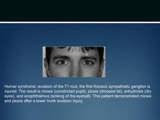
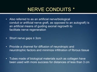
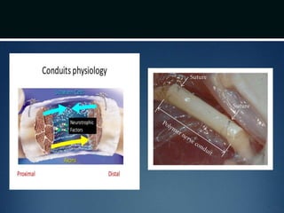
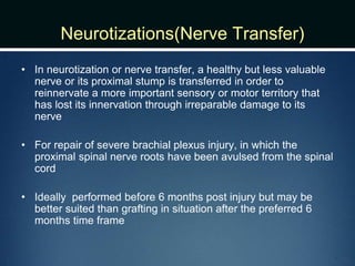
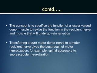
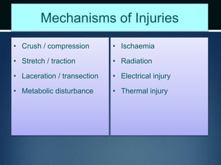
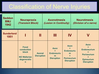
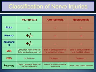
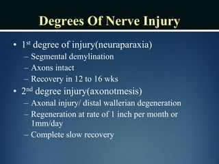
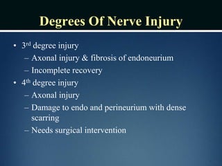
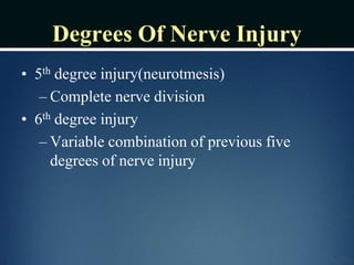
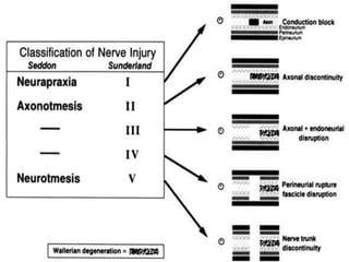
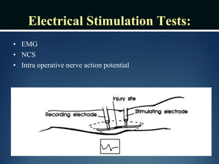
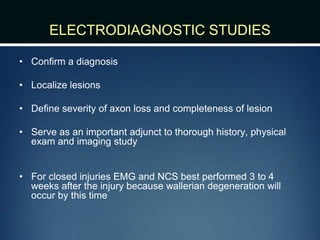
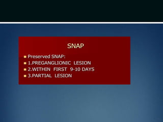
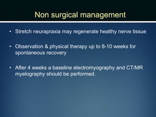
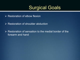
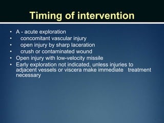
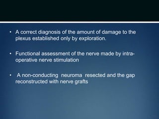
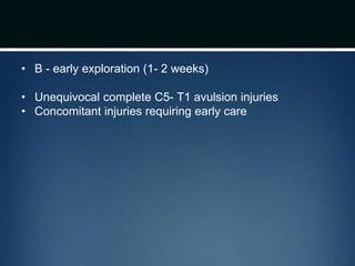
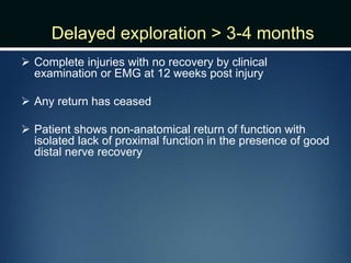
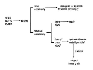
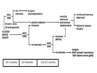
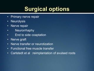
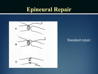
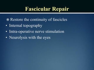
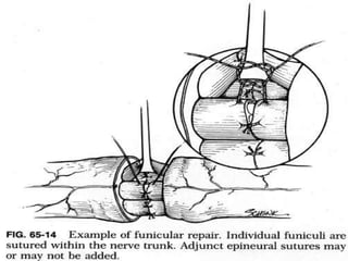
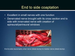
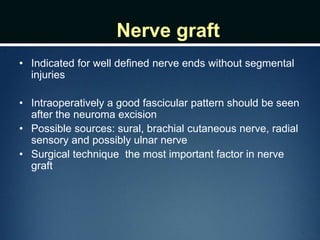
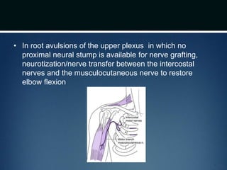
Basic concepts of Nerve injury
By
Dr.usman haqqani
Resident Neurosurgery
LRH
Nerve Anatomy
Mechanisms of Injuries
• Crush / compression
• Stretch / traction
• Laceration / transection
• Metabolic disturbance
• Ischaemia
• Radiation
• Electrical injury
• Thermal injury
Classification of Nerve Injuries
Seddon
BMJ
1942
Neurapraxia
(Transient Block)
Axonotmesis
(Lesion in Continuity)
Neurotmesis
(Division of a nerve)
Sunderland
1951 I II III IV V
Focal
conduction
block
NO Wallerian
degeneratio
n
Axonal
Disruption
Axon
+
Endoneuriu
m
Disruption
Axon
+
Endoneuriu
m
+
Perineurium
Disruption
Axon
+
Endoneurium
+
Perineurium
+
Epineurium
Disruption
Classification of Nerve Injuries
Neurapraxia Axonotmesis Neurotmesis
Motor
- - -
Sensory
+/- - -
Autonomi
c +/- - -
NCS
Conduction block at the site
Distal conduction preserved
Loss of conduction both at
and distal to the lesion
Loss of conduction both at
and distal to the lesion
EMG No fibrillation Fibrillation ++ Fibrillation ++
Recovery
Days to weeks provided the
cause is removed
Months provided the cause
is removed
No recovery unless repaired
Degrees Of Nerve Injury
• 1st degree of injury(neuraparaxia)
– Segmental demylination
– Axons intact
– Recovery in 12 to 16 wks
• 2nd degree injury(axonotmesis)
– Axonal injury/ distal wallerian degeneration
– Regeneration at rate of 1 inch per month or
1mm/day
– Complete slow recovery
Degrees Of Nerve Injury
• 3rd degree injury
– Axonal injury & fibrosis of endoneurium
– Incomplete recovery
• 4th degree injury
– Axonal injury
– Damage to endo and perineurium with dense
scarring
– Needs surgical intervention
Degrees Of Nerve Injury
• 5th degree injury(neurotmesis)
– Complete nerve division
• 6th degree injury
– Variable combination of previous five
degrees of nerve injury
Pre and post ganglionic injury
Horner syndrome; avulsion of the T1 root, the first thoracic sympathetic ganglion is
injured. The result is miosis (constricted pupil), ptosis (drooped lid), anhydrosis (dry
eyes), and enophthalmos (sinking of the eyeball). This patient demonstrated miosis
and ptosis after a lower trunk avulsion injury.
History and physical exam
• Obtain a good clinical hx
• High velocity injury is related to higher risk
of root avulsion
• Knowledge of evolution of pt motor and
sensory sensation over time is extremly
helpful.
• Physical exam begins with inspection
• Look for position of limb
• Examine from the back for
• asymmetry
• Droped shoulder
• Atrophy of rhomboid or supra spinatus
• winging of scapula
• Mechanism of abduction, internal/external rotation
• Diaphragmatic paralysis can be checked by percussing
from behind
• Elbow flextion extention,supination pronation are
checked
• Tendon reflexes ,sensory exam and Intrinsic muscles of
hand are examined
• Horner syndrome signs should also be looked for.
Tinel Sign
• Tinel sign: -
– peripheral tingling or dysaesthesia' provoked
by percussion of the nerve
– Positive in axonal injuries
Electrical and Imaging Studies
• Plain Radiographic
• Stretch injuries of brachial plexus have high
association with fractures
• In anteroposterior (AP) chest radiography, specific
attention directed to the distance between the
spinous processes of the thoracic spine and the
scapula
• Chest X ray would also suggest elevation of
hemidiaphragm in case of phrenic nerve injury.
CT Myelography
• Gold standard
• The most reliable indicator of root avulsion : an absent
root shadow on plain myelography
• A common sign of a root avulsion:absence of rootlet or
pseudo meningocele at the affected level
• Thining of root suggests partial avulsuion
• Delayed for 4 weeks so that any blood clot will not be
dislodged by the study and the meningocele can be
allowed to form
MRI
• MRI of cerviacal spine and brachial plexus
is fast replacing CT myelograph because
of great contrast resolution,multiplanar
imaging and non invasive nature.
Electrical Stimulation Tests:
• EMG
• NCS
• Intra operative nerve action potential
ELECTRODIAGNOSTIC STUDIES
• Confirm a diagnosis
• Localize lesions
• Define severity of axon loss and completeness of lesion
• Serve as an important adjunct to thorough history, physical
exam and imaging study
• For closed injuries EMG and NCS best performed 3 to 4
weeks after the injury because wallerian degeneration will
occur by this time
Non surgical management
• Stretch neurapraxia may regenerate healthy nerve tissue
• Observation & physical therapy up to 8-10 weeks for
spontaneous recovery
• After 4 weeks a baseline electromyography and CT/MR
myelography should be performed.
Surgical Goals
 Restoration of elbow flexion
 Restoration of shoulder abduction
 Restoration of sensation to the medial border of the
forearm and hand
Timing of intervention
• A - acute exploration
• concomitant vascular injury
• open injury by sharp laceration
• crush or contaminated wound
• Open injury with low-velocity missile
• Early exploration not indicated, unless injuries to
adjacent vessels or viscera make immediate treatment
necessary
• A correct diagnosis of the amount of damage to the
plexus established only by exploration.
• Functional assessment of the nerve made by intra-
operative nerve stimulation
• A non-conducting neuroma resected and the gap
reconstructed with nerve grafts
• B - early exploration (1- 2 weeks)
• Unequivocal complete C5- T1 avulsion injuries
• Concomitant injuries requiring early care
Delayed exploration > 3-4 months
 Complete injuries with no recovery by clinical
examination or EMG at 12 weeks post injury
 Any return has ceased
 Patient shows non-anatomical return of function with
isolated lack of proximal function in the presence of good
distal nerve recovery
 Evidence that the lesion is at the postganglionic level
 Anaesthetic limb, severe deafferentation pain, Horner’s
syndrome and pseudomeningoceles on imaging
• Postganglionic lesions :follow patients conservatively for
up to 3 months to watch for spontaneous motor
recovery. In upper-plexus injuries, if the biceps muscle is
not recovered within 3 months, then surgical exploration
Surgical options
• Primary nerve repair
• Neurolysis
• Nerve repair
• Neurorrhaphy
• End to side coaptation
• Nerve graft
• Nerve transfer or neurotization
• Functional free muscle transfer
• Carlstedt et al :reimplantation of avulsed roots
Neurolysis
◦ Removal of any scar or tethering attachments
to surroundings that obstruct nerve ability to
glide.
Neurolysis
• Effective only if scar tissue seen around nerve or inside
epineurium, preventing recovery or causing pain
• Pre and post neurolysis direct nerve stimulation is
mandatory to evaluate improvement in nerve conduction
Neurorraphy
◦ End-to-end repair.
◦ Resection of the proximal and distal nerve stumps and
then approximation.
◦ Sharp transection with excellent fascicular pattern and
minimal scar
• Primary nerve repair
– Epineural repair
– Grouped fascicular repair
Epineural Repair
Standard repair
Fascicular Repair
 Restore the continuity of fascicles
• Internal topography
• Intra-operative nerve stimulation
• Neurolysis with the eyes
End to side coaptation
• Excellent in small nerves with one function
• Denervated nerve brought with its cross section end to
side with innervated nerve with creation of
epineural/perineural windows
End-to-side neurorraphy ulnar nerve (distal stump) to median nerve (distal stump).
Nerve graft
• Indicated for well defined nerve ends without segmental
injuries
• Intraoperatively a good fascicular pattern should be seen
after the neuroma excision
• Possible sources: sural, brachial cutaneous nerve, radial
sensory and possibly ulnar nerve
• Surgical technique the most important factor in nerve
graft
• A tension free nerve graft better than a primary repair
under tension
• Thin cutaneous grafts (e.g. sural nerve) prepared
• Graft should be 20% longer than the length of the nerve
defect
NERVE ALLOGRAFTS
• Act as a temporary
scaffold across which
axons regenerate
• Ultimately, the allograft
tissue completely
replaced with host
materia
Ulnar nerve repair using allograft. Note
excellent return of intrinsic function.
Nerve fibrin glue *
• Nerve repairs performed with fibrin sealants produced
less inflammatory response and fibrosis, better axonal
regeneration, and better fiber alignment than the nerve
repairs performed with microsutures alone
• Fibrin sealant techniques are quicker and easier to use
NERVE CONDUITS *
• Also referred to as an artificial nerve/biological
conduit or artificial nerve graft, as opposed to an autograft) is
an artificial means of guiding axonal regrowth to
facilitate nerve regeneration
• Short nerve gaps ≤ 3cm
• Provide a channel for diffusion of neurotropic and
neurotrophic factors and minimize infiltration of fibrous tissue
• Tubes made of biological materials such as collagen have
been used with more success for distances of less than 3 cm
Neurotizations(Nerve Transfer)
• In neurotization or nerve transfer, a healthy but less valuable
nerve or its proximal stump is transferred in order to
reinnervate a more important sensory or motor territory that
has lost its innervation through irreparable damage to its
nerve
• For repair of severe brachial plexus injury, in which the
proximal spinal nerve roots have been avulsed from the spinal
cord
• Ideally performed before 6 months post injury but may be
better suited than grafting in situation after the preferred 6
months time frame
contd…..
• The concept is to sacrifice the function of a lesser valued
donor muscle to revive the function in the recipient nerve
and muscle that will undergo reinnervation
• Transferring a pure motor donor nerve to a motor
recipient nerve gives the best result of motor
neurotization, for example, spinal accessory to
suprascapular neurotization
contd….
• The more periphreal the recipient site the more better
the results are.
• Reinnervate the recipient nerve as close to the target
muscle as possible; e.g transfer of an ulnar nerve
fascicle directly to the biceps branch of the
musculocutaneous nerve in close proximity to its entry
into the muscle.
Note
• Direct intraoperative nerve stimulation and recording
required across damaged elements
• If nerve action potentials are obtained, simple neurolysis
indicated.
• If neural integrity completely lost, or if no nerve action
potentials recorded across a damaged element, excision
and nerve grafting are required
• In root avulsions of the upper plexus in which no
proximal neural stump is available for nerve grafting,
neurotization/nerve transfer between the intercostal
nerves and the musculocutaneous nerve to restore
elbow flexion
Summary
Brachial plexus surgery basic concepts

Brachial plexus surgery basic concepts

  • 1.
    Basic concepts ofNerve injury By Dr.usman haqqani Resident Neurosurgery LRH
  • 2.
  • 3.
    Mechanisms of Injuries •Crush / compression • Stretch / traction • Laceration / transection • Metabolic disturbance • Ischaemia • Radiation • Electrical injury • Thermal injury
  • 4.
    Classification of NerveInjuries Seddon BMJ 1942 Neurapraxia (Transient Block) Axonotmesis (Lesion in Continuity) Neurotmesis (Division of a nerve) Sunderland 1951 I II III IV V Focal conduction block NO Wallerian degeneratio n Axonal Disruption Axon + Endoneuriu m Disruption Axon + Endoneuriu m + Perineurium Disruption Axon + Endoneurium + Perineurium + Epineurium Disruption
  • 5.
    Classification of NerveInjuries Neurapraxia Axonotmesis Neurotmesis Motor - - - Sensory +/- - - Autonomi c +/- - - NCS Conduction block at the site Distal conduction preserved Loss of conduction both at and distal to the lesion Loss of conduction both at and distal to the lesion EMG No fibrillation Fibrillation ++ Fibrillation ++ Recovery Days to weeks provided the cause is removed Months provided the cause is removed No recovery unless repaired
  • 6.
    Degrees Of NerveInjury • 1st degree of injury(neuraparaxia) – Segmental demylination – Axons intact – Recovery in 12 to 16 wks • 2nd degree injury(axonotmesis) – Axonal injury/ distal wallerian degeneration – Regeneration at rate of 1 inch per month or 1mm/day – Complete slow recovery
  • 7.
    Degrees Of NerveInjury • 3rd degree injury – Axonal injury & fibrosis of endoneurium – Incomplete recovery • 4th degree injury – Axonal injury – Damage to endo and perineurium with dense scarring – Needs surgical intervention
  • 8.
    Degrees Of NerveInjury • 5th degree injury(neurotmesis) – Complete nerve division • 6th degree injury – Variable combination of previous five degrees of nerve injury
  • 10.
    Pre and postganglionic injury
  • 12.
    Horner syndrome; avulsionof the T1 root, the first thoracic sympathetic ganglion is injured. The result is miosis (constricted pupil), ptosis (drooped lid), anhydrosis (dry eyes), and enophthalmos (sinking of the eyeball). This patient demonstrated miosis and ptosis after a lower trunk avulsion injury.
  • 13.
    History and physicalexam • Obtain a good clinical hx • High velocity injury is related to higher risk of root avulsion • Knowledge of evolution of pt motor and sensory sensation over time is extremly helpful.
  • 14.
    • Physical exambegins with inspection • Look for position of limb • Examine from the back for • asymmetry • Droped shoulder • Atrophy of rhomboid or supra spinatus • winging of scapula • Mechanism of abduction, internal/external rotation • Diaphragmatic paralysis can be checked by percussing from behind
  • 15.
    • Elbow flextionextention,supination pronation are checked • Tendon reflexes ,sensory exam and Intrinsic muscles of hand are examined • Horner syndrome signs should also be looked for.
  • 16.
    Tinel Sign • Tinelsign: - – peripheral tingling or dysaesthesia' provoked by percussion of the nerve – Positive in axonal injuries
  • 17.
    Electrical and ImagingStudies • Plain Radiographic • Stretch injuries of brachial plexus have high association with fractures • In anteroposterior (AP) chest radiography, specific attention directed to the distance between the spinous processes of the thoracic spine and the scapula • Chest X ray would also suggest elevation of hemidiaphragm in case of phrenic nerve injury.
  • 18.
    CT Myelography • Goldstandard • The most reliable indicator of root avulsion : an absent root shadow on plain myelography • A common sign of a root avulsion:absence of rootlet or pseudo meningocele at the affected level • Thining of root suggests partial avulsuion • Delayed for 4 weeks so that any blood clot will not be dislodged by the study and the meningocele can be allowed to form
  • 21.
    MRI • MRI ofcerviacal spine and brachial plexus is fast replacing CT myelograph because of great contrast resolution,multiplanar imaging and non invasive nature.
  • 23.
    Electrical Stimulation Tests: •EMG • NCS • Intra operative nerve action potential
  • 24.
    ELECTRODIAGNOSTIC STUDIES • Confirma diagnosis • Localize lesions • Define severity of axon loss and completeness of lesion • Serve as an important adjunct to thorough history, physical exam and imaging study • For closed injuries EMG and NCS best performed 3 to 4 weeks after the injury because wallerian degeneration will occur by this time
  • 26.
    Non surgical management •Stretch neurapraxia may regenerate healthy nerve tissue • Observation & physical therapy up to 8-10 weeks for spontaneous recovery • After 4 weeks a baseline electromyography and CT/MR myelography should be performed.
  • 27.
    Surgical Goals  Restorationof elbow flexion  Restoration of shoulder abduction  Restoration of sensation to the medial border of the forearm and hand
  • 28.
    Timing of intervention •A - acute exploration • concomitant vascular injury • open injury by sharp laceration • crush or contaminated wound • Open injury with low-velocity missile • Early exploration not indicated, unless injuries to adjacent vessels or viscera make immediate treatment necessary
  • 29.
    • A correctdiagnosis of the amount of damage to the plexus established only by exploration. • Functional assessment of the nerve made by intra- operative nerve stimulation • A non-conducting neuroma resected and the gap reconstructed with nerve grafts
  • 30.
    • B -early exploration (1- 2 weeks) • Unequivocal complete C5- T1 avulsion injuries • Concomitant injuries requiring early care
  • 31.
    Delayed exploration >3-4 months  Complete injuries with no recovery by clinical examination or EMG at 12 weeks post injury  Any return has ceased  Patient shows non-anatomical return of function with isolated lack of proximal function in the presence of good distal nerve recovery
  • 32.
     Evidence thatthe lesion is at the postganglionic level  Anaesthetic limb, severe deafferentation pain, Horner’s syndrome and pseudomeningoceles on imaging • Postganglionic lesions :follow patients conservatively for up to 3 months to watch for spontaneous motor recovery. In upper-plexus injuries, if the biceps muscle is not recovered within 3 months, then surgical exploration
  • 35.
    Surgical options • Primarynerve repair • Neurolysis • Nerve repair • Neurorrhaphy • End to side coaptation • Nerve graft • Nerve transfer or neurotization • Functional free muscle transfer • Carlstedt et al :reimplantation of avulsed roots
  • 36.
    Neurolysis ◦ Removal ofany scar or tethering attachments to surroundings that obstruct nerve ability to glide.
  • 37.
    Neurolysis • Effective onlyif scar tissue seen around nerve or inside epineurium, preventing recovery or causing pain • Pre and post neurolysis direct nerve stimulation is mandatory to evaluate improvement in nerve conduction
  • 38.
    Neurorraphy ◦ End-to-end repair. ◦Resection of the proximal and distal nerve stumps and then approximation. ◦ Sharp transection with excellent fascicular pattern and minimal scar • Primary nerve repair – Epineural repair – Grouped fascicular repair
  • 39.
  • 40.
    Fascicular Repair  Restorethe continuity of fascicles • Internal topography • Intra-operative nerve stimulation • Neurolysis with the eyes
  • 42.
    End to sidecoaptation • Excellent in small nerves with one function • Denervated nerve brought with its cross section end to side with innervated nerve with creation of epineural/perineural windows End-to-side neurorraphy ulnar nerve (distal stump) to median nerve (distal stump).
  • 43.
    Nerve graft • Indicatedfor well defined nerve ends without segmental injuries • Intraoperatively a good fascicular pattern should be seen after the neuroma excision • Possible sources: sural, brachial cutaneous nerve, radial sensory and possibly ulnar nerve • Surgical technique the most important factor in nerve graft
  • 44.
    • A tensionfree nerve graft better than a primary repair under tension • Thin cutaneous grafts (e.g. sural nerve) prepared • Graft should be 20% longer than the length of the nerve defect
  • 45.
    NERVE ALLOGRAFTS • Actas a temporary scaffold across which axons regenerate • Ultimately, the allograft tissue completely replaced with host materia
  • 46.
    Ulnar nerve repairusing allograft. Note excellent return of intrinsic function.
  • 47.
    Nerve fibrin glue* • Nerve repairs performed with fibrin sealants produced less inflammatory response and fibrosis, better axonal regeneration, and better fiber alignment than the nerve repairs performed with microsutures alone • Fibrin sealant techniques are quicker and easier to use
  • 48.
    NERVE CONDUITS * •Also referred to as an artificial nerve/biological conduit or artificial nerve graft, as opposed to an autograft) is an artificial means of guiding axonal regrowth to facilitate nerve regeneration • Short nerve gaps ≤ 3cm • Provide a channel for diffusion of neurotropic and neurotrophic factors and minimize infiltration of fibrous tissue • Tubes made of biological materials such as collagen have been used with more success for distances of less than 3 cm
  • 50.
    Neurotizations(Nerve Transfer) • Inneurotization or nerve transfer, a healthy but less valuable nerve or its proximal stump is transferred in order to reinnervate a more important sensory or motor territory that has lost its innervation through irreparable damage to its nerve • For repair of severe brachial plexus injury, in which the proximal spinal nerve roots have been avulsed from the spinal cord • Ideally performed before 6 months post injury but may be better suited than grafting in situation after the preferred 6 months time frame
  • 51.
    contd….. • The conceptis to sacrifice the function of a lesser valued donor muscle to revive the function in the recipient nerve and muscle that will undergo reinnervation • Transferring a pure motor donor nerve to a motor recipient nerve gives the best result of motor neurotization, for example, spinal accessory to suprascapular neurotization
  • 52.
    contd…. • The moreperiphreal the recipient site the more better the results are. • Reinnervate the recipient nerve as close to the target muscle as possible; e.g transfer of an ulnar nerve fascicle directly to the biceps branch of the musculocutaneous nerve in close proximity to its entry into the muscle.
  • 54.
    Note • Direct intraoperativenerve stimulation and recording required across damaged elements • If nerve action potentials are obtained, simple neurolysis indicated. • If neural integrity completely lost, or if no nerve action potentials recorded across a damaged element, excision and nerve grafting are required
  • 56.
    • In rootavulsions of the upper plexus in which no proximal neural stump is available for nerve grafting, neurotization/nerve transfer between the intercostal nerves and the musculocutaneous nerve to restore elbow flexion
  • 57.

Editor's Notes

  • #41 In order to restore the sensory and motor modalities of a nerve..stitch the sensory & motor fascicles of proximal segment to those of distal segment if the internal topography of a nerve is clear..every nerve has a specific internal organization and u must know all of them before going into that micro repair..usually nerves r more monofascicular proximally and are polyfascicular distally and there is plexus formation in between these fascicles that diminish distally