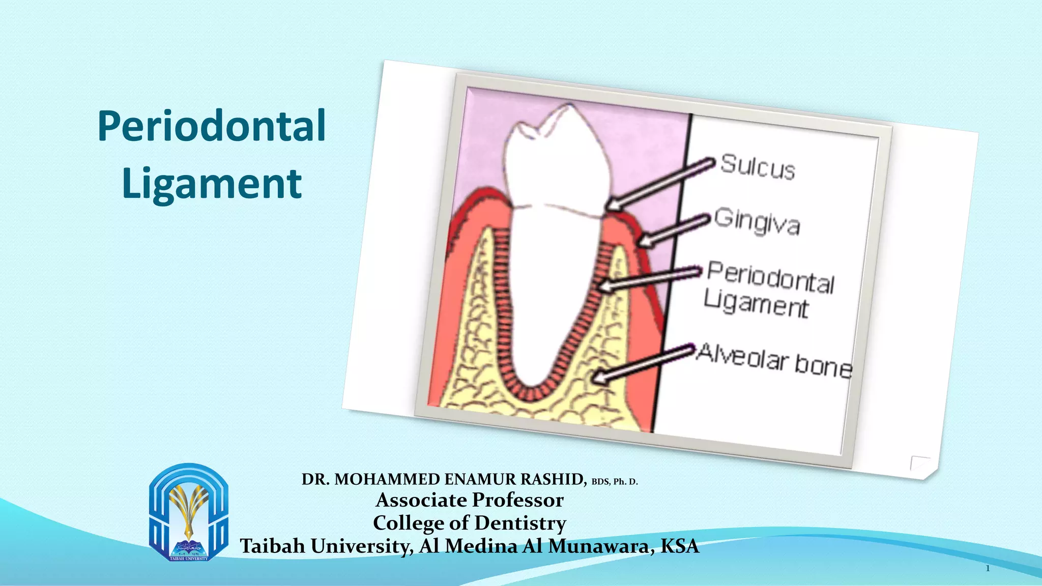
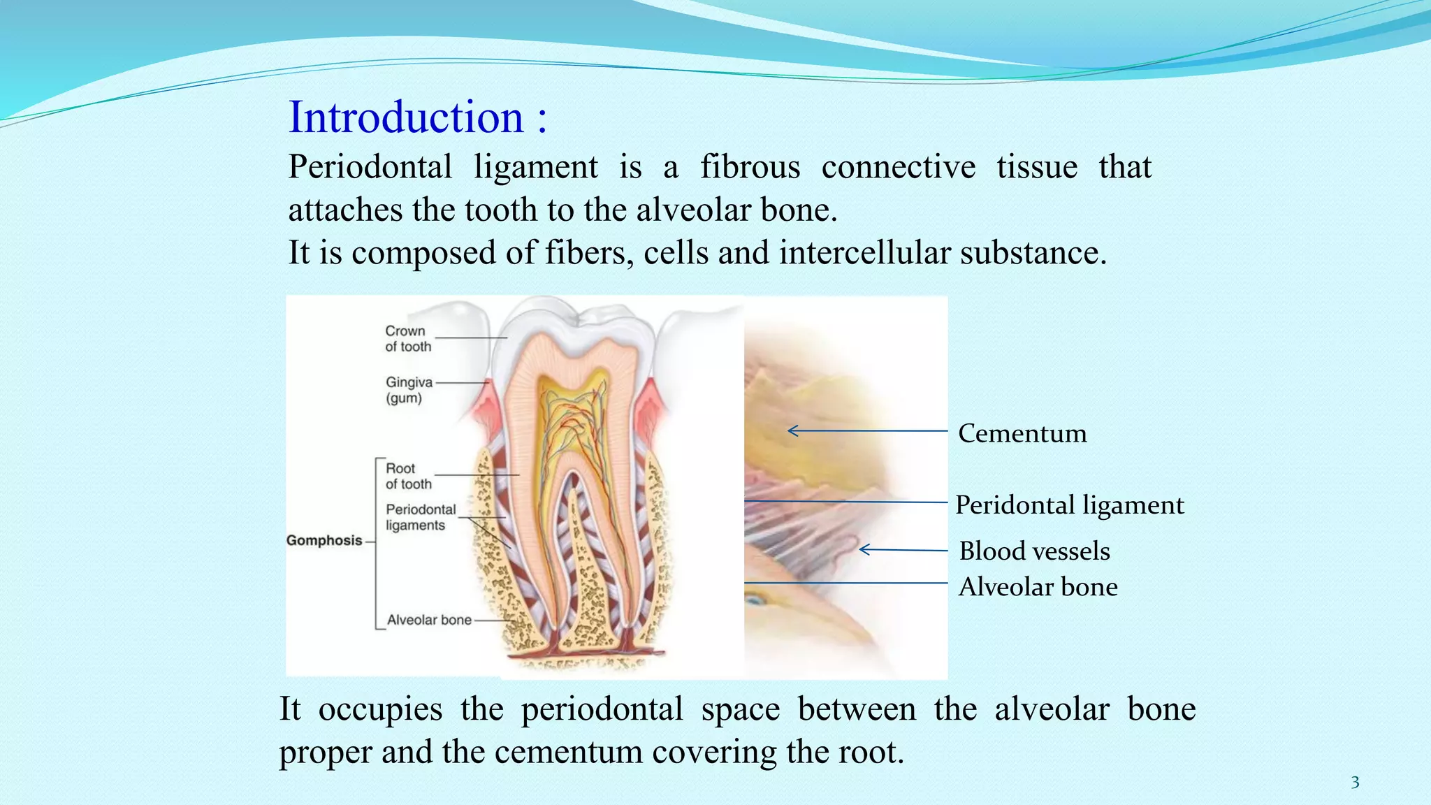
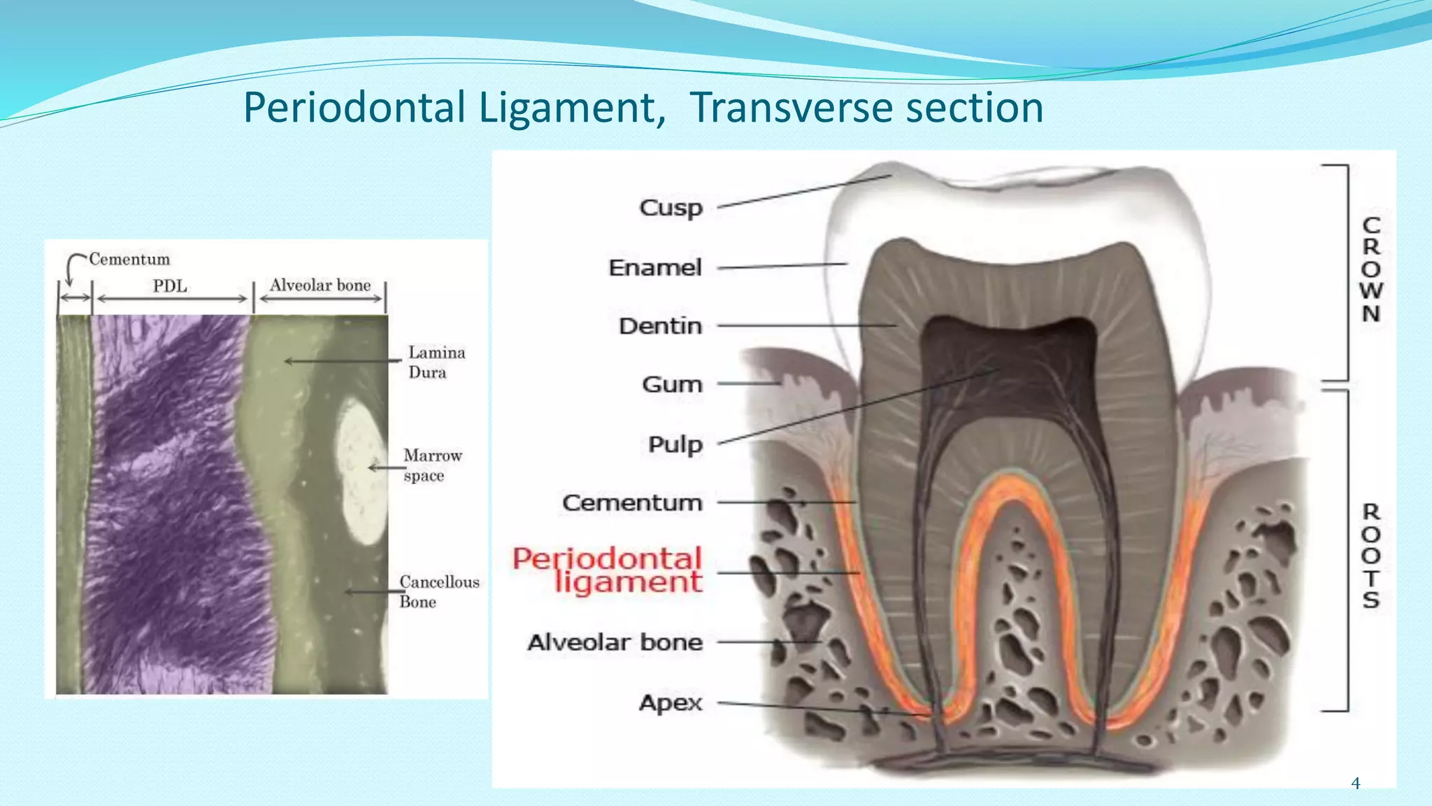
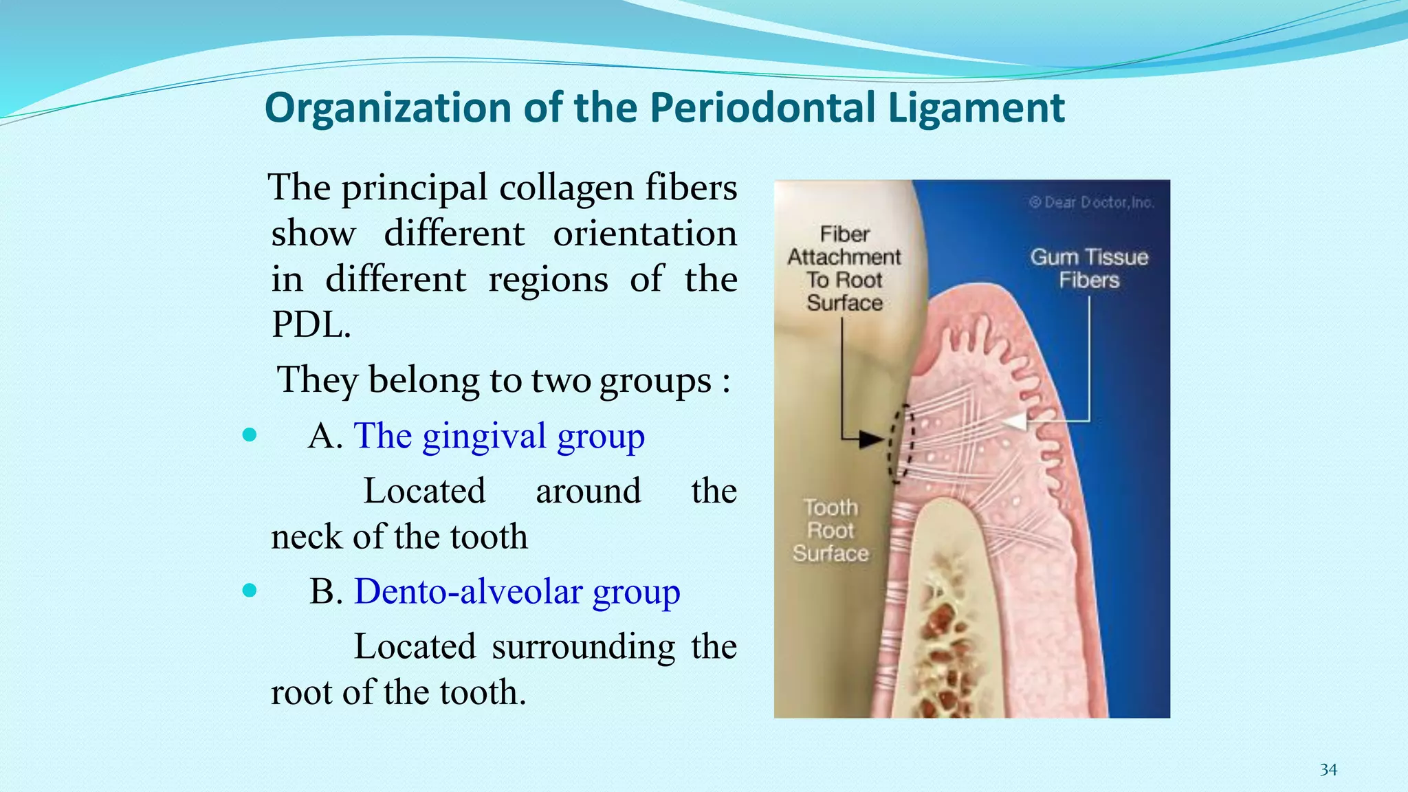
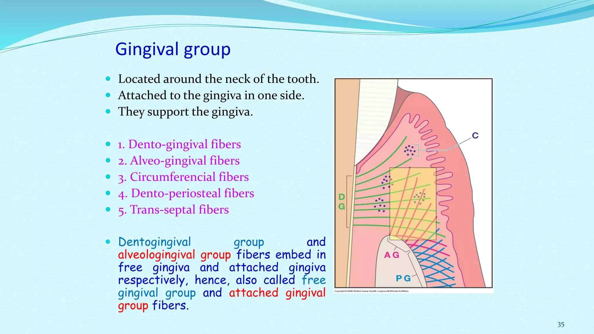
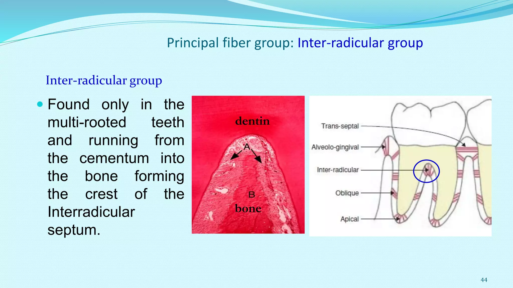
Periodontal
Ligament
DR. MOHAMMED ENAMUR RASHID, BDS, Ph. D.
Associate Professor
College of Dentistry
Taibah University, Al Medina Al Munawara, KSA
1
 Introduction
 Composition of PDL
 Organization of PDL Fibers
 Blood Supply
 Nerve Supply
 Function of PDL
 Tooth Support Mechanism by PDL
 Age Change of PDL
 Clinical Consideration
2
Peridontal ligament
Blood vessels
Alveolar bone
Cementum
Introduction :
Periodontal ligament is a fibrous connective tissue that
attaches the tooth to the alveolar bone.
It is composed of fibers, cells and intercellular substance.
It occupies the periodontal space between the alveolar bone
proper and the cementum covering the root.
3
Periodontal Ligament, Transverse section
4
The average width of the periodontal space is from 0.15-0.38 mm.
With its narrowest portion around the middle of the root.
 The width of the periodontal space varies as follows:
• At the cervical and apical regions.
• Tooth under excessive stress.
• Young tooth
• Deciduous teeth
Wide
• Around the Mid root.
• Functionless tooth
• Embedded tooth
• Aged tooth
• Permanent teeth
Narrow
average thickness of 0.22 mm
5
The PDL possesses high rate of turnover than virtually
all other connective tissues.
The half life of collagen fibers of the PDL has been
reported as being 3-23 days, but vary in different parts
of the tooth, being highest towards the root apex and
lowest towards the cervix .
6
cells Extracellular
substances
Synthetic
Resorptive
Progenitor
Defensive
Epithelial cells
Fibers
Ground substances
Blood vessels,
nerves & lymphatics
Composition of PDL
7
Histology
PDL is a fibrous CT formed of:
Cells Intercellular Substance
Synthetic cells Fibers Ground substance
Resorpative cells
Progenitor cells Collagen Elastic Proteoglycan
Epithelial cells Glycoproteins
Defense cells Glycosaminoglycans
In addition to blood vessels, nerves & lymphatics.
8
Histological Structure of PDL
 Cells:
 Connective tissue cells :
Synthetic cells : Fibroblast, Osteoblast, Cementoblast
(source : mesenchymal in origin)
Resorptive cells : Fibroblast, Osteoclast, Cementoclast
(monocyte / macrophage lineage from blood)
 Progenitor cell : Undifferentiated mesenchymal cell
 Defense cells : Macrophages, Mast cells, Eosinophils
 Epithelial cells : from epithelial rests of Malassez
9
Fibroblast are the
principal cell of PDL
and lies within the
ligament fibers.
They are aligned
along the general
direction of the fiber
bundles.
FIBROBLAST
10
FIBROBLAST
 Fibroblasts in PDL are large cell with an
extensive cytoplasm containing an
abundance of organelles (i.e. rough
endoplasmic reticulum, golgi complex,
secretory granules, mitochondria)
associated with protein synthesis and
secretion.
11
FIBROBLAST
 PDL fibroblasts are ectomesenchymal in origin and
derives from neural crest.
 Fibroblast of PDL have exceptional high rate of protein
turnover and capable of simultaneously synthesizing and
degradation of collagen.
 PDL fibroblasts have a well developed cytoskeleton with
prominent actin network.
 Shows high expression of alkaline phosphatase.
 Fibroblasts in PDL have cilia and many intercellular
contacts.*
*(which is not common in fibroblast in other connective tissue;
function of cilia is still un-known)
12
 They are found at the periodontal surface of the
alveolar bone in various stages of differentiation.
 It is an oval cell with eccentric nucleus and show
few cytoplasmic processes. Under the EM, it
shows the criteria of the protein synthetic cells.
 It is also rich in alkaline phosphatase and
pyrophosphatase enzymes which are concerned
with calcification.
Function:
-Synthesis of organic components of bone matrix to
(osteoid tissue ).
- Mineralization of bone matrix.
OSTEOBLAST
13
They are found
distributed on the
cementoid tissue of the
root surfaces in resting
state (described in the
cementum section).
CEMENTOBLAST
14
Resorptive cells :
Osteoclast and Cementoclast / Odontoclast
 Derives from blood cells of
Macrophage type.
 These cells are having same
cytoplasmic features and are
actively involved in resorption
process.
 Osteoclasts / odontoclast show
considerable variation in size and
shape : ranging from mononuclear
to multinuclear cells.
15
Osteoclast and Cementoclast / Odontoclast
 Surface of the alveolar
bone is lined by the
osteoblast & osteoclast.
 Surface of the cementum
is lined by cementoblast &
cementoclast.
 A: Alveolar bone B : Cementum
16
Osteoclast and Cementoclast / Odontoclast
 The resorption concavities
on the (alveolar) bone
surface is termed as
Howship’s lacinae.
17
Osteoclast and Cementoclast / Odontoclast
 The part of the cell that
lies adjacent to bone
has striated appearance
: so called Brush border.
 The brush border
comprises of many
tightly packed micro-villi,
coated with fine, bristle
like structures.
18
Progenitor cell :
Undifferentiated mesenchymal cell
 These are large, polyhedral cells with large, lightly
stained, centrally placed nucleus.
 Present abundant cytoplasm with peripheral cytoplasmic
extension.
 These cells may give
rise to osteoblasts
and cementoblasts.
19
Defense cell
 Defense cells in PDL includes :
Macrophages, Mast cells and
Eosonophills.
 Monocyte / Macrophage cells
constitute about 4% of the cell
population in PDL and located near
the blood vessels.
 Monocytes are oval shaped, ruffle
bordered cell, deriving from blood
vessels.
 Macrophages are derived from
monocytes.
20
Defense cell : Macrophage
 Macrophages are responsible
for phagocytosis of particulate
matter and invading organism.
 Also responsible for
synthesizing interferon
(antiviral), prostaglandin and
factors that enhance the growth
of fibroblast and endothelial
cells.
21
Defense cell : Mast cell
 Mast cells are associated with blood vessels and
produces Histamin, Heparin and factors associated
with anaphylaxis.
 Eosonophils are occasionally seen in the normal
PDL.
 Neutrophil and lymphocytes are also seen in PDL
under inflammatory condition.
22
Epithelial cell rests of Malassez
 Epithelial rest are normal
constituents of the PDL and seen
throughout the life.
 Each rest is an aggregation of
epithelial cells : some 4-6 in
number, closely packed cuboidal
cells and deeply stained than
adjacent fibroblast.
 The cells are believed to be
originated from the remnants of
Hertwig’s root sheath.
23
Epithelial cell rests of Malassez
 Distribution site:
 Scattered throughout the ligament.
 Up-to 20 years, more prevalent in the apical zone.
 From the third decay, majority of the cell rests are located
cervically in the gingiva above the alveolar crest.
 Clinical importance :
 Under chronic inflammation, they may proliferate to
form cysts or tumour.
24
25
Histological Structure of PDL
 Fibres:
 Connective tissue fibers are mainly collagenous (more
than 90%), but there is also small amount of Oxytalan and
Reticulin fibres.
 Main types of collagen fibers in the PDL are :
 Type I (about 80%)
 Type III (about 15%)
 (Type III collagen is not localized to any specific region of PDL, rather it is
covalently linked to type I collagen throughout the tissue)
 Type XII
(fibrils associated collagen, links other collagen together)
26
Histological Structure of PDL : Fibers
 Small amounts of :
 Type V, Type VI
(Type V : coats the cell surface
and other fibers)
(Type VI : Part of oxytallan fiber system
and promote proliferation of Fibroblast)
 Trace amount of :
 Type IV, Type VII
 (associated with epithelial cell
rests and blood vessels)
27
Histological Structure of PDL : Fiber
 Most collagen fibrils in the
PDL are arranged in
definite and distinct fiber
bundles.
 Each bundle resembles a
spliced rope.
 Individual strands can be
remodeled continuously
whereas overall fibers
maintains its architecture
and function.
28
II- Elastic fibers:
There are three types of elastic fibers : elastin,
oxytalan, and elaunin.
There is no elastic fibers in the human PDL
except around the walls of blood vessels.
However, the PDL of some animals may
contain such fibers.
Only oxytalan are present within PDL; however,
elaunin fibers (a variant of elastic fibers ) may be
found within fibers of gingival ligament (form a
meshwork extending from cementum to bone sheathing
the collagen fiber bundles.)
29
Insertion of PDL fiber into Bone & Cementum
 Sharpey’s Fiber :
 The principal fibres of the periodontal ligament that
are embeded into cementum and the alveolar bone
lining the tooth socket are termed as Sharpey’s Fiber.
A. alveolar bone B. cementum
30
 PDL is rich in ground substance.
 Even collagen fiber bundles are composed of mainly
ground substance (about 60%).
 70 % of the ground substance is water.
 Other constituents are:
 Glycose aminoglycans, Proteo-glycans and
Glycoprotiens
 All these are secreted by fibroblast.
Ground Substance
31
Ground Substance
 Function of ground
substance:
 Ion and water binding and
exchange.
 Control of collagen fibrino-
genesis and fiber
orientation.
 Binding of growth factors.
32
Interstitial tissue:
Between the bundles of the
oblique fibers there are round or
oval intervals where the
periodontal tissue is much less
dense.
In these areas it forms a reticular
framework of loose connective
tissue through which the blood
vessels, nerves and lymphatic run.
These are termed interstitial
tissue.
33
Organization of the Periodontal Ligament
The principal collagen fibers
show different orientation
in different regions of the
PDL.
They belong to two groups :
 A. The gingival group
Located around the
neck of the tooth
 B. Dento-alveolar group
Located surrounding the
root of the tooth.
34
Gingival group
 Located around the neck of the tooth.
 Attached to the gingiva in one side.
 They support the gingiva.
 1. Dento-gingival fibers
 2. Alveo-gingival fibers
 3. Circumferencial fibers
 4. Dento-periosteal fibers
 5. Trans-septal fibers
 Dentogingival group and
alveologingival group fibers embed in
free gingiva and attached gingiva
respectively, hence, also called free
gingival group and attached gingival
group fibers.
35
Organization of the PDL: Gingival Fibers
 1. Dento-Gingival Fibers :
Most numerous.
Arise from the surface of the
cementum in the cervical
region and pass into the
lamina propia of free gingiva.
2. Alveo-Gingival Fibers :
Arise from the alveolar crest
and pass into the lamina
propia of attached gingiva.
36
Organization of the PDL: Gingival Fibers
3. Circumferential
or Circular
Fibers:
These fibers are
continuous
around the neck
of the tooth and
resist gingival
displacement.
 4. Dentoperiosteul fiber:
 Runs apically from cementum over
the periosteum of the outer
cortical plates of the alveolar
process.
 Inserts into the alveolar process, or
the vestibular muscle and floor of
the mouth.
37
Organization of the PDL: Gingival Fibers
5. Trans-septal Fibers :
They originate in the
cervical cementum of each
tooth and extend to the similar
location on the mesial or distal
surface of the adjacent tooth.
38
Organization of the Periodontal Ligament :
PRINCIPAL FIBER GROUP
39
Principal Fiber bundle: The alveolar crest group
 These are attached to
the cementum just
below the cemento-
enamel junction and
running downward and
outward to insert into
the rim of the
alveolus.
40
Principal Fiber bundle: Horizontal Group
 These are just apical to
the alveolar crest fibers
and running at right
angles to the long axis of
the tooth from the
cementum to the bone
below the alveolar crest.
Horizontal
Group
41
Principal Fiber bundle: Oblique Group
 They are the most
numerous in the
PDL and running
from the cementum
in an oblique
direction to insert
into the bone
coronally.
c B
B : Bone , C: Cementum
42
Principal Fiber bundle : Apical group
 These are radiating from the
cementum around the apex of
the root to the bone forming the
base of the socket.
Apical group
dentin
bone
43
Principal fiber group: Inter-radicular group
 Found only in the
multi-rooted teeth
and running from
the cementum into
the bone forming
the crest of the
Interradicular
septum.
Inter-radicular group
dentin
bone
44
Organization of the Periodontal Ligament
 Eruptive tooth movement
modify the attachment of
the principal fiber bundles
of PDL.
 When the tooth erupts,
the crest of the alveolar
bone is above the CEJ
junction and the
developing fibers are
directed obliquely.
45
Organization of the Periodontal Ligament
 When the tooth moves
during eruption, the level of
the of the alveolar crest
coincides with the CEJ
junction and the oblique
fibers become horizontally
aligned.
 When the tooth finally comes
in function, the alveolar crest
fibers is positioned nearer
the apex.
46
Organization of the Periodontal Ligament
 The horizontal fibers termed as
alveolar crest fibers have
become oblique once again but
with the difference that now the
CEJ has reversed its relation to
the alveolar attachment and is
positioned in a coronal direction.
 Only after the teeth come into
function, the fiber bundles of the
PDL thicken appreciably.
47
Organization of the Periodontal Ligament
Development of the Principal fiber groups: Alveolar crest fiber in A,
Initially oblique in B, then horizontal in C and finally again oblique in D.
48
49
Interstitial space
There are spaces between the fibre bundles called the
interstitial spaces.
The interstitial spaces contain blood vessels, lymph
channels, and nerves.
These structures help maintain the vitality of the
periodontal ligament.
50
Blood Supply of Periodontal Ligament
Source:
1. Branches from Superior
and inferior alveolar
arteries.
2. Branches from gingival
vessels entering from a
coronal direction.
51
Blood Supply of Periodontal Ligament
 The main blood supply of PDL is from the
superior and inferior alveolar artery.
 The arteries supplying the PDL are not
derived from the arteries entering into the
pulp at the apex, rather
 They arise from a series of perforating
arteries passing through the alveolar bone.
 Branches from gingival vessels entering
from a coronal direction.
 This distinct source of blood supply allows
PDL to function following Endodontic
treatment.
52
Blood Supply of Periodontal Ligament
 Distribution :
 The superior and inferior alveolar artery pursue an
intra-osteal course and give off alveolar branches
that ascend within the bone as intra-alveolar arteries.
 Numerous branches arise from the intra-alveolar
vessels to run horizontally, penetrate the alveolar
bone and enter the PDL space as perforating
arteries.
 Within the PDL, the arteries occupy the areas of
loose connective tissue called interstitial space
between the principal fiber bundle and runs into both
apical and occlusal direction with numerous
transverse connection.
53
Blood Supply of Periodontal Ligament
 Perforating arteries are more abundant in the PDL of
posterior teeth than anterior teeth and greater
number in mandibular teeth than maxillary teeth.
 In single rooted tooth, these arteries are found most
frequently in the gingival third of the ligament
followed by the apical third.
 This pattern of distribution have clinical importance :
in healing extraction wounds, new tissue invade from
the perforation site.
 Formation of blood clot occupying the socket is more
rapid in the gingival and apical area.
54
Blood Supply of Periodontal Ligament
 Special feature of
periodontal ligament
vasculature are:
 1. They form a crevicular
plexus of capillary loop
that completely encircles
the tooth.
 2. Presence of large
number of fenestrations in
the capillaries (which is
unusual in fibrous connective
tissue).
These special features of the vasculature is related with the high metabolic
requirements of the periodontal ligament (i.e. high rate of turn over)
55
Nerve Supply of Periodontal Ligament
 Source : The nerve fibers
entering into the periodontal
ligament are derived from two
sources:
 1. Some nerve bundles enter the
periodontal space near the root
apex and pass up through the
periodontal ligament.
 2. Others enter the middle and
cervical portions of the ligament
as finer branches through the
openings in the alveolar walls.
56
Nerve Supply of Periodontal Ligament
57
Nerve Supply of Periodontal Ligament
 Types of nerve fiber :
 Functionally two types: Sensory and Autonomic
 Sensory :
 Associated with nociception and Mechanoreception.
 Myelinated, about 5 µ m. in diameter
 Un-myelinated, about 0.5 µ m. in diameter
 Autonomic :
 Associated with the supply of periodontal blood vessels.
 only un-myelinated fibers
58
Nerve Supply of Periodontal Ligament : Nerve Terminals
 The four types of nerve
endings found in the
human periodontal
ligament:
 A. Free nerve endings
(with tree like ramification)
 B. Ruffini’s endings
 C. Coiled endings
 D. Spindle type endings
(encapsulated)
59
Nerve Terminals : Free Nerve Endings
 1. The first and most frequent are
Free nerve endings that ramify
into a treelike configuration.
 They originate from the small,
unmyelinated fibres and carry with
them a schwann cell layer.
 They are located at regular
intervals along the length of the
root.
 This nerve terminals are thought to
be nociceptors or
mechanoreceptors and are
associated with pain.
60
Nerve Terminals : Ruffini,s corpuscles
 The large fibers end in a
variety of endings as
following:
 2.This type of nerve terminal
is found around the root apex
and resembles Ruffini’s
corpuscles. They appear
dendritic and end in terminal
expansions among the PDL
fiber bundles.
61
Nerve Terminals : Coiled
 3. The third type of nerve
terminal is a coiled
form.
 Found in the mid region
of the PDL.
 It’s function and
ultrastructures has not
yet been determined.
62
Nerve Terminals : Spindlelike
 4. The final and the lowest
frequency type consists of a
spindlelike endings
sorounded by a fibrous
capsule.
 This type of nerve terminal
is associated with the root
apex.
 The large diameter fibers
appear to be concerned with
discernment of pressure.
63
Functions of Periodontal Ligament
 A . Supportive
 1. The most important function of the periodontal
ligament is to attach the tooth into it’s socket and maintain
it’s functional position.
2. PDL permits teeth to withstand the considerable forces
of mastication.
3. During orthodontic tooth movement, periodontal
ligament responds as a co-ordinated unit and support the
tooth particularly during the recovery response after
loading.
4. It helps in the mechanism of tooth eruption.
64
Functions of Periodontal Ligament
 B. Sensory
 The mechanoreceptors present in the periodontal
ligament are involved in the neurological control of
mastication.
 The periodontal ligament is supplied with abundant
receptors and nerves that sense any movement of tooth
during function. When receptors sense any pressure, the
nerves send signals to the brain which activates the
masticatory apparatus and helps in the proper
positioning of the jaws during normal function.
65
Functions of Periodontal Ligament
C. Nutritive
 The blood vessels of the ligament provide the essential
nutrients for the ligament’s vitality and for the hard tissue
of cementum and alveolar bone. All the cells of the
periodontal ligament are also getting nutrients that is
carried out by the blood vessels of the ligament.
D. Protective
It absorbs the masticatory load and resist the displacing
forces and thereby protect the dental tissues from damage
caused by excessive occlusal loads.
E. Maintenance
 The cells in the periodontal ligament form, maintain and
repair the alveolar bone and cementum.
66
Tooth support mechanism by PDL
 Conventionally, it is believed that the PDL behaves as a
“suspensory ligament”during masticatory load.
 Oblique principal fibers dissipates the masticatory
load to the alveolar bone.
 Mechanism of tooth support should not be regarded as
a property of a single component, rather it should be
regarded as function of the tissue as a whole.
 Evidences from researches show that the collagen
fibers, vasculature and ground substance of the PDL,
all are involved in tooth support.
67
Age change of PDL
 Ageing alone leads to no critical loss of the periodontal
attachment in the healthy elderly person.
 In general number of fibroblast decreases and there is
decreased organic matrix production.
 Due to systemic endocrine alterations in the elderly,
the bio-molecular changes of the cells of periodontium
exacerbate bone loss in elderly patients with
periodontitis.
68
Clinical consideration
 Guided Tissue Regeneration :
 Following periodontal surgery, a blood clot is formed.
 To exclude the gingival tissue repopulation, some sort of
tissue barrier is placed over the alveolar crest.
This is called “guided tissue regeneration”.
 During orthodontic treatment, under controlled pressure,
there is resorption of the alveolar bone on the pressure side
and deposition of bone on the tension side.
 Uncontrolled large pressure may produce degenerative
changes, resulting into “hyalinization” (glassy appearance) of
PDL.
 In hyalinization, osteoclast is absent, there is oedema and
obliteration of blood vessels.
69
THANK YOU!!
70

Periodontal ligament

  • 1.
    Periodontal Ligament DR. MOHAMMED ENAMURRASHID, BDS, Ph. D. Associate Professor College of Dentistry Taibah University, Al Medina Al Munawara, KSA 1
  • 2.
     Introduction  Compositionof PDL  Organization of PDL Fibers  Blood Supply  Nerve Supply  Function of PDL  Tooth Support Mechanism by PDL  Age Change of PDL  Clinical Consideration 2
  • 3.
    Peridontal ligament Blood vessels Alveolarbone Cementum Introduction : Periodontal ligament is a fibrous connective tissue that attaches the tooth to the alveolar bone. It is composed of fibers, cells and intercellular substance. It occupies the periodontal space between the alveolar bone proper and the cementum covering the root. 3
  • 4.
  • 5.
    The average widthof the periodontal space is from 0.15-0.38 mm. With its narrowest portion around the middle of the root.  The width of the periodontal space varies as follows: • At the cervical and apical regions. • Tooth under excessive stress. • Young tooth • Deciduous teeth Wide • Around the Mid root. • Functionless tooth • Embedded tooth • Aged tooth • Permanent teeth Narrow average thickness of 0.22 mm 5
  • 6.
    The PDL possesseshigh rate of turnover than virtually all other connective tissues. The half life of collagen fibers of the PDL has been reported as being 3-23 days, but vary in different parts of the tooth, being highest towards the root apex and lowest towards the cervix . 6
  • 7.
  • 8.
    Histology PDL is afibrous CT formed of: Cells Intercellular Substance Synthetic cells Fibers Ground substance Resorpative cells Progenitor cells Collagen Elastic Proteoglycan Epithelial cells Glycoproteins Defense cells Glycosaminoglycans In addition to blood vessels, nerves & lymphatics. 8
  • 9.
    Histological Structure ofPDL  Cells:  Connective tissue cells : Synthetic cells : Fibroblast, Osteoblast, Cementoblast (source : mesenchymal in origin) Resorptive cells : Fibroblast, Osteoclast, Cementoclast (monocyte / macrophage lineage from blood)  Progenitor cell : Undifferentiated mesenchymal cell  Defense cells : Macrophages, Mast cells, Eosinophils  Epithelial cells : from epithelial rests of Malassez 9
  • 10.
    Fibroblast are the principalcell of PDL and lies within the ligament fibers. They are aligned along the general direction of the fiber bundles. FIBROBLAST 10
  • 11.
    FIBROBLAST  Fibroblasts inPDL are large cell with an extensive cytoplasm containing an abundance of organelles (i.e. rough endoplasmic reticulum, golgi complex, secretory granules, mitochondria) associated with protein synthesis and secretion. 11
  • 12.
    FIBROBLAST  PDL fibroblastsare ectomesenchymal in origin and derives from neural crest.  Fibroblast of PDL have exceptional high rate of protein turnover and capable of simultaneously synthesizing and degradation of collagen.  PDL fibroblasts have a well developed cytoskeleton with prominent actin network.  Shows high expression of alkaline phosphatase.  Fibroblasts in PDL have cilia and many intercellular contacts.* *(which is not common in fibroblast in other connective tissue; function of cilia is still un-known) 12
  • 13.
     They arefound at the periodontal surface of the alveolar bone in various stages of differentiation.  It is an oval cell with eccentric nucleus and show few cytoplasmic processes. Under the EM, it shows the criteria of the protein synthetic cells.  It is also rich in alkaline phosphatase and pyrophosphatase enzymes which are concerned with calcification. Function: -Synthesis of organic components of bone matrix to (osteoid tissue ). - Mineralization of bone matrix. OSTEOBLAST 13
  • 14.
    They are found distributedon the cementoid tissue of the root surfaces in resting state (described in the cementum section). CEMENTOBLAST 14
  • 15.
    Resorptive cells : Osteoclastand Cementoclast / Odontoclast  Derives from blood cells of Macrophage type.  These cells are having same cytoplasmic features and are actively involved in resorption process.  Osteoclasts / odontoclast show considerable variation in size and shape : ranging from mononuclear to multinuclear cells. 15
  • 16.
    Osteoclast and Cementoclast/ Odontoclast  Surface of the alveolar bone is lined by the osteoblast & osteoclast.  Surface of the cementum is lined by cementoblast & cementoclast.  A: Alveolar bone B : Cementum 16
  • 17.
    Osteoclast and Cementoclast/ Odontoclast  The resorption concavities on the (alveolar) bone surface is termed as Howship’s lacinae. 17
  • 18.
    Osteoclast and Cementoclast/ Odontoclast  The part of the cell that lies adjacent to bone has striated appearance : so called Brush border.  The brush border comprises of many tightly packed micro-villi, coated with fine, bristle like structures. 18
  • 19.
    Progenitor cell : Undifferentiatedmesenchymal cell  These are large, polyhedral cells with large, lightly stained, centrally placed nucleus.  Present abundant cytoplasm with peripheral cytoplasmic extension.  These cells may give rise to osteoblasts and cementoblasts. 19
  • 20.
    Defense cell  Defensecells in PDL includes : Macrophages, Mast cells and Eosonophills.  Monocyte / Macrophage cells constitute about 4% of the cell population in PDL and located near the blood vessels.  Monocytes are oval shaped, ruffle bordered cell, deriving from blood vessels.  Macrophages are derived from monocytes. 20
  • 21.
    Defense cell :Macrophage  Macrophages are responsible for phagocytosis of particulate matter and invading organism.  Also responsible for synthesizing interferon (antiviral), prostaglandin and factors that enhance the growth of fibroblast and endothelial cells. 21
  • 22.
    Defense cell :Mast cell  Mast cells are associated with blood vessels and produces Histamin, Heparin and factors associated with anaphylaxis.  Eosonophils are occasionally seen in the normal PDL.  Neutrophil and lymphocytes are also seen in PDL under inflammatory condition. 22
  • 23.
    Epithelial cell restsof Malassez  Epithelial rest are normal constituents of the PDL and seen throughout the life.  Each rest is an aggregation of epithelial cells : some 4-6 in number, closely packed cuboidal cells and deeply stained than adjacent fibroblast.  The cells are believed to be originated from the remnants of Hertwig’s root sheath. 23
  • 24.
    Epithelial cell restsof Malassez  Distribution site:  Scattered throughout the ligament.  Up-to 20 years, more prevalent in the apical zone.  From the third decay, majority of the cell rests are located cervically in the gingiva above the alveolar crest.  Clinical importance :  Under chronic inflammation, they may proliferate to form cysts or tumour. 24
  • 25.
  • 26.
    Histological Structure ofPDL  Fibres:  Connective tissue fibers are mainly collagenous (more than 90%), but there is also small amount of Oxytalan and Reticulin fibres.  Main types of collagen fibers in the PDL are :  Type I (about 80%)  Type III (about 15%)  (Type III collagen is not localized to any specific region of PDL, rather it is covalently linked to type I collagen throughout the tissue)  Type XII (fibrils associated collagen, links other collagen together) 26
  • 27.
    Histological Structure ofPDL : Fibers  Small amounts of :  Type V, Type VI (Type V : coats the cell surface and other fibers) (Type VI : Part of oxytallan fiber system and promote proliferation of Fibroblast)  Trace amount of :  Type IV, Type VII  (associated with epithelial cell rests and blood vessels) 27
  • 28.
    Histological Structure ofPDL : Fiber  Most collagen fibrils in the PDL are arranged in definite and distinct fiber bundles.  Each bundle resembles a spliced rope.  Individual strands can be remodeled continuously whereas overall fibers maintains its architecture and function. 28
  • 29.
    II- Elastic fibers: Thereare three types of elastic fibers : elastin, oxytalan, and elaunin. There is no elastic fibers in the human PDL except around the walls of blood vessels. However, the PDL of some animals may contain such fibers. Only oxytalan are present within PDL; however, elaunin fibers (a variant of elastic fibers ) may be found within fibers of gingival ligament (form a meshwork extending from cementum to bone sheathing the collagen fiber bundles.) 29
  • 30.
    Insertion of PDLfiber into Bone & Cementum  Sharpey’s Fiber :  The principal fibres of the periodontal ligament that are embeded into cementum and the alveolar bone lining the tooth socket are termed as Sharpey’s Fiber. A. alveolar bone B. cementum 30
  • 31.
     PDL isrich in ground substance.  Even collagen fiber bundles are composed of mainly ground substance (about 60%).  70 % of the ground substance is water.  Other constituents are:  Glycose aminoglycans, Proteo-glycans and Glycoprotiens  All these are secreted by fibroblast. Ground Substance 31
  • 32.
    Ground Substance  Functionof ground substance:  Ion and water binding and exchange.  Control of collagen fibrino- genesis and fiber orientation.  Binding of growth factors. 32
  • 33.
    Interstitial tissue: Between thebundles of the oblique fibers there are round or oval intervals where the periodontal tissue is much less dense. In these areas it forms a reticular framework of loose connective tissue through which the blood vessels, nerves and lymphatic run. These are termed interstitial tissue. 33
  • 34.
    Organization of thePeriodontal Ligament The principal collagen fibers show different orientation in different regions of the PDL. They belong to two groups :  A. The gingival group Located around the neck of the tooth  B. Dento-alveolar group Located surrounding the root of the tooth. 34
  • 35.
    Gingival group  Locatedaround the neck of the tooth.  Attached to the gingiva in one side.  They support the gingiva.  1. Dento-gingival fibers  2. Alveo-gingival fibers  3. Circumferencial fibers  4. Dento-periosteal fibers  5. Trans-septal fibers  Dentogingival group and alveologingival group fibers embed in free gingiva and attached gingiva respectively, hence, also called free gingival group and attached gingival group fibers. 35
  • 36.
    Organization of thePDL: Gingival Fibers  1. Dento-Gingival Fibers : Most numerous. Arise from the surface of the cementum in the cervical region and pass into the lamina propia of free gingiva. 2. Alveo-Gingival Fibers : Arise from the alveolar crest and pass into the lamina propia of attached gingiva. 36
  • 37.
    Organization of thePDL: Gingival Fibers 3. Circumferential or Circular Fibers: These fibers are continuous around the neck of the tooth and resist gingival displacement.  4. Dentoperiosteul fiber:  Runs apically from cementum over the periosteum of the outer cortical plates of the alveolar process.  Inserts into the alveolar process, or the vestibular muscle and floor of the mouth. 37
  • 38.
    Organization of thePDL: Gingival Fibers 5. Trans-septal Fibers : They originate in the cervical cementum of each tooth and extend to the similar location on the mesial or distal surface of the adjacent tooth. 38
  • 39.
    Organization of thePeriodontal Ligament : PRINCIPAL FIBER GROUP 39
  • 40.
    Principal Fiber bundle:The alveolar crest group  These are attached to the cementum just below the cemento- enamel junction and running downward and outward to insert into the rim of the alveolus. 40
  • 41.
    Principal Fiber bundle:Horizontal Group  These are just apical to the alveolar crest fibers and running at right angles to the long axis of the tooth from the cementum to the bone below the alveolar crest. Horizontal Group 41
  • 42.
    Principal Fiber bundle:Oblique Group  They are the most numerous in the PDL and running from the cementum in an oblique direction to insert into the bone coronally. c B B : Bone , C: Cementum 42
  • 43.
    Principal Fiber bundle: Apical group  These are radiating from the cementum around the apex of the root to the bone forming the base of the socket. Apical group dentin bone 43
  • 44.
    Principal fiber group:Inter-radicular group  Found only in the multi-rooted teeth and running from the cementum into the bone forming the crest of the Interradicular septum. Inter-radicular group dentin bone 44
  • 45.
    Organization of thePeriodontal Ligament  Eruptive tooth movement modify the attachment of the principal fiber bundles of PDL.  When the tooth erupts, the crest of the alveolar bone is above the CEJ junction and the developing fibers are directed obliquely. 45
  • 46.
    Organization of thePeriodontal Ligament  When the tooth moves during eruption, the level of the of the alveolar crest coincides with the CEJ junction and the oblique fibers become horizontally aligned.  When the tooth finally comes in function, the alveolar crest fibers is positioned nearer the apex. 46
  • 47.
    Organization of thePeriodontal Ligament  The horizontal fibers termed as alveolar crest fibers have become oblique once again but with the difference that now the CEJ has reversed its relation to the alveolar attachment and is positioned in a coronal direction.  Only after the teeth come into function, the fiber bundles of the PDL thicken appreciably. 47
  • 48.
    Organization of thePeriodontal Ligament Development of the Principal fiber groups: Alveolar crest fiber in A, Initially oblique in B, then horizontal in C and finally again oblique in D. 48
  • 49.
  • 50.
    Interstitial space There arespaces between the fibre bundles called the interstitial spaces. The interstitial spaces contain blood vessels, lymph channels, and nerves. These structures help maintain the vitality of the periodontal ligament. 50
  • 51.
    Blood Supply ofPeriodontal Ligament Source: 1. Branches from Superior and inferior alveolar arteries. 2. Branches from gingival vessels entering from a coronal direction. 51
  • 52.
    Blood Supply ofPeriodontal Ligament  The main blood supply of PDL is from the superior and inferior alveolar artery.  The arteries supplying the PDL are not derived from the arteries entering into the pulp at the apex, rather  They arise from a series of perforating arteries passing through the alveolar bone.  Branches from gingival vessels entering from a coronal direction.  This distinct source of blood supply allows PDL to function following Endodontic treatment. 52
  • 53.
    Blood Supply ofPeriodontal Ligament  Distribution :  The superior and inferior alveolar artery pursue an intra-osteal course and give off alveolar branches that ascend within the bone as intra-alveolar arteries.  Numerous branches arise from the intra-alveolar vessels to run horizontally, penetrate the alveolar bone and enter the PDL space as perforating arteries.  Within the PDL, the arteries occupy the areas of loose connective tissue called interstitial space between the principal fiber bundle and runs into both apical and occlusal direction with numerous transverse connection. 53
  • 54.
    Blood Supply ofPeriodontal Ligament  Perforating arteries are more abundant in the PDL of posterior teeth than anterior teeth and greater number in mandibular teeth than maxillary teeth.  In single rooted tooth, these arteries are found most frequently in the gingival third of the ligament followed by the apical third.  This pattern of distribution have clinical importance : in healing extraction wounds, new tissue invade from the perforation site.  Formation of blood clot occupying the socket is more rapid in the gingival and apical area. 54
  • 55.
    Blood Supply ofPeriodontal Ligament  Special feature of periodontal ligament vasculature are:  1. They form a crevicular plexus of capillary loop that completely encircles the tooth.  2. Presence of large number of fenestrations in the capillaries (which is unusual in fibrous connective tissue). These special features of the vasculature is related with the high metabolic requirements of the periodontal ligament (i.e. high rate of turn over) 55
  • 56.
    Nerve Supply ofPeriodontal Ligament  Source : The nerve fibers entering into the periodontal ligament are derived from two sources:  1. Some nerve bundles enter the periodontal space near the root apex and pass up through the periodontal ligament.  2. Others enter the middle and cervical portions of the ligament as finer branches through the openings in the alveolar walls. 56
  • 57.
    Nerve Supply ofPeriodontal Ligament 57
  • 58.
    Nerve Supply ofPeriodontal Ligament  Types of nerve fiber :  Functionally two types: Sensory and Autonomic  Sensory :  Associated with nociception and Mechanoreception.  Myelinated, about 5 µ m. in diameter  Un-myelinated, about 0.5 µ m. in diameter  Autonomic :  Associated with the supply of periodontal blood vessels.  only un-myelinated fibers 58
  • 59.
    Nerve Supply ofPeriodontal Ligament : Nerve Terminals  The four types of nerve endings found in the human periodontal ligament:  A. Free nerve endings (with tree like ramification)  B. Ruffini’s endings  C. Coiled endings  D. Spindle type endings (encapsulated) 59
  • 60.
    Nerve Terminals :Free Nerve Endings  1. The first and most frequent are Free nerve endings that ramify into a treelike configuration.  They originate from the small, unmyelinated fibres and carry with them a schwann cell layer.  They are located at regular intervals along the length of the root.  This nerve terminals are thought to be nociceptors or mechanoreceptors and are associated with pain. 60
  • 61.
    Nerve Terminals :Ruffini,s corpuscles  The large fibers end in a variety of endings as following:  2.This type of nerve terminal is found around the root apex and resembles Ruffini’s corpuscles. They appear dendritic and end in terminal expansions among the PDL fiber bundles. 61
  • 62.
    Nerve Terminals :Coiled  3. The third type of nerve terminal is a coiled form.  Found in the mid region of the PDL.  It’s function and ultrastructures has not yet been determined. 62
  • 63.
    Nerve Terminals :Spindlelike  4. The final and the lowest frequency type consists of a spindlelike endings sorounded by a fibrous capsule.  This type of nerve terminal is associated with the root apex.  The large diameter fibers appear to be concerned with discernment of pressure. 63
  • 64.
    Functions of PeriodontalLigament  A . Supportive  1. The most important function of the periodontal ligament is to attach the tooth into it’s socket and maintain it’s functional position. 2. PDL permits teeth to withstand the considerable forces of mastication. 3. During orthodontic tooth movement, periodontal ligament responds as a co-ordinated unit and support the tooth particularly during the recovery response after loading. 4. It helps in the mechanism of tooth eruption. 64
  • 65.
    Functions of PeriodontalLigament  B. Sensory  The mechanoreceptors present in the periodontal ligament are involved in the neurological control of mastication.  The periodontal ligament is supplied with abundant receptors and nerves that sense any movement of tooth during function. When receptors sense any pressure, the nerves send signals to the brain which activates the masticatory apparatus and helps in the proper positioning of the jaws during normal function. 65
  • 66.
    Functions of PeriodontalLigament C. Nutritive  The blood vessels of the ligament provide the essential nutrients for the ligament’s vitality and for the hard tissue of cementum and alveolar bone. All the cells of the periodontal ligament are also getting nutrients that is carried out by the blood vessels of the ligament. D. Protective It absorbs the masticatory load and resist the displacing forces and thereby protect the dental tissues from damage caused by excessive occlusal loads. E. Maintenance  The cells in the periodontal ligament form, maintain and repair the alveolar bone and cementum. 66
  • 67.
    Tooth support mechanismby PDL  Conventionally, it is believed that the PDL behaves as a “suspensory ligament”during masticatory load.  Oblique principal fibers dissipates the masticatory load to the alveolar bone.  Mechanism of tooth support should not be regarded as a property of a single component, rather it should be regarded as function of the tissue as a whole.  Evidences from researches show that the collagen fibers, vasculature and ground substance of the PDL, all are involved in tooth support. 67
  • 68.
    Age change ofPDL  Ageing alone leads to no critical loss of the periodontal attachment in the healthy elderly person.  In general number of fibroblast decreases and there is decreased organic matrix production.  Due to systemic endocrine alterations in the elderly, the bio-molecular changes of the cells of periodontium exacerbate bone loss in elderly patients with periodontitis. 68
  • 69.
    Clinical consideration  GuidedTissue Regeneration :  Following periodontal surgery, a blood clot is formed.  To exclude the gingival tissue repopulation, some sort of tissue barrier is placed over the alveolar crest. This is called “guided tissue regeneration”.  During orthodontic treatment, under controlled pressure, there is resorption of the alveolar bone on the pressure side and deposition of bone on the tension side.  Uncontrolled large pressure may produce degenerative changes, resulting into “hyalinization” (glassy appearance) of PDL.  In hyalinization, osteoclast is absent, there is oedema and obliteration of blood vessels. 69
  • 70.