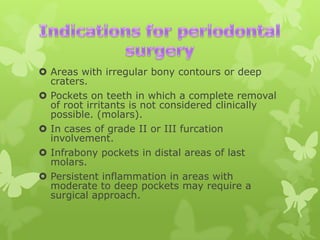
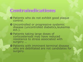
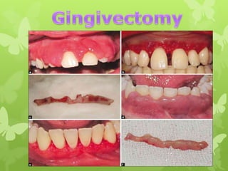
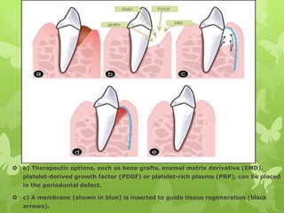
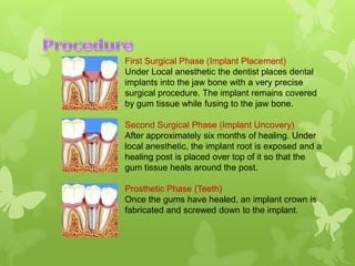
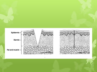
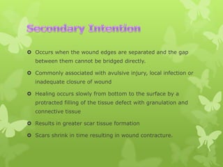
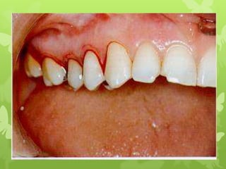
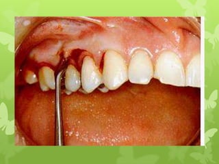
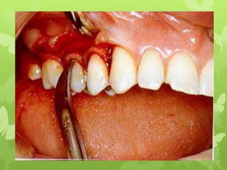
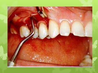
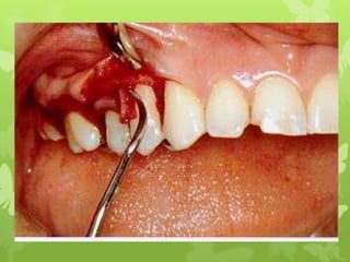
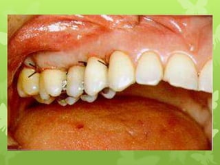
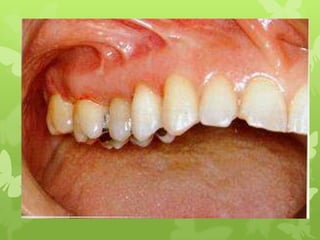
ENDO-PERIO SEMINAR
 Areas with irregular bony contours or deep
craters.
 Pockets on teeth in which a complete removal
of root irritants is not considered clinically
possible. (molars).
 In cases of grade II or III furcation
involvement.
 Infrabony pockets in distal areas of last
molars.
 Persistent inflammation in areas with
moderate to deep pockets may require a
surgical approach.
 Patients who do not exhibit good plaque
control.
 Uncontrolled or progressive systemic
disease (uncontrolled diabetics,leukemia
ect.).
 Patients taking large doses of
corticosteriods may have reduced
resistance to stress associated with
surgery ..
 Patients with imminent terminal disease
who are debilitated are not candidates for
surgery.
• Resective Procedures.
• New attachment procedures.
• Regeneration procedures.
• It is the procedure that means to
eliminate or reduce the pocket, by

excising or amputating the tissue
constricting the pocket wall.

(in this case we remove bone).
Gingivectomy, Gingivoplasty.
Apically positioned flap without
osseous surgery.
Apically positioned flap with osseous
surgery (Osteoplasty, Osteoctomy).
Root resection.
It is the reunion of connective
tissue by formation of new
cementum with inserting
collagen fibers on root surface
that has been deprived of its
periodontal ligament.
 Gingivectomy: Excision of soft tissue wall of
periodontal pocket.
 Basic rational is pocket elimination to allow access
for root instrumentation.
 Gingivoplasty: To restore gingival contours.(not
commonly used now days).
 External bevel incision is done to remove excess
gingiva and healing is by secondary intention.
 Are surgical procedures aimed at
Reproduction or reconstruction of lost or
injured periodontium.

 Aim is to restore the periodontium to the
normal physiologic levels.


b) Therapeutic options, such as bone grafts, enamel matrix derivative (EMD),
platelet-derived growth factor (PDGF) or platelet-rich plasma (PRP), can be placed
in the periodontal defect.



c) A membrane (shown in blue) is inserted to guide tissue regeneration (black
arrows).
 Swelling and bruising
 Pain, excessive bleeding, exposing the apex,
damage to flap.
 infection, secondary to bleeding and pain.

 Possible nerve injury may follow depending on site
 Infection
 Pain killer
 Keep pack in place.
 Avoid hot food.
 Use ice pack on the face.
 Do not brush the area.
 Use mouth rinse after one day.
 Do not smoke, follow normal activity, however avoid
excessive exertion.
 Come back to your next appointment.
Measure recession
from the
cementoenamel
junction to the
gingival margin
Class I - Incipient furcal involvement
Class II - Patent furcal involvement
Class III - Communicating furcal involvement
Class IV - Clinically visible furcation
 Dental implant is an artificial
titanium fixture which is placed
surgically into the jaw bone to
substitute for a missing tooth and

its root(s).
Partial and Full Dentures

Crowns

Bridges
Screw Implants
(Left to Right: TPS screw,
Ledermann screw,
Branemark screw, ITI
Bonefit screw)

Cylinder Implants
(Left to Right: IMZ, Integral,
Frialit-1 step-cylinder,
Frialit-2 step-cylinder)
First Surgical Phase (Implant Placement)
Under Local anesthetic the dentist places dental
implants into the jaw bone with a very precise
surgical procedure. The implant remains covered
by gum tissue while fusing to the jaw bone.
Second Surgical Phase (Implant Uncovery)
After approximately six months of healing. Under
local anesthetic, the implant root is exposed and a
healing post is placed over top of it so that the
gum tissue heals around the post.
Prosthetic Phase (Teeth)
Once the gums have healed, an implant crown is
fabricated and screwed down to the implant.
 Quality of healing response is also influenced by the
nature of tissue disruption and circumstances
surrounding wound closure.
 Categorized into:



Healing by First Intention

 Healing by Second Intention
 Healing by Third intention
 This occurs when a clean laceration or surgical incision is
closed primarily with sutures/clips with the edges in
apposition.

 Healing proceeds rapidly with no dehiscence and
minimal scar formation
 Soundly united within 2weeks and dense scar tissue is
laid down within 1 month.
 Occurs when the wound edges are separated and the gap
between them cannot be bridged directly.
 Commonly associated with avulsive injury, local infection or
inadequate closure of wound
 Healing occurs slowly from bottom to the surface by a
protracted filling of the tissue defect with granulation and
connective tissue
 Results in greater scar tissue formation
 Scars shrink in time resulting in wound contracture.
 Occurs through a staged procedure that combines
secondary healing with delayed primary closure.
 Avulsive or contaminated wound are repeatedly
debrided, along with antibiotic therapy and allowed to
granulate and heal by secondary intention for 5-7 days.
 Once adequate granulation tissue has formed and risk of
infection minimal, the wound is then sutured close to
heal by primary intention.
surgical procedure for periodontal diseases

surgical procedure for periodontal diseases

  • 1.
  • 2.
     Areas withirregular bony contours or deep craters.  Pockets on teeth in which a complete removal of root irritants is not considered clinically possible. (molars).  In cases of grade II or III furcation involvement.  Infrabony pockets in distal areas of last molars.  Persistent inflammation in areas with moderate to deep pockets may require a surgical approach.
  • 3.
     Patients whodo not exhibit good plaque control.  Uncontrolled or progressive systemic disease (uncontrolled diabetics,leukemia ect.).  Patients taking large doses of corticosteriods may have reduced resistance to stress associated with surgery ..  Patients with imminent terminal disease who are debilitated are not candidates for surgery.
  • 4.
    • Resective Procedures. •New attachment procedures. • Regeneration procedures.
  • 5.
    • It isthe procedure that means to eliminate or reduce the pocket, by excising or amputating the tissue constricting the pocket wall. (in this case we remove bone).
  • 6.
    Gingivectomy, Gingivoplasty. Apically positionedflap without osseous surgery. Apically positioned flap with osseous surgery (Osteoplasty, Osteoctomy). Root resection.
  • 9.
    It is thereunion of connective tissue by formation of new cementum with inserting collagen fibers on root surface that has been deprived of its periodontal ligament.
  • 11.
     Gingivectomy: Excisionof soft tissue wall of periodontal pocket.  Basic rational is pocket elimination to allow access for root instrumentation.  Gingivoplasty: To restore gingival contours.(not commonly used now days).  External bevel incision is done to remove excess gingiva and healing is by secondary intention.
  • 20.
     Are surgicalprocedures aimed at Reproduction or reconstruction of lost or injured periodontium.  Aim is to restore the periodontium to the normal physiologic levels.
  • 21.
     b) Therapeutic options,such as bone grafts, enamel matrix derivative (EMD), platelet-derived growth factor (PDGF) or platelet-rich plasma (PRP), can be placed in the periodontal defect.  c) A membrane (shown in blue) is inserted to guide tissue regeneration (black arrows).
  • 22.
     Swelling andbruising  Pain, excessive bleeding, exposing the apex, damage to flap.  infection, secondary to bleeding and pain.  Possible nerve injury may follow depending on site  Infection
  • 23.
     Pain killer Keep pack in place.  Avoid hot food.  Use ice pack on the face.  Do not brush the area.  Use mouth rinse after one day.  Do not smoke, follow normal activity, however avoid excessive exertion.  Come back to your next appointment.
  • 24.
  • 28.
    Class I -Incipient furcal involvement Class II - Patent furcal involvement Class III - Communicating furcal involvement Class IV - Clinically visible furcation
  • 29.
     Dental implantis an artificial titanium fixture which is placed surgically into the jaw bone to substitute for a missing tooth and its root(s).
  • 30.
    Partial and FullDentures Crowns Bridges
  • 31.
    Screw Implants (Left toRight: TPS screw, Ledermann screw, Branemark screw, ITI Bonefit screw) Cylinder Implants (Left to Right: IMZ, Integral, Frialit-1 step-cylinder, Frialit-2 step-cylinder)
  • 32.
    First Surgical Phase(Implant Placement) Under Local anesthetic the dentist places dental implants into the jaw bone with a very precise surgical procedure. The implant remains covered by gum tissue while fusing to the jaw bone. Second Surgical Phase (Implant Uncovery) After approximately six months of healing. Under local anesthetic, the implant root is exposed and a healing post is placed over top of it so that the gum tissue heals around the post. Prosthetic Phase (Teeth) Once the gums have healed, an implant crown is fabricated and screwed down to the implant.
  • 33.
     Quality ofhealing response is also influenced by the nature of tissue disruption and circumstances surrounding wound closure.  Categorized into:  Healing by First Intention  Healing by Second Intention  Healing by Third intention
  • 34.
     This occurswhen a clean laceration or surgical incision is closed primarily with sutures/clips with the edges in apposition.  Healing proceeds rapidly with no dehiscence and minimal scar formation  Soundly united within 2weeks and dense scar tissue is laid down within 1 month.
  • 36.
     Occurs whenthe wound edges are separated and the gap between them cannot be bridged directly.  Commonly associated with avulsive injury, local infection or inadequate closure of wound  Healing occurs slowly from bottom to the surface by a protracted filling of the tissue defect with granulation and connective tissue  Results in greater scar tissue formation  Scars shrink in time resulting in wound contracture.
  • 37.
     Occurs througha staged procedure that combines secondary healing with delayed primary closure.  Avulsive or contaminated wound are repeatedly debrided, along with antibiotic therapy and allowed to granulate and heal by secondary intention for 5-7 days.  Once adequate granulation tissue has formed and risk of infection minimal, the wound is then sutured close to heal by primary intention.