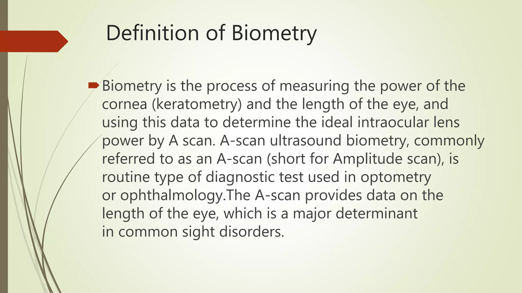
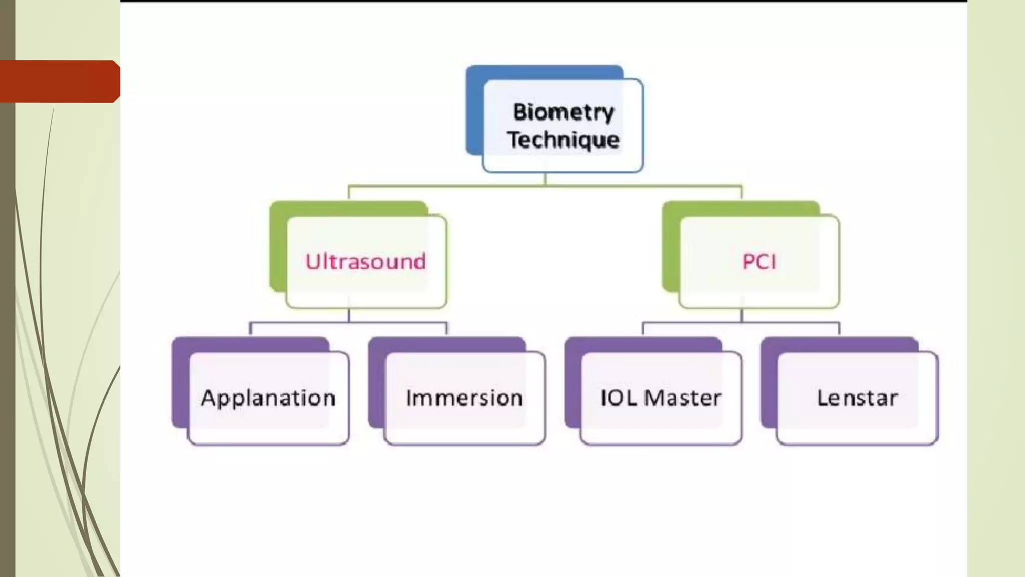
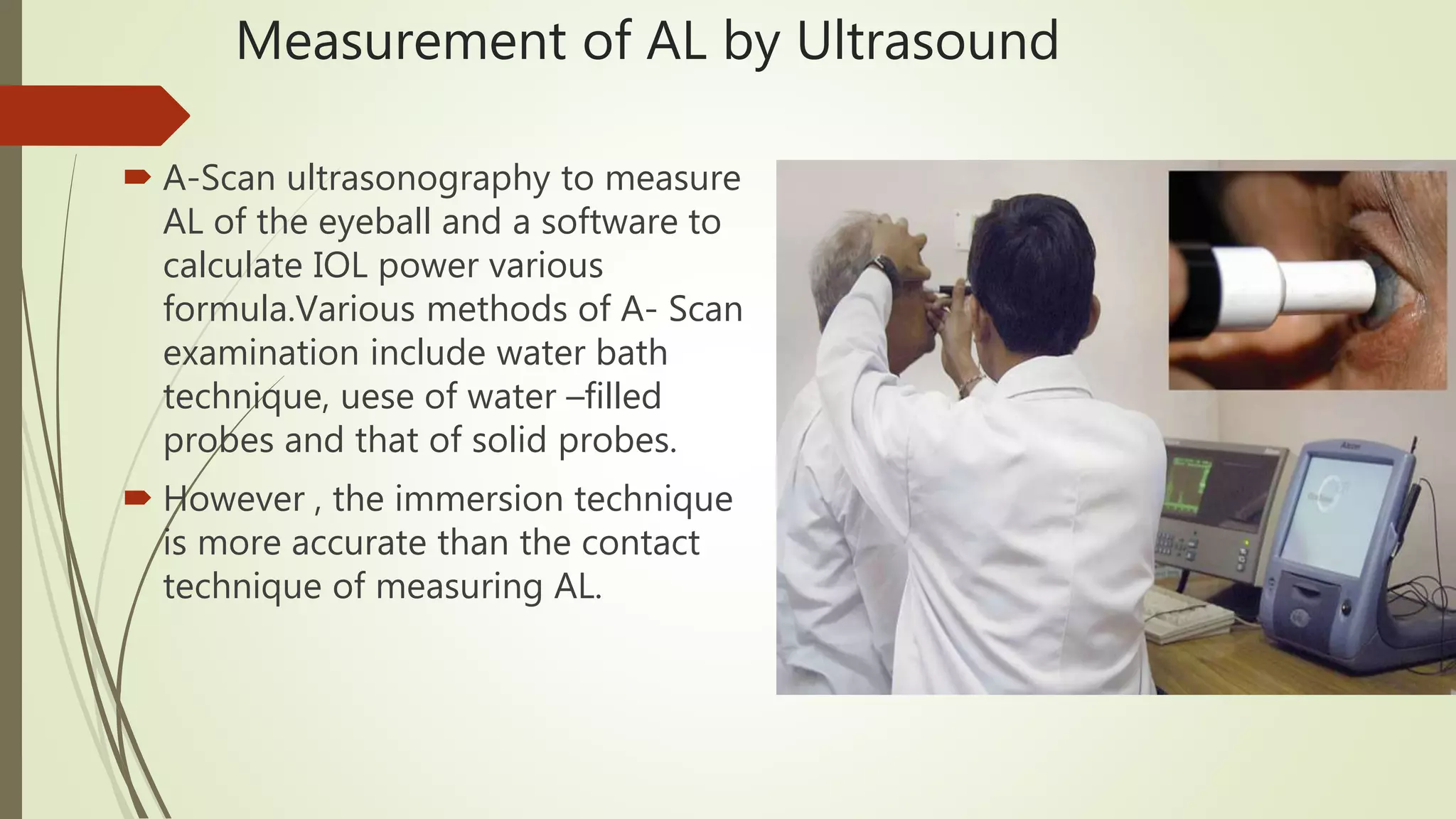
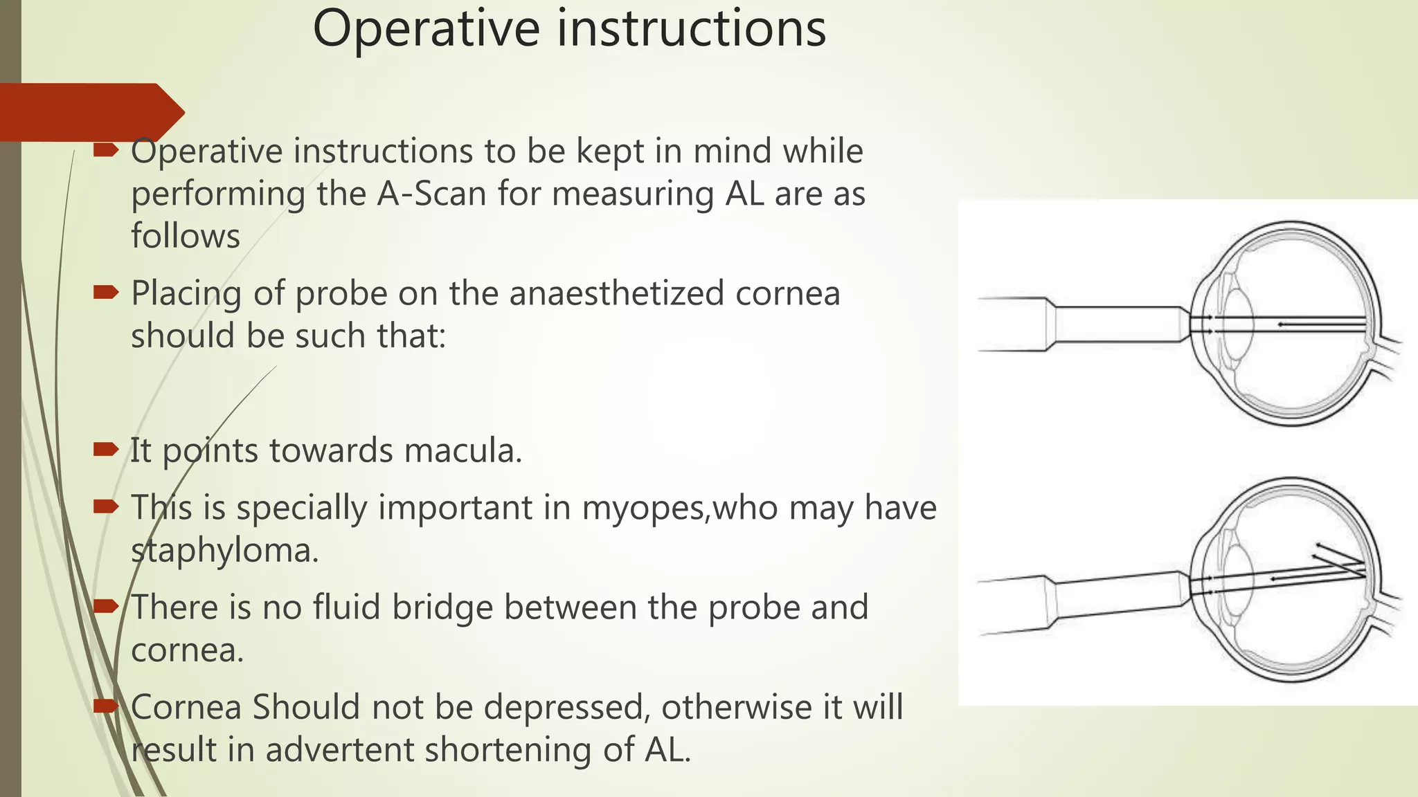
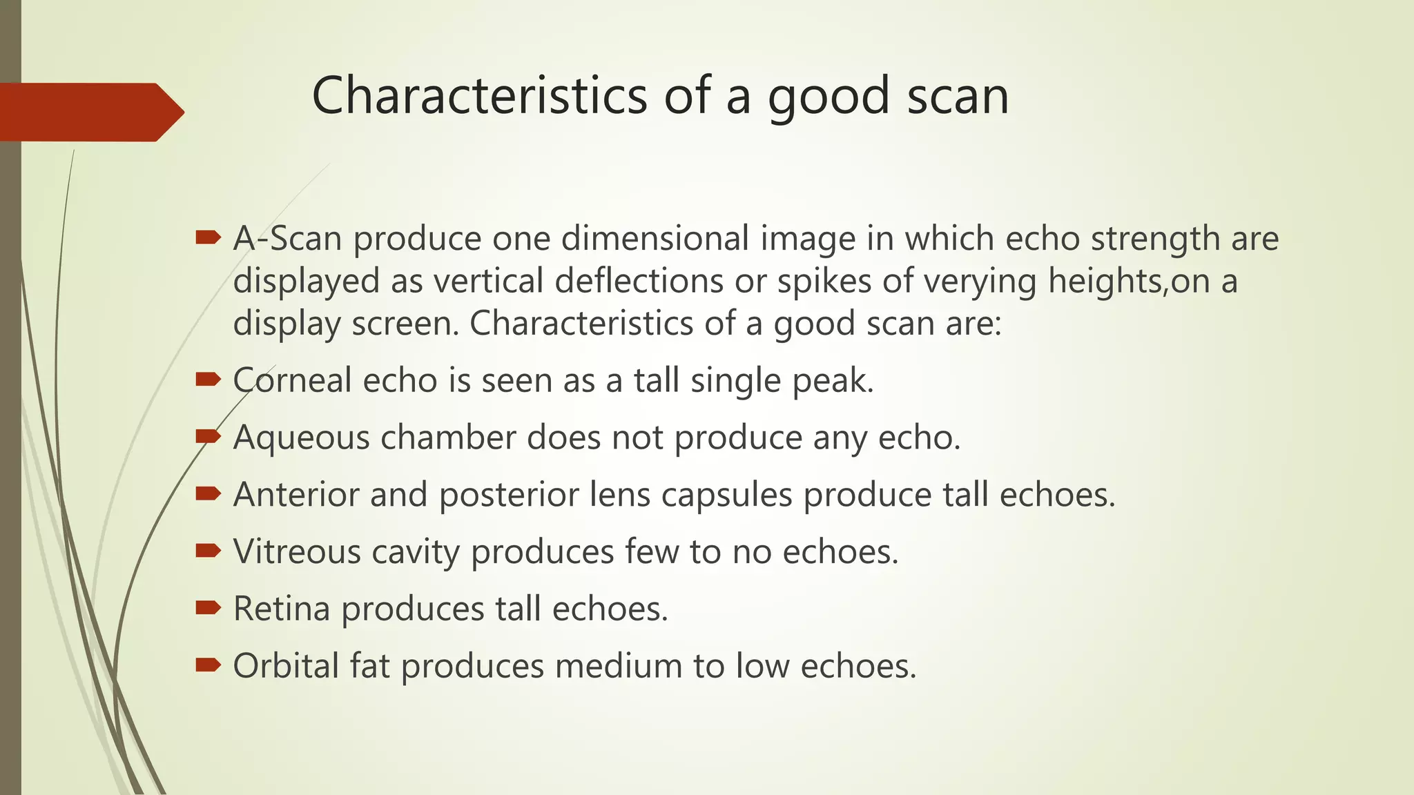
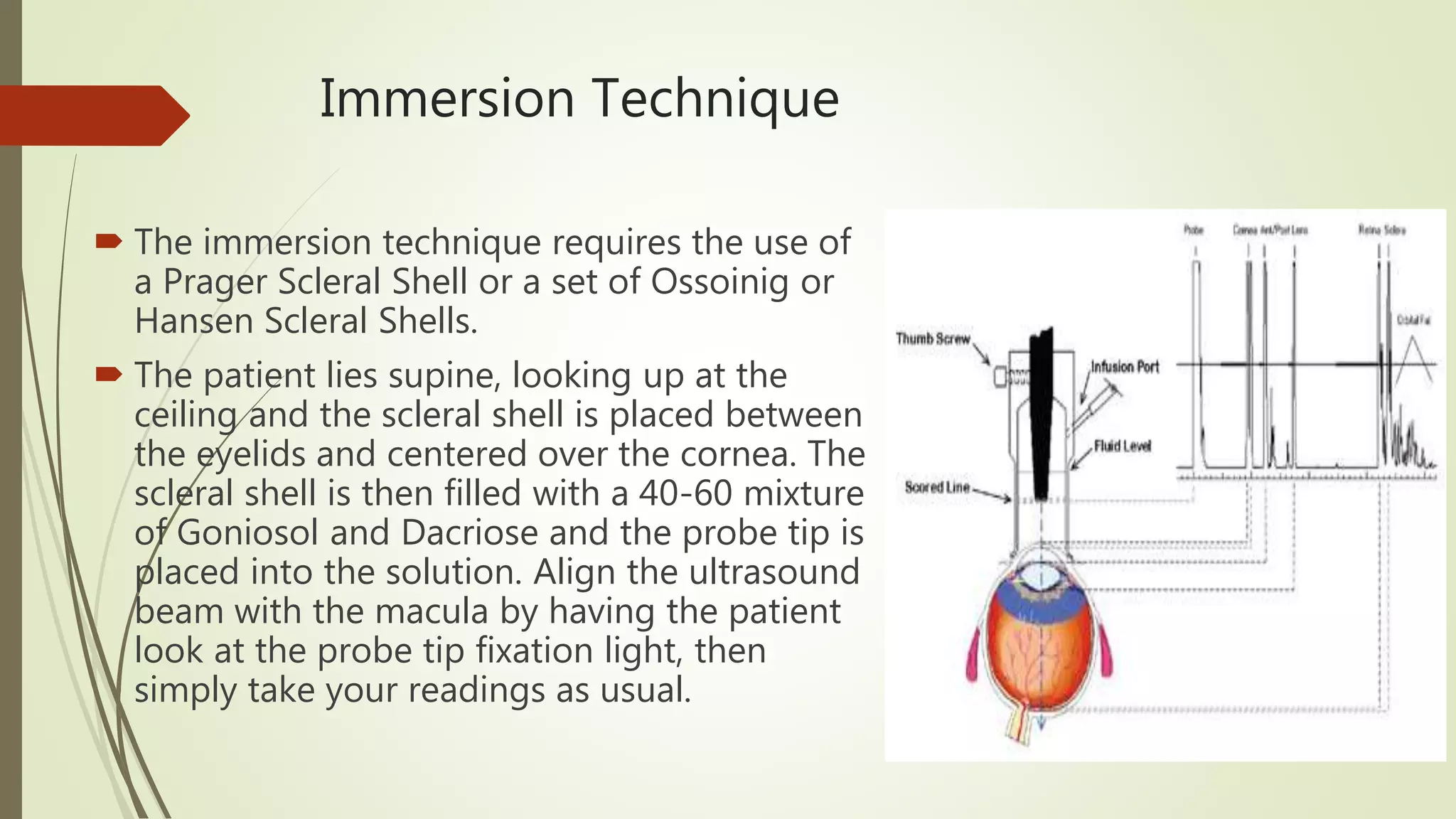
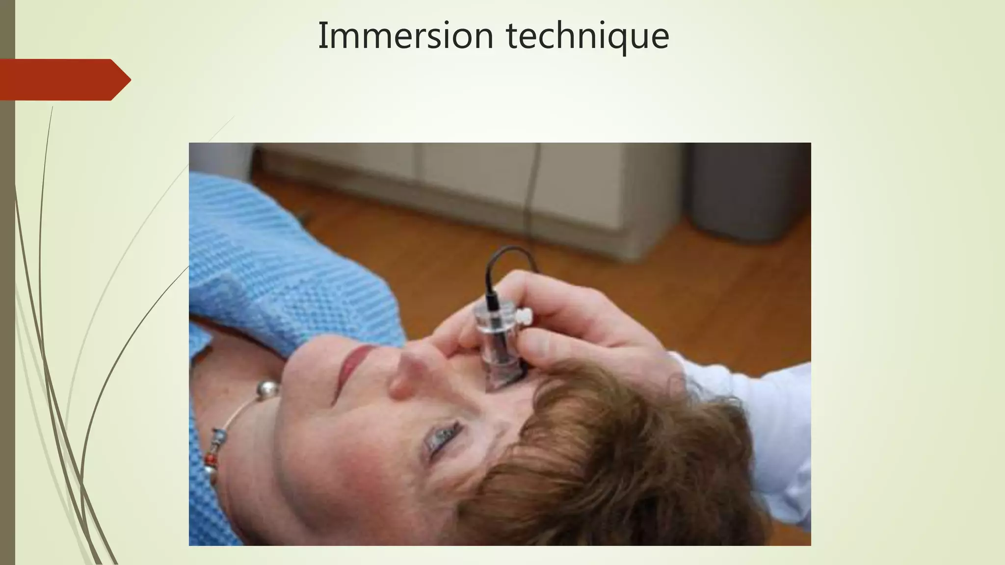
Biometry or A-Scan
Procedure
Md.Asaduzzaman
Associate Optometrist
Definition of Biometry
Biometry is the process of measuring the power of the
cornea (keratometry) and the length of the eye, and
using this data to determine the ideal intraocular lens
power by A scan. A-scan ultrasound biometry, commonly
referred to as an A-scan (short for Amplitude scan), is
routine type of diagnostic test used in optometry
or ophthalmology.The A-scan provides data on the
length of the eye, which is a major determinant
in common sight disorders.
Examination Procedure
 History taking
 Patient preparation
 Biometry technique or types
Measurement of AL by Ultrasound
 A-Scan ultrasonography to measure
AL of the eyeball and a software to
calculate IOL power various
formula.Various methods of A- Scan
examination include water bath
technique, uese of water –filled
probes and that of solid probes.
 However , the immersion technique
is more accurate than the contact
technique of measuring AL.
Gain or sensitivity Setting
 Gain refers to the electronic
amplification of the sound waves
received by the transducer.
 Increase in gain may be required when
the height of echoes achieved is
inadequate as in very dense
cataracts,other ocular opacities and high
myopia.Increase in the gain produces
taller echoes.
 Decrease in the gain is done when
artefacts are seen near the retinal
echoes, e.g.in slicone filled eyes and
pseudophakic eyes.
Sound velocity setting
 Sound velocity setting should be done
appropriately according to the type of
eye, i.e.
 Normal
 Cataractous (mild,moderate or dense)
 Aphakic
 Pseudophakic (Sliicone or acrylic)
Note :Change in the sound velocity due
to materials like silicone oil in the eye
can produce an error of 3-4D
Operative instructions
 Operative instructions to be kept in mind while
performing the A-Scan for measuring AL are as
follows
 Placing of probe on the anaesthetized cornea
should be such that:
 It points towards macula.
 This is specially important in myopes,who may have
staphyloma.
 There is no fluid bridge between the probe and
cornea.
 Cornea Should not be depressed, otherwise it will
result in advertent shortening of AL.
Characteristics of a good scan
 A-Scan produce one dimensional image in which echo strength are
displayed as vertical deflections or spikes of verying heights,on a
display screen. Characteristics of a good scan are:
 Corneal echo is seen as a tall single peak.
 Aqueous chamber does not produce any echo.
 Anterior and posterior lens capsules produce tall echoes.
 Vitreous cavity produces few to no echoes.
 Retina produces tall echoes.
 Orbital fat produces medium to low echoes.
Immersion Technique
 The immersion technique requires the use of
a Prager Scleral Shell or a set of Ossoinig or
Hansen Scleral Shells.
 The patient lies supine, looking up at the
ceiling and the scleral shell is placed between
the eyelids and centered over the cornea. The
scleral shell is then filled with a 40-60 mixture
of Goniosol and Dacriose and the probe tip is
placed into the solution. Align the ultrasound
beam with the macula by having the patient
look at the probe tip fixation light, then
simply take your readings as usual.
Immersion technique
IOL Master
 A non-contact optical device that
measures the distance from the
corneal vertex to the retinal
pigment epithelium by partial
coherence interferometry, the IOL
Master is consistently accurate to
within ±0.02 mm or better.
The IOL Master is the first such
device to be widely used in clinical
ophthalmology.
Home Messages
 Increase the patient comfort.
 See refractive status before doing
biometry.
 Biometry is an important step in
successful IOL implantation.So
you will do it carefully.
Any Questions
Biometry procedure

Biometry procedure

  • 1.
  • 2.
    Definition of Biometry Biometryis the process of measuring the power of the cornea (keratometry) and the length of the eye, and using this data to determine the ideal intraocular lens power by A scan. A-scan ultrasound biometry, commonly referred to as an A-scan (short for Amplitude scan), is routine type of diagnostic test used in optometry or ophthalmology.The A-scan provides data on the length of the eye, which is a major determinant in common sight disorders.
  • 3.
    Examination Procedure  Historytaking  Patient preparation  Biometry technique or types
  • 5.
    Measurement of ALby Ultrasound  A-Scan ultrasonography to measure AL of the eyeball and a software to calculate IOL power various formula.Various methods of A- Scan examination include water bath technique, uese of water –filled probes and that of solid probes.  However , the immersion technique is more accurate than the contact technique of measuring AL.
  • 6.
    Gain or sensitivitySetting  Gain refers to the electronic amplification of the sound waves received by the transducer.  Increase in gain may be required when the height of echoes achieved is inadequate as in very dense cataracts,other ocular opacities and high myopia.Increase in the gain produces taller echoes.  Decrease in the gain is done when artefacts are seen near the retinal echoes, e.g.in slicone filled eyes and pseudophakic eyes.
  • 7.
    Sound velocity setting Sound velocity setting should be done appropriately according to the type of eye, i.e.  Normal  Cataractous (mild,moderate or dense)  Aphakic  Pseudophakic (Sliicone or acrylic) Note :Change in the sound velocity due to materials like silicone oil in the eye can produce an error of 3-4D
  • 8.
    Operative instructions  Operativeinstructions to be kept in mind while performing the A-Scan for measuring AL are as follows  Placing of probe on the anaesthetized cornea should be such that:  It points towards macula.  This is specially important in myopes,who may have staphyloma.  There is no fluid bridge between the probe and cornea.  Cornea Should not be depressed, otherwise it will result in advertent shortening of AL.
  • 9.
    Characteristics of agood scan  A-Scan produce one dimensional image in which echo strength are displayed as vertical deflections or spikes of verying heights,on a display screen. Characteristics of a good scan are:  Corneal echo is seen as a tall single peak.  Aqueous chamber does not produce any echo.  Anterior and posterior lens capsules produce tall echoes.  Vitreous cavity produces few to no echoes.  Retina produces tall echoes.  Orbital fat produces medium to low echoes.
  • 10.
    Immersion Technique  Theimmersion technique requires the use of a Prager Scleral Shell or a set of Ossoinig or Hansen Scleral Shells.  The patient lies supine, looking up at the ceiling and the scleral shell is placed between the eyelids and centered over the cornea. The scleral shell is then filled with a 40-60 mixture of Goniosol and Dacriose and the probe tip is placed into the solution. Align the ultrasound beam with the macula by having the patient look at the probe tip fixation light, then simply take your readings as usual.
  • 11.
  • 12.
    IOL Master  Anon-contact optical device that measures the distance from the corneal vertex to the retinal pigment epithelium by partial coherence interferometry, the IOL Master is consistently accurate to within ±0.02 mm or better. The IOL Master is the first such device to be widely used in clinical ophthalmology.
  • 13.
    Home Messages  Increasethe patient comfort.  See refractive status before doing biometry.  Biometry is an important step in successful IOL implantation.So you will do it carefully.
  • 14.