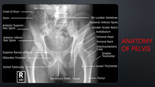
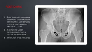
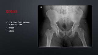
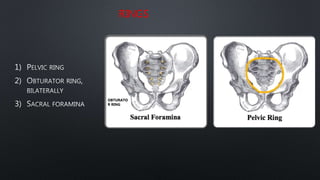
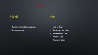
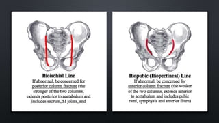
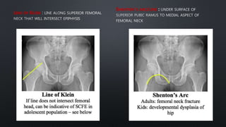
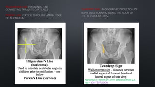
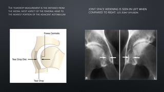
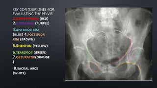
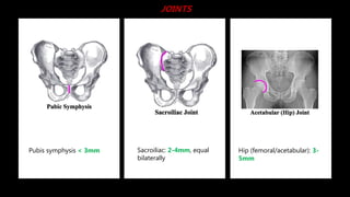
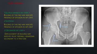
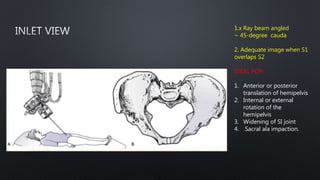
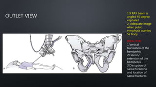
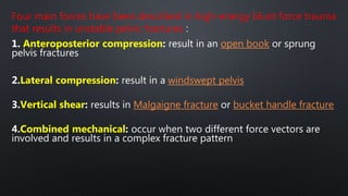
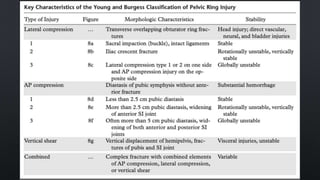
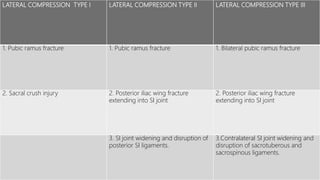
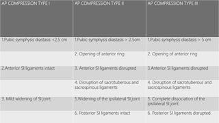
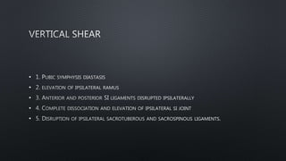
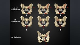
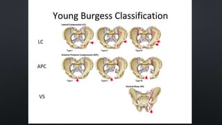
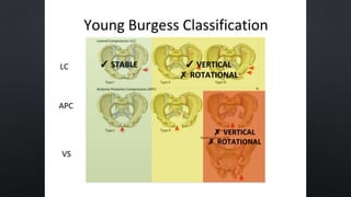
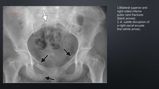
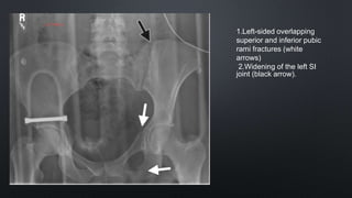
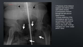
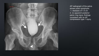
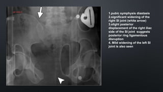
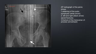
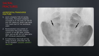
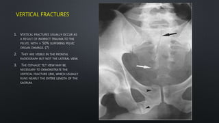
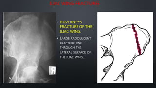
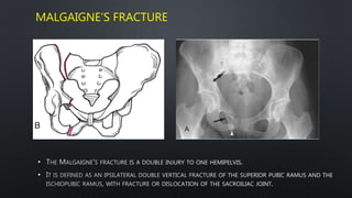
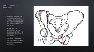
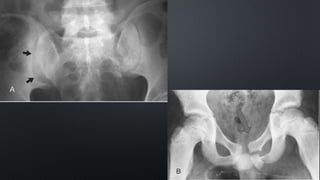
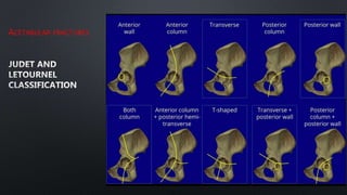
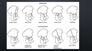
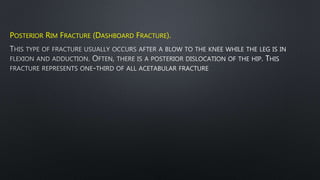
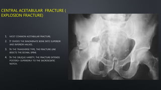
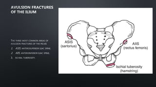
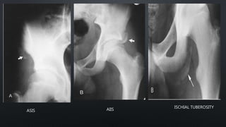
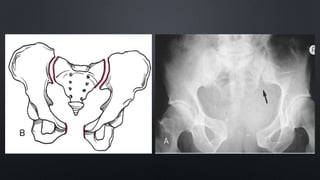
By
Dr. Lohith Varma
ANATOMY
OF PELVIS
•1.POSITIONING
•2.BONES
•3.JOINTS
•4.SOFT TISSUE
POSITIONING
1.
2.
3.
BONES
•
•
•
RINGS
1)
2)
3)
OBTURATO
R RING
LINES
PELVIS
•
•
HIP
•
•
•
•
•
LINE OF KLEIN
SHENTON’S ARC/LINE
HILGENREINER LINE
PERKIN’S LINE
TEARDROP SIGN
Abnormal if >11mm or >2mm difference from C/L
hip – JOINT EFFUSION
1.ILIOPECTINEAL
ILIOISCHIAL
3.ANTERIOR RIM
POSTERIOR
RIM
5.SHENTON
6.TEARDROP
OBTURATOR
SACRAL ARCS
Sacroiliac: 2-4mm, equal
bilaterally
Pubis symphysis < 3mm Hip (femoral/acetabular): 3-
5mm
JOINTS
SOFT TISSUES
1.GLUTEUS MINIMUS FAT STRIPE
BULGING OF THIS PAD MAY INDICATE
PRESENCE OF EFFUSION IN HIP JOINT
2.ILIOPSOAS
BULGING OF THIS PAD MAY INDICATE
PRESENCE OF EFFUSION IN HIP JOINT
3.OBTURATOR FAT STRIP
DISPLACEMENT OR BULGING MAY
INDICATE PELVIC SIDEWALL HEMATOMA
SECONDARY TO A FRACTURE
•
•
•
1.x Ray beam angled
~ 45-degree cauda
2. Adequate image when S1
overlaps S2
IDEAL FOR:
1. Anterior or posterior
translation of hemipelvis
2. Internal or external
rotation of the
hemipelvis
3. Widening of SI joint
4. Sacral ala impaction.
1.X RAY beam is
angled 45 degree
cephalad
2. Adequate image
when pubic
symphysis overlies
S2 body.
IDEAL FOR:
1.Vertical
translation of the
hemipelvis
2.Flexion/
extension of the
hemipelvis
3.Disruption of
sacral foramina
and location of
sacral fractures
Four main forces have been described in high-energy blunt force trauma
that results in unstable pelvic fractures :
1. Anteroposterior compression: result in an open book or sprung
pelvis fractures
2.Lateral compression: result in a windswept pelvis
3.Vertical shear: results in Malgaigne fracture or bucket handle fracture
4.Combined mechanical: occur when two different force vectors are
involved and results in a complex fracture pattern
LATERAL COMPRESSION TYPE I LATERAL COMPRESSION TYPE II LATERAL COMPRESSION TYPE III
1. Pubic ramus fracture 1. Pubic ramus fracture 1. Bilateral pubic ramus fracture
2. Sacral crush injury 2. Posterior iliac wing fracture
extending into SI joint
2. Posterior iliac wing fracture
extending into SI joint
3. SI joint widening and disruption of
posterior SI ligaments.
3.Contralateral SI joint widening and
disruption of sacrotuberous and
sacrospinous ligaments.
AP COMPRESSION TYPE I AP COMPRESSION TYPE II AP COMPRESSION TYPE III
1.Pubic symphysis diastasis <2.5 cm 1.Pubic symphysis diastasis > 2.5cm 1.Pubic symphysis diastasis > 5 cm
2. Opening of anterior ring 2. Opening of anterior ring
2.Anterior SI ligaments intact 3. Anterior SI ligaments disrupted 3.Anterior SI ligaments disrupted
4. Disruption of sacrotuberous and
sacrospinous ligaments
4. Disruption of sacrotuberous and
sacrospinous ligaments
3. Mild widening of SI joint. 5.Widening of the ipsilateral SI joint 5. Complete dissociation of the
ipsilateral SI joint.
6. Posterior SI ligaments intact 6. Posterior SI ligaments disrupted.
•
•
•
•
•
1.Bilateral superior and
right-sided inferior
pubic rami fractures
(black arrows)
2. A subtle disruption of
a right sacral arcuate
line (white arrow).
1.Left-sided overlapping
superior and inferior pubic
rami fractures (white
arrows)
2.Widening of the left SI
joint (black arrow).
1.Fractures of the bilateral
superior and inferior pubic
rami (white arrows)
2.compression fracture
through the left sacrum
(black arrow),
3.Gross widening of the
contralateral right SI joint
(arrowhead) with external
rotation of the right iliac
wing is also seen, findings
indicative of a “wind- swept
pelvis” injury.
AP radiograph of the pelvis
shows pubic symphysis
1. diastasis of 1.5 cm
2. no apparent posterior
pelvic ring injury, findings
consistent with an AP
compression type 1 injury.
1.pubic symphysis diastasis
2.significant widening of the
right SI joint (white arrow)
3.slight posterior
displacement of the right iliac
side of the SI joint suggests
posterior ring ligamentous
disruption
4. Mild widening of the left SI
joint is also seen
AP radiograph of the pelvis
shows
1.widening of the pubic
symphysis (white arrow),
2. left and right (black arrow)
sacral fractures.
3.fracture of the transverse L5
process (arrowhead)
SACRAL
FRACTURES
•HORIZONTAL/TRANSVERSE
FRACTURE:
1.
2.
3.
SUICIDAL JUMPER’S
FRACTURE
VERTICAL FRACTURES
1.
2.
3.
ILIAC WING FRACTURES
• DUVERNEY’S
FRACTURE OF THE
ILIAC WING.
•
MALGAIGNE’S FRACTURE
•
•
1.
2.
3.
BUCKET HANDLE
FRACTURE
ACETABULAR FRACTURES
POSTERIOR RIM FRACTURE (DASHBOARD FRACTURE).
CENTRAL ACETABULAR FRACTURE (
EXPLOSION FRACTURE)
1.
2.
3.
4.
1.
2.
3.
ASIS AIIS
ISCHIAL TUBEROSITY
•
•
•
•
•
•
•
Pelvis fractures
Pelvis fractures

Pelvis fractures