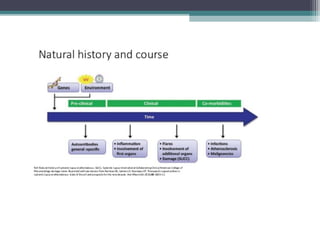
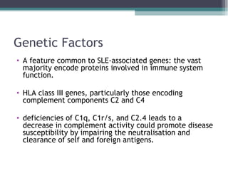
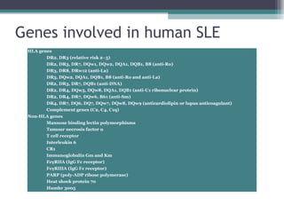
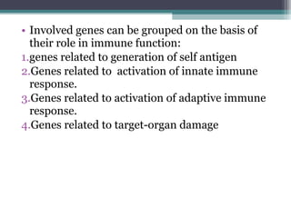
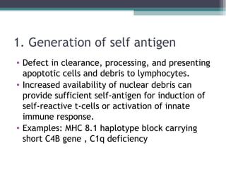
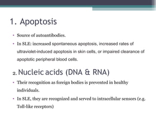
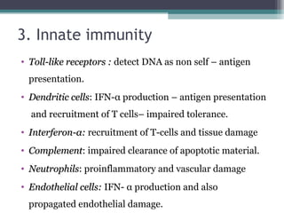
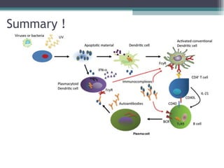
Systemic Lupus Erythematosus
Pathogenesis and pathophysiology
We will talk about:
Pathogenesis
aetiology or
contributing
factors
Pathophysiology
Key events in
development of
the disease
In one sentence!
SLE results from recurrent activation of immune
system with production of antibodies and other
protein products contributing to inflammation and
tissue damage.
Aetiology & Pathogenesis
Contributing factors
• Environmental factors
• Genetic factors
• Hormonal factors
Environmental factors
1. EBV infection
• Epstein–Barr virus (EBV) has been identifed as
a possible factor in
the development of lupus.
• EBV may reside in and interact with B cells and
promotes (IFNα) production by plasmacytoid
dendritic cells, suggesting that elevated IFNα in
lupus may be—at least in part—due to aberrantly
controlled chronic viral infection.
2.Drugs
• Over 100 drugs have been reported to
causedrug-induced lupus (DIL), including a
number of the newer biologics and antiviral
agents.
• Autoimmunity not hypersensitivity!
• t is believed that the development of DILE
requires a certain degree of genetic
susceptibility.
Theories proposed in the pathogenesis
of DIL
• Theory 1: altering T-cell cell DNA methylation,
which has a primordial role in the regulation and
expression of genes and cell differentiation.
• Theory 2: autoreactive B cells are activated by
drug-specific T cells.
• Theory 3:the culprit drug subverts de novo
acquisition of B-cell tolerance if it is present in
the thymus as these cells develop.
3. UV light
• UV light may induce breaks in in DNA that
might alter gene expression or lead to apoptotic
or necrotic cell death.
Genetic factors
• Concordance rate in monozygotic twins is 10
times more frequent than in dizygotic twins.
• Siblings of SLE patients are approximately 30
times more likely to develop SLE compared
with individuals without an a ected sibling.ff
Genetic Factors
• A feature common to SLE-associated genes: the vast
majority encode proteins involved in immune system
function.
• HLA class III genes, particularly those encoding
complement components C2 and C4
• deficiencies of C1q, C1r/s, and C2.4 leads to a
decrease in complement activity could promote disease
susceptibility by impairing the neutralisation and
clearance of self and foreign antigens.
Genes involved in human SLE
HLA genes
    DR2, DR3 (relative risk 2–5)
    DR2, DR3, DR7, DQw1, DQw2, DQA1, DQB1, B8 (anti-Ro)
    DR3, DR8, DRw12 (anti-La)
    DR3, DQw2, DQA1, DQB1, B8 (anti-Ro and anti-La)
    DR2, DR3, DR7, DQB1 (anti-DNA)
    DR2, DR4, DQw5, DQw8, DQA1, DQB1 (anti-U1 ribonuclear protein)
    DR2, DR4, DR7, DQw6, B61 (anti-Sm)
    DR4, DR7, DQ6, DQ7, DQw7, DQw8, DQw9 (anticardiolipin or lupus anticoagulant)
    Complement genes (C2, C4, C1q)
Non-HLA genes
    Mannose binding lectin polymorphisms
    Tumour necrosis factor α
    T cell receptor
    Interleukin 6
    CR1
    Immunoglobulin Gm and Km
    FcγRIIA (IgG Fc receptor)
    FcγRIIIA (IgG Fc receptor)
    PARP (poly-ADP ribose polymerase)
    Heat shock protein 70
    Humhr 3005
• Involved genes can be grouped on the basis of
their role in immune function:
1.genes related to generation of self antigen
2.Genes related to activation of innate immune
response.
3.Genes related to activation of adaptive immune
response.
4.Genes related to target-organ damage
1. Generation of self antigen
• Defect in clearance, processing, and presenting
apoptotic cells and debris to lymphocytes.
• Increased availability of nuclear debris can
provide sufficient self-antigen for induction of
self-reactive t-cells or activation of innate
immune response.
• Examples: MHC 8.1 haplotype block carrying
short C4B gene , C1q deficiency
2. Activation of innate immunity
• Large numbers of single nucleotide
polymorphism (SNPs) are found in the genes
that encode proteins inducing type I interferon
(IFN).
• single nucleotide polymorphisms (SNPs) of
genes encoding TLR7, TLR8,and TLR9
Type I interferon (IFN I)
• Type I interferons (IFNs) are polypeptides that are
secreted by infected cells.
• They have 3 functions:
▫ they induce cell-intrinsic antimicrobial states in infected
and neighbouring cells that limit the spread of infectious
agents
▫ promotes antigen presentation and natural killer cell
functions while restraining pro-inflammatory pathways
and cytokine production (balanced innate immunity).
▫ activate the adaptive immune system,(antigen-specific T-
cells and B-cells and immunological memory
• increase in IFN-I-related genes was identified in
the peripheral blood monocytes (PBMCs) from
patients with SLE.
• overproduction of type I interferon can
promote the expression of proinflammatory
cytokines and chemokines, the maturation of
dendritic cells, the activation of autoreactive B
and T cells, the production of autoantibodies, and
loss of self-tolerance.
Toll-like receptors
• a class of proteins that that activate the innate immune
system in response to a variety of pathogen-associated
molecular patterns (PAMPs).
• Recognition of microbial components by TLRs initiates
signal transduction pathways, which triggers expression
of genes. These gene products control innate immune
responses and further instruct development of antigen-
specific acquired immunity.
Toll-like receptors
• The TLRs that act as nucleic acid receptors are
TLR3, TLR7 (and TLR8 in humans), and TLR9.
• TLR9 is expressed in the endoplasmic reticulum
of macrophages and dendritic cells
Toll-like receptors
3. Activation of adaptive immunity
• T-cell abnormalities
▫ Decreased proliferation of T-cell in response to
allogenic (non-self) antigens
▫ Produce less IL-2, that contributes to T-cell
regulation
▫ Hypomethylation of DNA
▫ Though lymphopenia, expansion of T-helper
population that mediates differentiation of
autoantigen – specific B-cells.
• B-cell abnormalities
▫ Polyclonal B-cells overactivity
▫ Pssible mechanisms:
 intrinsic hyper-reactivity leading to polyclonal B-
cell activation with disturbed activation thresholds
and ineffective negative selection
 lack of immunoregulatory functions
 Secondary to overactivation of T-helper cells
 Disturbed cytokine production
SLE pathogenesis
Key events
Key events in the pathophysiology
of SLE
1. Apoptosis
2. Nucleic acids
3. Innate immunity
4. Adaptive immunity
1. Apoptosis
• Source of autoantibodies.
• In SLE: increased spontaneous apoptosis, increased rates of
ultraviolet-induced apoptosis in skin cells, or impaired clearance of
apoptotic peripheral blood cells.
2. Nucleic acids (DNA & RNA)
• Their recognition as foreign bodies is prevented in healthy
individuals.
• In SLE, they are recognized and served to intracellular sensors (e.g.
Toll-like receptors)
3. Innate immunity
• Toll-like receptors :Toll-like receptors : detect DNA as non self – antigen
presentation.
• Dendritic cellsDendritic cells: IFN-α production – antigen presentation
and recruitment of T cells– impaired tolerance.
• Interferon-Interferon-αα:: recruitment of T-cells and tissue damage
• ComplementComplement: impaired clearance of apoptotic material.
• NeutrophilsNeutrophils: proinflammatory and vascular damage
• Endothelial cells:Endothelial cells: IFN- α production and also
propagated endothelial damage.
4. Adaptive immunity
• Activation of T-helper cells by the previous events.
• B-cell differentiation to antibody-producing plasma
cells.
• Cytokines and chemokines produced by T and B cells
also shape the immune response and promote tissue
damage.
• Excess production of immune complexes, with impaired
clearance leads to deposition and tissue damage.
Summary !
Thanks
“He who issues forth in search of
knowledge is busy in the cause of Allah
till he returns from his quest.”
Prophet Muhammad (PBUH)
Reference
• Bertasias G., Cervera R., Boumpas D. T., systemic lupus erythematosus : pathogenesis and
clinical features. EULAR Textbook on Rheumatic Diseases (2012).
• CROW, M. etiology and pathogenesis of systemic lupus erythematosus. In: FIERSTEIN, G.,
et al. Kelly's textbook of rheumatology. 9th. ed. philadelphia: Elsevier Saunders, v. 3, 2013.
Cap. 79, p. 1269.

pathogenesis and pathophysiology of SLE

  • 1.
  • 2.
    We will talkabout: Pathogenesis aetiology or contributing factors Pathophysiology Key events in development of the disease
  • 3.
    In one sentence! SLEresults from recurrent activation of immune system with production of antibodies and other protein products contributing to inflammation and tissue damage.
  • 5.
  • 6.
    Contributing factors • Environmentalfactors • Genetic factors • Hormonal factors
  • 7.
  • 8.
    1. EBV infection •Epstein–Barr virus (EBV) has been identifed as a possible factor in the development of lupus. • EBV may reside in and interact with B cells and promotes (IFNα) production by plasmacytoid dendritic cells, suggesting that elevated IFNα in lupus may be—at least in part—due to aberrantly controlled chronic viral infection.
  • 9.
    2.Drugs • Over 100drugs have been reported to causedrug-induced lupus (DIL), including a number of the newer biologics and antiviral agents. • Autoimmunity not hypersensitivity! • t is believed that the development of DILE requires a certain degree of genetic susceptibility.
  • 10.
    Theories proposed inthe pathogenesis of DIL • Theory 1: altering T-cell cell DNA methylation, which has a primordial role in the regulation and expression of genes and cell differentiation. • Theory 2: autoreactive B cells are activated by drug-specific T cells. • Theory 3:the culprit drug subverts de novo acquisition of B-cell tolerance if it is present in the thymus as these cells develop.
  • 11.
    3. UV light •UV light may induce breaks in in DNA that might alter gene expression or lead to apoptotic or necrotic cell death.
  • 12.
  • 13.
    • Concordance ratein monozygotic twins is 10 times more frequent than in dizygotic twins. • Siblings of SLE patients are approximately 30 times more likely to develop SLE compared with individuals without an a ected sibling.ff
  • 14.
    Genetic Factors • Afeature common to SLE-associated genes: the vast majority encode proteins involved in immune system function. • HLA class III genes, particularly those encoding complement components C2 and C4 • deficiencies of C1q, C1r/s, and C2.4 leads to a decrease in complement activity could promote disease susceptibility by impairing the neutralisation and clearance of self and foreign antigens.
  • 15.
    Genes involved inhuman SLE HLA genes     DR2, DR3 (relative risk 2–5)     DR2, DR3, DR7, DQw1, DQw2, DQA1, DQB1, B8 (anti-Ro)     DR3, DR8, DRw12 (anti-La)     DR3, DQw2, DQA1, DQB1, B8 (anti-Ro and anti-La)     DR2, DR3, DR7, DQB1 (anti-DNA)     DR2, DR4, DQw5, DQw8, DQA1, DQB1 (anti-U1 ribonuclear protein)     DR2, DR4, DR7, DQw6, B61 (anti-Sm)     DR4, DR7, DQ6, DQ7, DQw7, DQw8, DQw9 (anticardiolipin or lupus anticoagulant)     Complement genes (C2, C4, C1q) Non-HLA genes     Mannose binding lectin polymorphisms     Tumour necrosis factor α     T cell receptor     Interleukin 6     CR1     Immunoglobulin Gm and Km     FcγRIIA (IgG Fc receptor)     FcγRIIIA (IgG Fc receptor)     PARP (poly-ADP ribose polymerase)     Heat shock protein 70     Humhr 3005
  • 17.
    • Involved genescan be grouped on the basis of their role in immune function: 1.genes related to generation of self antigen 2.Genes related to activation of innate immune response. 3.Genes related to activation of adaptive immune response. 4.Genes related to target-organ damage
  • 18.
    1. Generation ofself antigen • Defect in clearance, processing, and presenting apoptotic cells and debris to lymphocytes. • Increased availability of nuclear debris can provide sufficient self-antigen for induction of self-reactive t-cells or activation of innate immune response. • Examples: MHC 8.1 haplotype block carrying short C4B gene , C1q deficiency
  • 19.
    2. Activation ofinnate immunity • Large numbers of single nucleotide polymorphism (SNPs) are found in the genes that encode proteins inducing type I interferon (IFN). • single nucleotide polymorphisms (SNPs) of genes encoding TLR7, TLR8,and TLR9
  • 20.
    Type I interferon(IFN I) • Type I interferons (IFNs) are polypeptides that are secreted by infected cells. • They have 3 functions: ▫ they induce cell-intrinsic antimicrobial states in infected and neighbouring cells that limit the spread of infectious agents ▫ promotes antigen presentation and natural killer cell functions while restraining pro-inflammatory pathways and cytokine production (balanced innate immunity). ▫ activate the adaptive immune system,(antigen-specific T- cells and B-cells and immunological memory
  • 21.
    • increase inIFN-I-related genes was identified in the peripheral blood monocytes (PBMCs) from patients with SLE. • overproduction of type I interferon can promote the expression of proinflammatory cytokines and chemokines, the maturation of dendritic cells, the activation of autoreactive B and T cells, the production of autoantibodies, and loss of self-tolerance.
  • 22.
    Toll-like receptors • aclass of proteins that that activate the innate immune system in response to a variety of pathogen-associated molecular patterns (PAMPs). • Recognition of microbial components by TLRs initiates signal transduction pathways, which triggers expression of genes. These gene products control innate immune responses and further instruct development of antigen- specific acquired immunity.
  • 23.
    Toll-like receptors • TheTLRs that act as nucleic acid receptors are TLR3, TLR7 (and TLR8 in humans), and TLR9. • TLR9 is expressed in the endoplasmic reticulum of macrophages and dendritic cells
  • 24.
  • 25.
    3. Activation ofadaptive immunity • T-cell abnormalities ▫ Decreased proliferation of T-cell in response to allogenic (non-self) antigens ▫ Produce less IL-2, that contributes to T-cell regulation ▫ Hypomethylation of DNA ▫ Though lymphopenia, expansion of T-helper population that mediates differentiation of autoantigen – specific B-cells.
  • 26.
    • B-cell abnormalities ▫Polyclonal B-cells overactivity ▫ Pssible mechanisms:  intrinsic hyper-reactivity leading to polyclonal B- cell activation with disturbed activation thresholds and ineffective negative selection  lack of immunoregulatory functions  Secondary to overactivation of T-helper cells  Disturbed cytokine production
  • 27.
  • 28.
    Key events inthe pathophysiology of SLE 1. Apoptosis 2. Nucleic acids 3. Innate immunity 4. Adaptive immunity
  • 29.
    1. Apoptosis • Sourceof autoantibodies. • In SLE: increased spontaneous apoptosis, increased rates of ultraviolet-induced apoptosis in skin cells, or impaired clearance of apoptotic peripheral blood cells. 2. Nucleic acids (DNA & RNA) • Their recognition as foreign bodies is prevented in healthy individuals. • In SLE, they are recognized and served to intracellular sensors (e.g. Toll-like receptors)
  • 30.
    3. Innate immunity •Toll-like receptors :Toll-like receptors : detect DNA as non self – antigen presentation. • Dendritic cellsDendritic cells: IFN-α production – antigen presentation and recruitment of T cells– impaired tolerance. • Interferon-Interferon-αα:: recruitment of T-cells and tissue damage • ComplementComplement: impaired clearance of apoptotic material. • NeutrophilsNeutrophils: proinflammatory and vascular damage • Endothelial cells:Endothelial cells: IFN- α production and also propagated endothelial damage.
  • 31.
    4. Adaptive immunity •Activation of T-helper cells by the previous events. • B-cell differentiation to antibody-producing plasma cells. • Cytokines and chemokines produced by T and B cells also shape the immune response and promote tissue damage. • Excess production of immune complexes, with impaired clearance leads to deposition and tissue damage.
  • 32.
  • 33.
    Thanks “He who issuesforth in search of knowledge is busy in the cause of Allah till he returns from his quest.” Prophet Muhammad (PBUH)
  • 34.
    Reference • Bertasias G.,Cervera R., Boumpas D. T., systemic lupus erythematosus : pathogenesis and clinical features. EULAR Textbook on Rheumatic Diseases (2012). • CROW, M. etiology and pathogenesis of systemic lupus erythematosus. In: FIERSTEIN, G., et al. Kelly's textbook of rheumatology. 9th. ed. philadelphia: Elsevier Saunders, v. 3, 2013. Cap. 79, p. 1269.