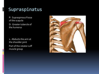
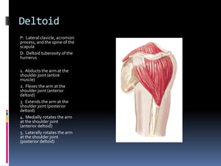
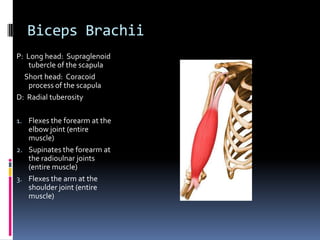
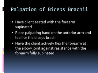
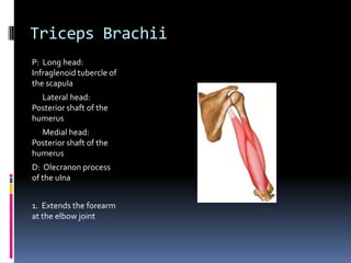
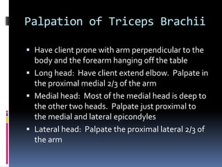
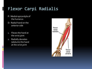
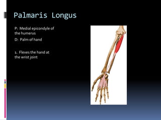
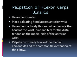
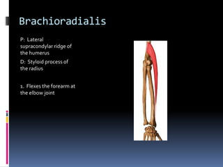
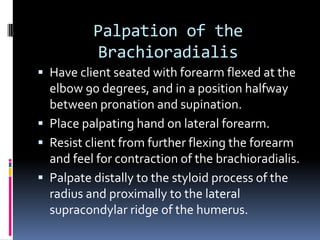
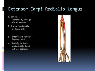
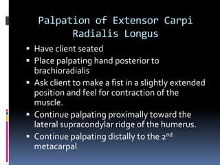
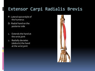
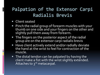
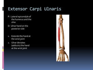
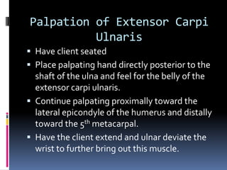
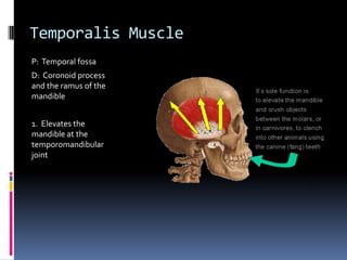
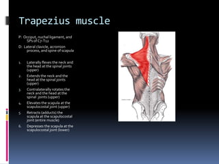
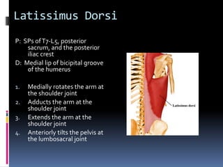
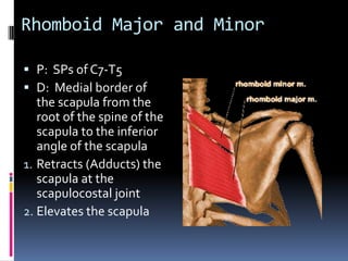
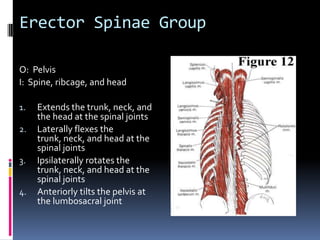
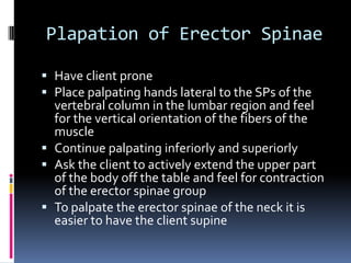
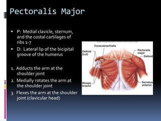
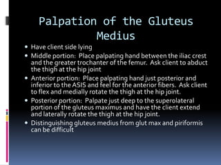
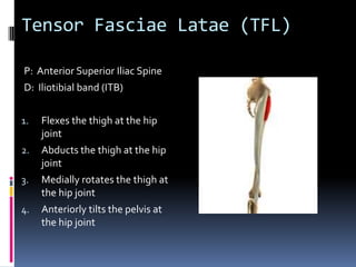
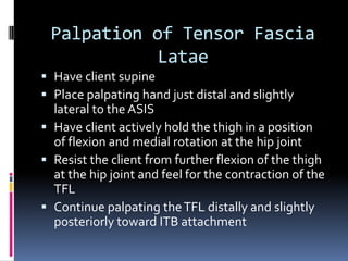
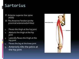
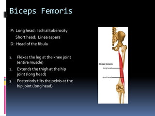
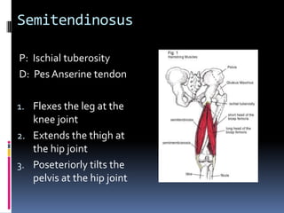
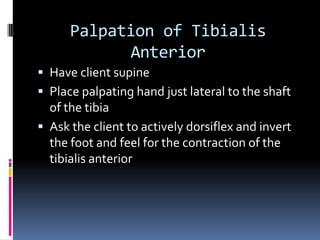
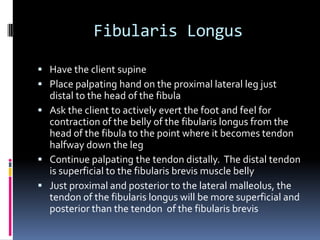
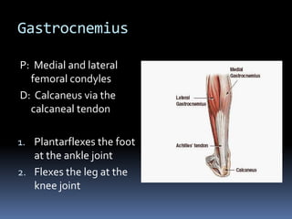
Palpation Review for Final ExamMuscles from Weeks One, Two, And Three
SupraspinatusP:  Supraspinous Fossa of the scapulaD:  Greater tubercle of the humerus1.  Abducts the arm at the shoulder jointPart of the rotator cuff muscle group
Palpation of SupraspinatusClient seated with arm medially rotated and hanging at the sidePlace palpating hand just superior to the spine of the scapulaAsk the client to perform a short, quick range of motion of active abduction of the arm at the shoulder joint
DeltoidP:  Lateral clavicle, acromion process, and the spine of the scapulaD:  Deltoid tuberosity of the humerus1.  Abducts the arm at the shoulder joint (entire muscle)2.  Flexes the arm at the shoulder joint (anterior deltoid)3.  Extends the arm at the shoulder joint (posterior deltoid)4.  Medially rotates the arm at the shoulder joint (anterior deltoid)5.  Laterally rotates the arm at the shoulder joint (posterior deltoid)
Palpation of the DeltoidHave client seatedMiddle deltoid:  Place palpating hand just proximal to the deltoid tuberosity.  Have client actively abduct the armAnterior deltoid:  Place hand on anterior shoulder.  Have client actively horizontally flex the arm at the shoulder jointPosterior deltoid:  Place palpating hand just inferior to the spine of the scapula.  Have client actively horizontally extend the arm at the shoulder joint
Biceps BrachiiP:  Long head:  Supraglenoid tubercle of the scapula     Short head:  Coracoid process of the scapulaD:  Radial tuberosityFlexes the forearm at the elbow joint (entire muscle)Supinates the forearm at the radioulnar joints (entire muscle)Flexes the arm at the shoulder joint (entire muscle)
Palpation of Biceps BrachiiHave client seated with the forearm supinatedPlace palpating hand on the anterior arm and feel for the biceps brachiiHave the client actively flex the forearm at the elbow joint against resistance with the forearm fully supinated
Triceps Brachii	P:  Long head:  Infraglenoid tubercle of the scapula      Lateral head:  Posterior shaft of the humerus      Medial head:  Posterior shaft of the humerusD:  Olecranon process of the ulna1.  Extends the forearm at the elbow joint
Palpation of Triceps BrachiiHave client prone with arm perpendicular to the body and the forearm hanging off the tableLong head:  Have client extend elbow.  Palpate in the proximal medial 2/3 of the armMedial head:  Most of the medial head is deep to the other two heads.  Palpate just proximal to the medial and lateral epicondylesLateral head:  Palpate the proximal lateral 2/3 of the arm
Flexor Carpi RadialisP:  Medial epicondyle of the humerusD:  Radial hand on the anterior sideFlexes the hand at the wrist jointRadially deviates (abducts) the hand at the wrist joint
Plapation of Flexor Carpi RadialisClient seatedPlace palpating hand across anterior wristHave client actively flex and radially deviate the hand at the wrist joint and feel for the distal tendon on the radial side just lateral to the palmaris longusPalpate proximally toward the medial epicondyle It is difficult to palpate the proximal tendon because it blends with the common flexor tendon of the elbow
Palmaris LongusP:  Medial epicondyle of the humerusD:  Palm of hand1.  Flexes the hand at the wrist joint
Palpation of Palmaris Longus	Have client seatedPlace palpating hand on anterior wristAsk the client to cup the hand (bring thenar and hypothenar eminence together)Ask client to flex the hand at the  wrist joint against resistance with the fingers fully extended; feel for the distal tendon of the palmaris longus in the center of the wrist.Palpate distally to the common flexor tendon of the elbow
Flexor Carpi Ulnaris	P:  Medial epicondyle of the humerusD:  Ulnar hand on the anterior sideFlexes the hand at the wrist jointUlnar deviates  (adducts) the hand at the wrist joint
Palpation of Flexor Carpi UlnarisHave client seatedPlace palpating hand across anterior wristHave client actively flex and ulnar deviate the hand at the wrist joint and feel for the distal tendon on the medial side of the anterior wrist.Palpate proximally toward the medial epicondyle and the common flexor tendon of the elbow.
BrachioradialisP:  Lateral supracondylar ridge of the humerusD:  Styloid process of the radius1.  Flexes the forearm at the elbow joint
Palpation of the BrachioradialisHave client seated with forearm flexed at the elbow 90 degrees, and in a position halfway between pronation and supination.Place palpating hand on lateral forearm.Resist client from further flexing the forearm and feel for contraction of the brachioradialis.Palpate distally to the styloid process of the radius and proximally to the lateral supracondylar ridge of the humerus.
Extensor Carpi Radialis LongusP:  Lateral supracondylar ridge of the humerusD:  Radial hand on the posterior sideExtends the hand at the wrist jointRadially deviates (abducts) the hand at the wrist joint
Palpation of Extensor Carpi Radialis LongusHave client seated Place palpating hand posterior to brachioradialisAsk client to make a fist in a slightly extended position and feel for contraction of the muscle.Continue palpating proximally toward the lateral supracondylar ridge of the humerus.Continue palpating distally to the 2nd metacarpal
Extensor Carpi Radialis BrevisP:  Lateral epicondyle of the humerusD:  Radial hand on the posterior sideExtends the hand at the wrist jointRadially deviates (abducts) the hand at the wrist joint
Palpation of the Extensor Carpi Radialis BrevisClient seatedPinch the radial group of forearm muscles with your thumb on one side and your fingers on the other and slightly pull them away from forearm.The fingers on the posterior aspect of the radial group are on the extensor carpi radialis brevisHave client actively extend and/or radially deviate the hand at the wrist to feel for contraction of the muscle.The distal tendon can be palpated by having the client make a fist with the wrist slightly extended.  Attaches to 3rd metacarpal.
Extensor Carpi UlnarisP:  Lateral epicondyle of the humerus and the ulnaD:  Ulnar hand on the posterior sideExtends the hand at the wrist jointUlnar deviates (adducts) the hand at the wrist joint
Palpation of Extensor Carpi UlnarisHave client seatedPlace palpating hand directly posterior to the shaft of the ulna and feel for the belly of the extensor carpi ulnaris.Continue palpating proximally toward the lateral epicondyle of the humerus and distally toward the 5th metacarpal.Have the client extend and ulnar deviate the wrist to further bring out this muscle.
Temporalis MuscleP:  Temporal fossaD:  Coronoid process and the ramus of the mandible1.  Elevates the mandible at the temporomandibular joint
Palpation of TemporalisHave client supinePlace palpating fingers on the temporal fossa.Ask client to clench and relax the teeth.Feel for the contraction and relaxation of the muscle.The inferior attachment on the coronoid process of the mandible can be palpated if the client opens mouth widely.  Difficult to distinguish from the masseter.
Trapezius muscleP:  Occiput, nuchal ligament, and SPs of C7-T12D:  Lateral clavicle, acromion process, and spine of scapulaLaterally flexes the neck and the head at the spinal joints (upper)Extends the neck and the head at the spinal joints (upper)Contralaterally rotates the neck and the head at the spinal  joints (upper)Elevates the scapula at the scapulocostal joint (upper)Retracts (adducts) the scapula at the scapulocostal joint (entire muscle)Depresses the scapula at the scapulocostal joint (lower)
Palpation of the TrapeziusHave the client pronePlace palpating hand over the upper trapezius.Have client actively abduct the arm at the shoulder joint and retract the scapulaRepeat above to palpate the middle and lower trapeziusTo further bring out the upper trapezius, have the client slightly extend the neck
Levator scapulaeP:  TPs of C1-C4D:  Medial border of the scapula, from the superior angle to the root of the spine of the scapulaElevates the scapula at the scapulocostal jointExtends the neck at the spinal jointsLaterally flexes the neck at the spinal joints
Palpation of the Levator ScapulaeClient seated with the forearm in the small of the backPlace palpating hand just superior to the superior angle of the scapulaHave client perform short, quick range of motion of active elevation of the scapula and feel for the contraction of the levator scapulae.Continue palpating superiorly into the posterior triangle of the neck
Sternocleidomastoid P:  Sternal head:  Manubrium of the sternum       Clavicular head:  Medial clavicleD:  Mastoid process of the temporal boneFlexes the head at the spinal jointsLaterally flexes the neck and the head at the spinal jointsContralaterally rotates the neck and the head at the spinal joints
Palpation of the SternocleidomastoidHave client seated with the neck and head rotated to one side.Stand on the opposite side.Place palpating hand on the anterolateral neck of the side where you are standing.Now resist the client from laterally flexing the neck and head toward the side you are standing and feel for contraction of the SCM.
Week 5, 6, and 7Palpation Review For Final Exam
Latissimus DorsiP:  SPs of T7-L5, posterior sacrum, and the posterior iliac crestD:  Medial lip of bicipital groove of the humerusMedially rotates the arm at the shoulder jointAdducts the arm at the shoulder jointExtends the arm at the shoulder jointAnteriorly tilts the pelvis at the lumbosacral joint
Have client seated with hands at their sides
Place palpating hand on the client’s posterior axillary fold
Ask the client to push down on the table with both hands, attempting to elevate the pelvis and trunk off the table.  Feel for the contraction of the latissimusdorsi
Continue palpating the muscle toward its humeral attachment.  It can be difficult to distinguish the tendon of the latissimusdorsi from the tendon of the teres major because they often blend together.Palpation of LatissimusDorsi
Rhomboid Major and MinorP:  SPs of C7-T5D:  Medial border of the scapula from the root of the spine of the scapula to the inferior angle of the scapulaRetracts (Adducts) the scapula at the scapulocostal jointElevates the scapula
Have client seated with hand placed in the small of the backPlace palpating hand between the scapula at a level that is between the inferior angle and the root of the spine of the scapula and the spineAsk the client to lift the hand away from the back and feel for the contraction of the rhomboidsPalpation of Rhomboid Major and Minor
Erector Spinae GroupO:  PelvisI:  Spine, ribcage, and headExtends the trunk, neck, and the head at the spinal jointsLaterally flexes the trunk, neck, and head at the spinal jointsIpsilaterally rotates the trunk, neck, and head at the spinal jointsAnteriorly tilts the pelvis at the lumbosacral joint
Have client pronePlace palpating hands lateral to the SPs of the vertebral column in the lumbar region and feel for the vertical orientation of the fibers of the muscleContinue palpating inferiorly and superiorlyAsk the client to actively extend the upper part of the body off the table and feel for contraction of the erector spinae groupTo palpate the erector spinae of the neck it is easier to have the client supine Plapation of Erector Spinae
Pectoralis MajorP:  Medial clavicle, sternum, and the costal cartilages of ribs 1-7D:  Lateral lip of the bicipital groove of the humerus1.  Adducts the arm at the shoulder joint2.  Medially rotates the arm at the shoulder joint3.  Flexes the arm at the shoulder joint (clavicular head)
Have client seatedHave client raise arm to 90 degrees of abduction at the shoulder jointPlace palpating hand on the belly of the muscleAsk the client to actively horizontally flex the arm at the shoulder joint against resistance and feel for the contraction of the pectoralis majorContinue palpating the muscle medially and superiorly toward its attachment s on the trunk and distally toward its humeral attachmentPalpation of the Pectoralis Major
Rectus AbdominisP:  Pubis D:  Xiphoid process and the cartilage of ribs 5-7Flexes the trunk at the spinal jointsPosteriorly tilts the pelvis at the lumbosacral joint
Have the client supine with a pillow under the knees Place palpating hands between the xiphoid process of the sternum and the adjacent ribs superiorly and the pubis inferiorlyAsk the client to alternate between mild flexion of the trunk and relaxation so that you can feel for the contraction of the musclePalpate the rectus abdominis superiorly toward its ribcage attachment and inferiorly toward its pelvic attachmentPalpation of the Rectus Abdominis
Gluteus MaximusP:  Posterior iliac crest, posterolateral sacrum, and the coccyxD:  Iliotibial band (ITB) and glutealtuberosity of the femurExtends the thigh at the hip jointLaterally rotates the thigh at the hip jointAbducts the thigh at the hip joint (upper 1/3)Adducts the thigh at the hip joint (lower 2/3)Posteriorly tilts the pelvis at the hip joint
Have client lay prone
Place palpating hand on the lateral sacrum
Palpate the gluteus maximus from its medial attachment (lateral sacrum) to its lateral attachment (ITB)
Have the client actively extend and laterally rotate the thigh at the hip joint to further bring out the muscle.
The gluteus maximus does not cover the entire buttocks;  the gluteus medius is just inferior to most of the posterior iliac crestPalpation of Gluteus Maximus
Gluteus MediusP:  External IliumD:  Greater trochanter of the femurAbducts the thigh at the hip joint (entire muscle)Flexes the thigh at the hip joint (anterior fibers)Medially rotates the thigh at the hip joint (anterior fibers)Extends the thigh at the hip joint (posterior fibers)Laterally rotates the thigh at the hip joint (posterior fibers)Posteriorly tilts the pelvis at the hip joint (posterior fibers)Anteriorly tilts the pelvis at the hip joint (anterior fibers)
Have client side lying
Middle portion:  Place palpating hand between the iliac crest and the greater trochanter of the femur.  Ask client to abduct the thigh at the hip joint
Anterior portion:  Place palpating hand just posterior and inferior to the ASIS and feel for the anterior fibers.  Ask client to flex and medially rotate the thigh at the hip joint.
Posterior portion:  Palpate just deep to the superolateral portion of the gluteus maximus and have the client extend and laterally rotate the thigh at the hip joint.
Distinguishing gluteus medius from glut max and piriformis can be difficultPalpation of the Gluteus Medius
Tensor Fasciae Latae (TFL)P:  Anterior Superior Iliac SpineD:  Iliotibial band (ITB)Flexes the thigh at the hip jointAbducts the thigh at the hip jointMedially rotates the thigh at the hip jointAnteriorly tilts the pelvis at the hip joint
Have client supinePlace palpating hand just distal and slightly lateral to the ASISHave client actively hold the thigh in a position of flexion and medial rotation at the hip jointResist the client from further flexion of the thigh at the hip joint and feel for the contraction of the TFLContinue palpating the TFL distally and slightly posteriorly toward ITB attachmentPalpation of Tensor Fascia Latae
SartoriusP:  Anterior superior iliac spine (ASIS)D:  Pes Anserine Tendon (at the proximal anteromedial tibia)Flexes the thigh at the hip jointAbducts the thigh at the hip jointLaterally flexes the thigh at the hip jointFlexes the leg at the knee jointAnteriorly tilts the pelvis at the hip joint
Have client supinePlace palpating hand just distal and slightly medial to the ASISAsk client to actively flex and laterally  rotate the thigh at the hip joint and feel for the contraction of the sartoriusContinue palpating the sartorius from the ASIS to the pes anserine tendonTo further bring out the muscle give the client resistancePalpation of the Sartorius
Rectus FemorisP:  Anterior Inferior iliac spine (AIIS)D:  TibialTuberosityExtends the leg at the knee jointFlexes the thigh at the hip jointAnteriorly tilts the pelvis at the hip joint
Have client supine with a pillow under their kneesPlace palpating hand just proximal to the patellaResist the client from actively extending the leg at the knee joint and feel for the contraction of the rectus femorisContinue palpating the muscle toward the ASISTo palpate the proximal attachment passively flex the thigh at the hip joint and palpate slightly distal to the ASISPalpation of the Rectus Femoris
Vastus LateralisP:  Linea Aspera of the femur	D:  Tibial tuberostiy1.  Extends the leg at the knee joint
Have client supinePlace palpating hand just distal to the greater trochanterAsk the client to contract the quadriceps femoris group and feel for contraction of the vastus lateralisContinue palpating distally toward the patellaPalpation of the Vastus Lateralis
Vastus MedialisP:  Linea Aspera of the femurD:  Tibial tuberosity1.  Extends the leg at the knee joint
Have client supinePlace palpating hand just proximal and medial to the patellaAsk the client to contract the quadriceps femoris group and feel for the contraction of the vastus medialisContinue palpating the vastus medialis proximally as far as possibleThe vastus medialis is best palpated medially in the distal 1/3 of the thighPalpation of the Vastus Medialis
Vastus IntermediusP:  Anterior shaft and the linea aspera of the femurD:  Tibial tuberosity1.  Extends the leg at the knee joint
Deep and difficult to palpateHave client supinePlace palpating hand just proximal to the patellaIf the rectus femoris can be lifted and/or moved aside, the distal vastus intermedius may be palpated deep to the rectus femoris when approached from either the medial or lateral sideTo feel the vastus intermedius deep to the rectus femoris, make sure that the direction of your pressure is directed toward the middle of the femurPalpation of Vastus Intermedius
Biceps FemorisP:  Long head:  Ischialtuberosity      Short head:  Linea asperaD:  Head of the fibulaFlexes the leg at the knee joint (entire muscle)Extends the thigh at the hip joint (long head)Posteriorly tilts the pelvis at the hip joint (long head)
The biceps femoris is superficial for most of its course in the posterolateral thigh and is easy to palpateHave the client prone with the leg partially flexed at the knee jointPlace palpating hand on the distal posterolateral thighResist the client from performing further flexion of the leg at the knee joint and feel for the distal tendon of the biceps femorisContinue palpating the biceps femoris toward  the ischial tuberosityPalpation of the Biceps Femoris
SemitendinosusP:  IschialtuberosityD:  Pes Anserine tendonFlexes the leg at the knee jointExtends the thigh at the hip jointPoseteriorly tilts the pelvis at the hip joint

Palpation Review For Final Exam

  • 1.
    Palpation Review forFinal ExamMuscles from Weeks One, Two, And Three
  • 2.
    SupraspinatusP: SupraspinousFossa of the scapulaD: Greater tubercle of the humerus1. Abducts the arm at the shoulder jointPart of the rotator cuff muscle group
  • 3.
    Palpation of SupraspinatusClientseated with arm medially rotated and hanging at the sidePlace palpating hand just superior to the spine of the scapulaAsk the client to perform a short, quick range of motion of active abduction of the arm at the shoulder joint
  • 4.
    DeltoidP: Lateralclavicle, acromion process, and the spine of the scapulaD: Deltoid tuberosity of the humerus1. Abducts the arm at the shoulder joint (entire muscle)2. Flexes the arm at the shoulder joint (anterior deltoid)3. Extends the arm at the shoulder joint (posterior deltoid)4. Medially rotates the arm at the shoulder joint (anterior deltoid)5. Laterally rotates the arm at the shoulder joint (posterior deltoid)
  • 5.
    Palpation of theDeltoidHave client seatedMiddle deltoid: Place palpating hand just proximal to the deltoid tuberosity. Have client actively abduct the armAnterior deltoid: Place hand on anterior shoulder. Have client actively horizontally flex the arm at the shoulder jointPosterior deltoid: Place palpating hand just inferior to the spine of the scapula. Have client actively horizontally extend the arm at the shoulder joint
  • 6.
    Biceps BrachiiP: Long head: Supraglenoid tubercle of the scapula Short head: Coracoid process of the scapulaD: Radial tuberosityFlexes the forearm at the elbow joint (entire muscle)Supinates the forearm at the radioulnar joints (entire muscle)Flexes the arm at the shoulder joint (entire muscle)
  • 7.
    Palpation of BicepsBrachiiHave client seated with the forearm supinatedPlace palpating hand on the anterior arm and feel for the biceps brachiiHave the client actively flex the forearm at the elbow joint against resistance with the forearm fully supinated
  • 8.
    Triceps Brachii P: Long head: Infraglenoid tubercle of the scapula Lateral head: Posterior shaft of the humerus Medial head: Posterior shaft of the humerusD: Olecranon process of the ulna1. Extends the forearm at the elbow joint
  • 9.
    Palpation of TricepsBrachiiHave client prone with arm perpendicular to the body and the forearm hanging off the tableLong head: Have client extend elbow. Palpate in the proximal medial 2/3 of the armMedial head: Most of the medial head is deep to the other two heads. Palpate just proximal to the medial and lateral epicondylesLateral head: Palpate the proximal lateral 2/3 of the arm
  • 10.
    Flexor Carpi RadialisP: Medial epicondyle of the humerusD: Radial hand on the anterior sideFlexes the hand at the wrist jointRadially deviates (abducts) the hand at the wrist joint
  • 11.
    Plapation of FlexorCarpi RadialisClient seatedPlace palpating hand across anterior wristHave client actively flex and radially deviate the hand at the wrist joint and feel for the distal tendon on the radial side just lateral to the palmaris longusPalpate proximally toward the medial epicondyle It is difficult to palpate the proximal tendon because it blends with the common flexor tendon of the elbow
  • 12.
    Palmaris LongusP: Medial epicondyle of the humerusD: Palm of hand1. Flexes the hand at the wrist joint
  • 13.
    Palpation of PalmarisLongus Have client seatedPlace palpating hand on anterior wristAsk the client to cup the hand (bring thenar and hypothenar eminence together)Ask client to flex the hand at the wrist joint against resistance with the fingers fully extended; feel for the distal tendon of the palmaris longus in the center of the wrist.Palpate distally to the common flexor tendon of the elbow
  • 14.
    Flexor Carpi Ulnaris P: Medial epicondyle of the humerusD: Ulnar hand on the anterior sideFlexes the hand at the wrist jointUlnar deviates (adducts) the hand at the wrist joint
  • 15.
    Palpation of FlexorCarpi UlnarisHave client seatedPlace palpating hand across anterior wristHave client actively flex and ulnar deviate the hand at the wrist joint and feel for the distal tendon on the medial side of the anterior wrist.Palpate proximally toward the medial epicondyle and the common flexor tendon of the elbow.
  • 16.
    BrachioradialisP: Lateralsupracondylar ridge of the humerusD: Styloid process of the radius1. Flexes the forearm at the elbow joint
  • 17.
    Palpation of theBrachioradialisHave client seated with forearm flexed at the elbow 90 degrees, and in a position halfway between pronation and supination.Place palpating hand on lateral forearm.Resist client from further flexing the forearm and feel for contraction of the brachioradialis.Palpate distally to the styloid process of the radius and proximally to the lateral supracondylar ridge of the humerus.
  • 18.
    Extensor Carpi RadialisLongusP: Lateral supracondylar ridge of the humerusD: Radial hand on the posterior sideExtends the hand at the wrist jointRadially deviates (abducts) the hand at the wrist joint
  • 19.
    Palpation of ExtensorCarpi Radialis LongusHave client seated Place palpating hand posterior to brachioradialisAsk client to make a fist in a slightly extended position and feel for contraction of the muscle.Continue palpating proximally toward the lateral supracondylar ridge of the humerus.Continue palpating distally to the 2nd metacarpal
  • 20.
    Extensor Carpi RadialisBrevisP: Lateral epicondyle of the humerusD: Radial hand on the posterior sideExtends the hand at the wrist jointRadially deviates (abducts) the hand at the wrist joint
  • 21.
    Palpation of theExtensor Carpi Radialis BrevisClient seatedPinch the radial group of forearm muscles with your thumb on one side and your fingers on the other and slightly pull them away from forearm.The fingers on the posterior aspect of the radial group are on the extensor carpi radialis brevisHave client actively extend and/or radially deviate the hand at the wrist to feel for contraction of the muscle.The distal tendon can be palpated by having the client make a fist with the wrist slightly extended. Attaches to 3rd metacarpal.
  • 22.
    Extensor Carpi UlnarisP: Lateral epicondyle of the humerus and the ulnaD: Ulnar hand on the posterior sideExtends the hand at the wrist jointUlnar deviates (adducts) the hand at the wrist joint
  • 23.
    Palpation of ExtensorCarpi UlnarisHave client seatedPlace palpating hand directly posterior to the shaft of the ulna and feel for the belly of the extensor carpi ulnaris.Continue palpating proximally toward the lateral epicondyle of the humerus and distally toward the 5th metacarpal.Have the client extend and ulnar deviate the wrist to further bring out this muscle.
  • 24.
    Temporalis MuscleP: Temporal fossaD: Coronoid process and the ramus of the mandible1. Elevates the mandible at the temporomandibular joint
  • 25.
    Palpation of TemporalisHaveclient supinePlace palpating fingers on the temporal fossa.Ask client to clench and relax the teeth.Feel for the contraction and relaxation of the muscle.The inferior attachment on the coronoid process of the mandible can be palpated if the client opens mouth widely. Difficult to distinguish from the masseter.
  • 26.
    Trapezius muscleP: Occiput, nuchal ligament, and SPs of C7-T12D: Lateral clavicle, acromion process, and spine of scapulaLaterally flexes the neck and the head at the spinal joints (upper)Extends the neck and the head at the spinal joints (upper)Contralaterally rotates the neck and the head at the spinal joints (upper)Elevates the scapula at the scapulocostal joint (upper)Retracts (adducts) the scapula at the scapulocostal joint (entire muscle)Depresses the scapula at the scapulocostal joint (lower)
  • 27.
    Palpation of theTrapeziusHave the client pronePlace palpating hand over the upper trapezius.Have client actively abduct the arm at the shoulder joint and retract the scapulaRepeat above to palpate the middle and lower trapeziusTo further bring out the upper trapezius, have the client slightly extend the neck
  • 28.
    Levator scapulaeP: TPs of C1-C4D: Medial border of the scapula, from the superior angle to the root of the spine of the scapulaElevates the scapula at the scapulocostal jointExtends the neck at the spinal jointsLaterally flexes the neck at the spinal joints
  • 29.
    Palpation of theLevator ScapulaeClient seated with the forearm in the small of the backPlace palpating hand just superior to the superior angle of the scapulaHave client perform short, quick range of motion of active elevation of the scapula and feel for the contraction of the levator scapulae.Continue palpating superiorly into the posterior triangle of the neck
  • 30.
    Sternocleidomastoid P: Sternal head: Manubrium of the sternum Clavicular head: Medial clavicleD: Mastoid process of the temporal boneFlexes the head at the spinal jointsLaterally flexes the neck and the head at the spinal jointsContralaterally rotates the neck and the head at the spinal joints
  • 31.
    Palpation of theSternocleidomastoidHave client seated with the neck and head rotated to one side.Stand on the opposite side.Place palpating hand on the anterolateral neck of the side where you are standing.Now resist the client from laterally flexing the neck and head toward the side you are standing and feel for contraction of the SCM.
  • 32.
    Week 5, 6,and 7Palpation Review For Final Exam
  • 33.
    Latissimus DorsiP: SPs of T7-L5, posterior sacrum, and the posterior iliac crestD: Medial lip of bicipital groove of the humerusMedially rotates the arm at the shoulder jointAdducts the arm at the shoulder jointExtends the arm at the shoulder jointAnteriorly tilts the pelvis at the lumbosacral joint
  • 34.
    Have client seatedwith hands at their sides
  • 35.
    Place palpating handon the client’s posterior axillary fold
  • 36.
    Ask the clientto push down on the table with both hands, attempting to elevate the pelvis and trunk off the table. Feel for the contraction of the latissimusdorsi
  • 37.
    Continue palpating themuscle toward its humeral attachment. It can be difficult to distinguish the tendon of the latissimusdorsi from the tendon of the teres major because they often blend together.Palpation of LatissimusDorsi
  • 38.
    Rhomboid Major andMinorP: SPs of C7-T5D: Medial border of the scapula from the root of the spine of the scapula to the inferior angle of the scapulaRetracts (Adducts) the scapula at the scapulocostal jointElevates the scapula
  • 39.
    Have client seatedwith hand placed in the small of the backPlace palpating hand between the scapula at a level that is between the inferior angle and the root of the spine of the scapula and the spineAsk the client to lift the hand away from the back and feel for the contraction of the rhomboidsPalpation of Rhomboid Major and Minor
  • 40.
    Erector Spinae GroupO: PelvisI: Spine, ribcage, and headExtends the trunk, neck, and the head at the spinal jointsLaterally flexes the trunk, neck, and head at the spinal jointsIpsilaterally rotates the trunk, neck, and head at the spinal jointsAnteriorly tilts the pelvis at the lumbosacral joint
  • 41.
    Have client pronePlacepalpating hands lateral to the SPs of the vertebral column in the lumbar region and feel for the vertical orientation of the fibers of the muscleContinue palpating inferiorly and superiorlyAsk the client to actively extend the upper part of the body off the table and feel for contraction of the erector spinae groupTo palpate the erector spinae of the neck it is easier to have the client supine Plapation of Erector Spinae
  • 42.
    Pectoralis MajorP: Medial clavicle, sternum, and the costal cartilages of ribs 1-7D: Lateral lip of the bicipital groove of the humerus1. Adducts the arm at the shoulder joint2. Medially rotates the arm at the shoulder joint3. Flexes the arm at the shoulder joint (clavicular head)
  • 43.
    Have client seatedHaveclient raise arm to 90 degrees of abduction at the shoulder jointPlace palpating hand on the belly of the muscleAsk the client to actively horizontally flex the arm at the shoulder joint against resistance and feel for the contraction of the pectoralis majorContinue palpating the muscle medially and superiorly toward its attachment s on the trunk and distally toward its humeral attachmentPalpation of the Pectoralis Major
  • 44.
    Rectus AbdominisP: Pubis D: Xiphoid process and the cartilage of ribs 5-7Flexes the trunk at the spinal jointsPosteriorly tilts the pelvis at the lumbosacral joint
  • 45.
    Have the clientsupine with a pillow under the knees Place palpating hands between the xiphoid process of the sternum and the adjacent ribs superiorly and the pubis inferiorlyAsk the client to alternate between mild flexion of the trunk and relaxation so that you can feel for the contraction of the musclePalpate the rectus abdominis superiorly toward its ribcage attachment and inferiorly toward its pelvic attachmentPalpation of the Rectus Abdominis
  • 46.
    Gluteus MaximusP: Posterior iliac crest, posterolateral sacrum, and the coccyxD: Iliotibial band (ITB) and glutealtuberosity of the femurExtends the thigh at the hip jointLaterally rotates the thigh at the hip jointAbducts the thigh at the hip joint (upper 1/3)Adducts the thigh at the hip joint (lower 2/3)Posteriorly tilts the pelvis at the hip joint
  • 47.
  • 48.
    Place palpating handon the lateral sacrum
  • 49.
    Palpate the gluteusmaximus from its medial attachment (lateral sacrum) to its lateral attachment (ITB)
  • 50.
    Have the clientactively extend and laterally rotate the thigh at the hip joint to further bring out the muscle.
  • 51.
    The gluteus maximusdoes not cover the entire buttocks; the gluteus medius is just inferior to most of the posterior iliac crestPalpation of Gluteus Maximus
  • 52.
    Gluteus MediusP: External IliumD: Greater trochanter of the femurAbducts the thigh at the hip joint (entire muscle)Flexes the thigh at the hip joint (anterior fibers)Medially rotates the thigh at the hip joint (anterior fibers)Extends the thigh at the hip joint (posterior fibers)Laterally rotates the thigh at the hip joint (posterior fibers)Posteriorly tilts the pelvis at the hip joint (posterior fibers)Anteriorly tilts the pelvis at the hip joint (anterior fibers)
  • 53.
  • 54.
    Middle portion: Place palpating hand between the iliac crest and the greater trochanter of the femur. Ask client to abduct the thigh at the hip joint
  • 55.
    Anterior portion: Place palpating hand just posterior and inferior to the ASIS and feel for the anterior fibers. Ask client to flex and medially rotate the thigh at the hip joint.
  • 56.
    Posterior portion: Palpate just deep to the superolateral portion of the gluteus maximus and have the client extend and laterally rotate the thigh at the hip joint.
  • 57.
    Distinguishing gluteus mediusfrom glut max and piriformis can be difficultPalpation of the Gluteus Medius
  • 58.
    Tensor Fasciae Latae(TFL)P: Anterior Superior Iliac SpineD: Iliotibial band (ITB)Flexes the thigh at the hip jointAbducts the thigh at the hip jointMedially rotates the thigh at the hip jointAnteriorly tilts the pelvis at the hip joint
  • 59.
    Have client supinePlacepalpating hand just distal and slightly lateral to the ASISHave client actively hold the thigh in a position of flexion and medial rotation at the hip jointResist the client from further flexion of the thigh at the hip joint and feel for the contraction of the TFLContinue palpating the TFL distally and slightly posteriorly toward ITB attachmentPalpation of Tensor Fascia Latae
  • 60.
    SartoriusP: Anteriorsuperior iliac spine (ASIS)D: Pes Anserine Tendon (at the proximal anteromedial tibia)Flexes the thigh at the hip jointAbducts the thigh at the hip jointLaterally flexes the thigh at the hip jointFlexes the leg at the knee jointAnteriorly tilts the pelvis at the hip joint
  • 61.
    Have client supinePlacepalpating hand just distal and slightly medial to the ASISAsk client to actively flex and laterally rotate the thigh at the hip joint and feel for the contraction of the sartoriusContinue palpating the sartorius from the ASIS to the pes anserine tendonTo further bring out the muscle give the client resistancePalpation of the Sartorius
  • 62.
    Rectus FemorisP: Anterior Inferior iliac spine (AIIS)D: TibialTuberosityExtends the leg at the knee jointFlexes the thigh at the hip jointAnteriorly tilts the pelvis at the hip joint
  • 63.
    Have client supinewith a pillow under their kneesPlace palpating hand just proximal to the patellaResist the client from actively extending the leg at the knee joint and feel for the contraction of the rectus femorisContinue palpating the muscle toward the ASISTo palpate the proximal attachment passively flex the thigh at the hip joint and palpate slightly distal to the ASISPalpation of the Rectus Femoris
  • 64.
    Vastus LateralisP: Linea Aspera of the femur D: Tibial tuberostiy1. Extends the leg at the knee joint
  • 65.
    Have client supinePlacepalpating hand just distal to the greater trochanterAsk the client to contract the quadriceps femoris group and feel for contraction of the vastus lateralisContinue palpating distally toward the patellaPalpation of the Vastus Lateralis
  • 66.
    Vastus MedialisP: Linea Aspera of the femurD: Tibial tuberosity1. Extends the leg at the knee joint
  • 67.
    Have client supinePlacepalpating hand just proximal and medial to the patellaAsk the client to contract the quadriceps femoris group and feel for the contraction of the vastus medialisContinue palpating the vastus medialis proximally as far as possibleThe vastus medialis is best palpated medially in the distal 1/3 of the thighPalpation of the Vastus Medialis
  • 68.
    Vastus IntermediusP: Anterior shaft and the linea aspera of the femurD: Tibial tuberosity1. Extends the leg at the knee joint
  • 69.
    Deep and difficultto palpateHave client supinePlace palpating hand just proximal to the patellaIf the rectus femoris can be lifted and/or moved aside, the distal vastus intermedius may be palpated deep to the rectus femoris when approached from either the medial or lateral sideTo feel the vastus intermedius deep to the rectus femoris, make sure that the direction of your pressure is directed toward the middle of the femurPalpation of Vastus Intermedius
  • 70.
    Biceps FemorisP: Long head: Ischialtuberosity Short head: Linea asperaD: Head of the fibulaFlexes the leg at the knee joint (entire muscle)Extends the thigh at the hip joint (long head)Posteriorly tilts the pelvis at the hip joint (long head)
  • 71.
    The biceps femorisis superficial for most of its course in the posterolateral thigh and is easy to palpateHave the client prone with the leg partially flexed at the knee jointPlace palpating hand on the distal posterolateral thighResist the client from performing further flexion of the leg at the knee joint and feel for the distal tendon of the biceps femorisContinue palpating the biceps femoris toward the ischial tuberosityPalpation of the Biceps Femoris
  • 72.
    SemitendinosusP: IschialtuberosityD: Pes Anserine tendonFlexes the leg at the knee jointExtends the thigh at the hip jointPoseteriorly tilts the pelvis at the hip joint