This document summarizes the origin, insertion, action, synergists, and antagonists of several muscles in the pelvis, thigh, and lower leg regions. Key muscles discussed include the iliopsoas, gluteus maximus, medius and minimus, sartorius, quadriceps femoris group (rectus femoris, vastus lateralis, medialis and intermedius), and hamstrings. These muscles act to flex, extend, abduct, rotate and stabilize the hip, knee and ankle joints during movements like walking, running, sitting and climbing stairs. Understanding muscle origins and insertions is important for comprehending their functional actions.
manual muscle testing by K Adhi lakshmi vapms copvrkv2007
Manual muscle testing (MMT) involves grading the strength of individual muscles or muscle groups on a scale based on their ability to perform movements against gravity or resistance. Key aspects of MMT include positioning and stabilizing the patient, demonstrating the movement, applying the appropriate grade of resistance, and documenting the results objectively. MMT is useful for assessing muscle weakness from various neuromuscular and musculoskeletal conditions and monitoring the effectiveness of treatment over time. Contraindications include certain neurological or orthopedic injuries or diseases that could be exacerbated by strength testing.
The document summarizes key information about the femoral nerve including its origin from spinal nerves L2-L4, branches within the thigh that innervate muscles like the sartorius and rectus femoris, cutaneous branches that include the medial and intermediate cutaneous nerves, and its role in supplying the hip and knee joints according to Hilton's Law. It also notes the relationship of the femoral nerve to the femoral artery as it courses through the thigh.
The document discusses the anatomy and biomechanics of the hip joint. It describes the ball and socket structure of the hip joint formed by the acetabulum and femoral head. It details the angles of the hip joint including the central edge angle and angle of anteversion. It discusses the muscles, ligaments, biomechanics including ranges of motion, and forces across the hip joint during activities like standing, walking, and squatting. Pathomechanics of conditions like hip fractures and dislocations are also mentioned.
2. shoulder joint & its applied anatomy 07[1]MBBS IMS MSU
The shoulder joint is a synovial ball-and-socket joint between the head of the humerus bone and the shallow glenoid cavity of the scapula. It has a thin, lax capsule strengthened by rotator cuff muscles and ligaments. The joint allows wide range of movement including flexion, extension, abduction, adduction, medial and lateral rotation, and circumduction. It is prone to dislocations, especially anterior dislocations, due to its anatomy. Recurrent dislocations often require surgical repair to reattach the capsular ligaments.
This document describes the anatomy of the front of the thigh. It details the cutaneous nerves, muscles in the anterior compartment including the pectineus, sartorius and quadriceps femoris. It describes the femoral triangle containing the femoral vessels and nerve. The adductor canal containing the continuation of the femoral artery and vein is also summarized. Finally, the courses and branches of the femoral artery and vein are outlined.
3. biomechanics of Patellofemoral jointSaurab Sharma
The patellofemoral joint is one of the most incongruent joints in the body. It depends on static structures like the lateral lip of the femoral condyle and the length of the patellar tendon for stability. Forces through the joint increase significantly during activities like squatting or ascending stairs. Pathologies of the patellofemoral joint can include osteoarthritis, ligament injuries, meniscal tears, and patellofemoral pain syndrome resulting from an imbalance of forces through the joint.
The document discusses the range of motion of muscle work, specifically focusing on the quadriceps and hamstrings muscles. It defines the full range of motion as the muscle changing from full stretch to maximal shortening. This full range is divided into the outer, middle, and inner ranges. It then provides details on the specific ranges of motion for the quadriceps and hamstrings muscles, and how weaknesses in certain ranges can impact functions like stair climbing or cause pain. Physiotherapy exercises often target strengthening the quadriceps muscle throughout its full range of motion.
GONIOMETRY FOR UPPER LIMB DISCUSSES IN CONCISE THE DIFFERENT TYPES OF GONIOMETERS AVAILABLE FOR MEASURING VARIOUS JOINT ROM, PRINCIPLES OF GONIOMETRY AND PLACEMENT OF GONIOMETER FOR MEASURING RANGE OF MOTION IN UPPER LIMB (SHOULDER, ELBOW, FOREARM AND WRIST JOINT).
The document discusses the scapulohumeral rhythm, which is the coordinated movement between the glenohumeral joint and scapulothoracic joint during shoulder movement. Specifically, it notes that for every 2 degrees of shoulder abduction or flexion, the scapula upwardly rotates approximately 1 degree. This ratio maintains proper shoulder range of motion and prevents impingement. Clinical issues like frozen shoulder and scapular winging can result from impairments affecting the scapulothoracic joint.
The document discusses static and dynamic stability of the glenohumeral joint. Statically, the joint is stabilized by the humeral head resting in the glenoid fossa, creating negative pressure. The rotator cuff muscles and deltoid provide a vertical force to counteract gravity. Dynamically, the deltoid, rotator cuff, biceps and scapulohumeral rhythm work together to precisely guide humeral movement and stabilize the joint throughout its range of motion. Scapulohumeral rhythm involves greater scapular movement in the first 90 degrees of arm elevation compared to humeral movement.
The document discusses the arches of the foot and their functions. It describes that there are three arches - the longitudinal medial and lateral arches, and the transverse arch. The medial longitudinal arch is the highest and most important arch, composed of several tarsal bones and supported by the tibialis anterior muscle. The lateral longitudinal arch is flatter and rests on the ground during standing. The transverse arch runs side to side across the foot. These arches are maintained through both passive factors like the shape of the bones and ligaments, as well as dynamic supports from the intrinsic foot muscles and muscles with long tendons.
The popliteal fossa is a diamond shaped space behind the knee with several important structures passing through it. It is bounded above and medially by the semimembranosus and semitendinosus muscles, above and laterally by the biceps femoris muscle, below and medially by the medial head of the gastrocnemius, and below and laterally by the lateral head of the gastrocnemius with the plantaris. The popliteal vessels and tibial nerve pass through the fossa, along with lymph nodes, fat, and branches of surrounding nerves. The roof of the fossa contains skin, fascia, and vessels like the short saphenous vein, while the floor is
The posterior compartment of the thigh contains the hamstring muscles (biceps femoris, semitendinosus, semimembranosus, and part of the adductor magnus), which are supplied by branches from the sciatic nerve. It also contains cutaneous nerves that innervate the skin (medial, posterior, and lateral cutaneous nerves) and veins that drain into the great and small saphenous veins. The blood supply comes from branches of the profunda femoris artery and drains into the profunda femoris vein.
It consist of Rib Cage:Sternum Thoracic vertebrae Ribs KINEMATICS
Ribs and manubriosternum
Ribs and thoracic vertebrae
MUSCLES ASSOCIATED WITH RIB CAGE
Primary muscles of ventilation
Secondary muscles of ventilation
PATHO-MECHANICS
The document discusses pelvic tilt, which is an anteroposterior motion of the pelvis around an imaginary axis in the frontal plane. There are three main types of pelvic tilt: anterior, posterior, and lateral. Anterior pelvic tilt involves the ASIS moving anteroinferiorly, dropping the front of the pelvis and raising the back. Posterior pelvic tilt is the opposite, with the ASIS moving posteriorly, raising the front of the pelvis and dropping the back. Lateral pelvic tilt describes tilting to either side and can occur with conditions like scoliosis. The document also discusses measuring the angle of pelvic tilt and using exercises to improve postural control of the pel
A summary for learning the muscles of the hip including their attachments, innervation, etc., without having to have too many books open. Resources: "Grey's anatomy", "Taschenatlas Anatomie", "McMinn's Clinical Atlas of Human Anatomy" and Wikipedia. Awaiting further proof-reading!
This document discusses prehension, or gripping, which is made possible by the opposable thumb in humans. It describes two main types of grip: power grip, which involves the whole hand and is used to hold cylindrical or spherical objects, and precision grip, which requires finer motor control and pad-to-pad, tip-to-tip, or pad-to-side contact between the thumb and fingers. Specific grips like hook, spherical, and lateral grips are subtypes of power grip. Precision grips depend on intact sensation and muscles like the flexor pollicis brevis and opponens pollicis. The functional position of the wrist and fingers optimizes power and efficiency of grip.
It is a direct current named after inventor Dr.L. Galvani. 2. Its direction of polarity is constant and passing continuously in one direction only, so termed as constant direct current. 3. Because of its unidirectional property, when applied to a muscle tend to contract and remain in that position till it is brought to zero, which seems to be very painful. GALVANIC CURRENT
The document summarizes the biomechanics of the ankle joint complex. It describes the anatomy and function of the talocrural joint (ankle joint), subtalar joint, and transverse tarsal joint. The ankle-foot complex consists of 28 bones and 25 joints that allow the foot to meet stability and mobility demands through dorsiflexion, plantarflexion, pronation, and supination movements. Key bones include the talus, tibia, and fibula. Ligaments such as the deltoid and tibiofibular ligaments provide stability to the ankle mortise.
This document describes the anatomy of the popliteal fossa and its contents. It discusses the structures that form the boundaries of the popliteal fossa including the popliteal surface of the femur, knee joint capsule, oblique popliteal ligament, and popliteus muscle. It provides details on the popliteal artery, vein, tibial nerve and common fibular nerve within the fossa. It also mentions varicose veins, popliteal artery entrapment syndrome, and Baker's cysts as conditions that can affect the popliteal fossa.
The document defines and describes various types of human movement, including osteokinematics and arthokinematics. It explains the differences between kinematics and kinetics, and defines the three planes of motion (sagittal, frontal, transverse) and three axes of rotation (X, Y, Z). Finally, it provides definitions and examples for various terms used to describe types of motion, such as flexion, extension, abduction, rotation, and others.
The document summarizes the major muscles of the gluteal region, thigh, and leg. It describes the origin, insertion, innervation, and function of muscles that act on the hip, knee, and ankle joints. Key muscles discussed include the gluteus maximus, tensor fascia lata, gluteus medius and minimus, piriformis, obturator internus, semitendinosus, semimembranosus, biceps femoris, iliopsoas, sartorius, quadriceps femoris, gracilis, gastrocnemius, soleus, tibialis anterior, extensor digitorum longus, fibularis long
This document discusses strategies to reduce force on the hip joint for individuals with hip osteoarthritis or weak hip abductor muscles. It analyzes using a lateral lean, cane on the same side, or cane on the opposite side. A lateral lean reduces gravitational torque but increases energy expenditure. A cane on the same side provides some relief but a cane on the opposite side may offset gravity's torque, reducing the need for abductor muscle force and joint compression to just body weight. However, the full distance between hand and hip may overestimate the cane's effectiveness.
Therapeutic Ultrasound for Physiotherapy studentsSaurab Sharma
This lecture intends to provide general outline about the uses, parameters, precautions and contraindications of therapeutic ultrasound for undergraduate physiotherapy students at Kathmandu University School of Medical Sciences, Nepal. After the lecture, students will explore the evidences about current practices of therapeutic ultrasound in various musculoskeletal pain conditions, critically appraise them and present the evidences to the class.
The median nerve arises from the brachial plexus and provides motor innervation to the flexor muscles of the anterior forearm and intrinsic hand muscles like the thenar muscles. It provides sensory innervation to the palmar surfaces of the lateral three and a half digits. Damage to the median nerve can occur in different locations and results in characteristic motor and sensory deficits, with carpal tunnel syndrome being a common cause of injury within the carpal tunnel.
This document summarizes the muscles of the shoulder, arm, and forearm. It provides the name, origin, insertion, innervation, and function for each muscle. Some key shoulder muscles include the deltoid, trapezius, pectoralis major, latissimus dorsi, and rotator cuff muscles. Key arm muscles are the biceps brachii, triceps brachii, and brachialis. Important forearm muscles include the pronator teres, flexor carpi radialis, extensor digitorum, and flexor digitorum profundis.
A summary for learning the muscles of the upper limb including their attachments, innervation, etc., without having to have too many books open. Resources: "Gray’s Anatomy", "Taschenatlas der Anatomie" and Wikipedia. Awaiting further proof-reading!
The anterior and posterior muscles of the thigh are summarized in 3 sentences:
Anterior: The quadriceps femoris muscle is located on the anterior thigh and extends the knee. The sartorius muscle runs obliquely across the anterior thigh. The gracilis and semitendinosus muscles make up the pes anserinus on the medial thigh.
Posterior: The hamstring muscles - the biceps femoris, semitendinosus, and semimembranosus muscles - are located on the posterior thigh and flex the knee. The gluteal muscles originating from the pelvis insert on the posterior thigh and extend and laterally rotate the thigh.
A summary for learning the muscles of the shoulder including their attachments, innervation, etc., without having to have too many books open. Resources: "Grey’s anatomy", "Taschenatlas Anatomie", "McMinn’s Clinical Atlas of Human Anatomy" and Wikipedia. Awaiting further proof-reading!
The brachial plexus is formed by the ventral rami of cervical nerves C5-C8 and thoracic nerve T1. It provides motor innervation to the muscles of the upper limb and sensory innervation to the upper limb. The roots combine to form three trunks, which each divide into anterior and posterior divisions. The divisions then branch out to form the nerves that innervate specific muscles and skin areas of the arm and hand. The brachial plexus travels from the neck, behind the clavicle, and down the arm, surrounding blood vessels along the way.
The skeletal system consists of 206 bones that provide structure, protect organs, allow movement, and store minerals. Bones are formed from osteoblasts that initially create cartilage templates then deposit minerals. Long bones like those in the arms and legs have a hollow shaft and solid ends. The axial skeleton includes the skull, spine, ribs and sternum, while the appendicular skeleton comprises the shoulders, hips, arms, legs, hands and feet.
This document classifies and describes the main types of joints in the human body. It divides joints into four categories: head, trunk, upper limb, and lower limb. For each category it lists the specific joints and indicates whether they are fibrous, cartilaginous, or synovial joints. Synovial joints are further divided into typical and atypical types. Brief definitions are provided for the different joint types mentioned.
This document provides information about the 12 pairs of cranial nerves, including their names, exit points from the skull, branches, functions and potential defects or test findings if damaged. It lists the nerve name, exit point from the skull, branches, motor or sensory functions and visceral or somatic innervation. Colour coding indicates the different functional categories of the nerves.
This document lists various anatomical structures and their relationships to specific vertebral levels in the body. Some key points include:
- The bifurcation of the common carotid artery occurs at C4, while the beginning of the esophagus and trachea occur at C6.
- The brachiocephalic trunk bifurcates at the sternoclavicular joint.
- The aortic arch begins at T3/4 and ends at T4/5.
- The thoracic duct crosses from right to left at T5.
- The inferior vena cava and right phrenic nerve enter the diaphragm at T8.
- The abdominal aorta bifurcates into the common i
General principles of surgery - medical finals revision notesChristiane Riedinger
1) This document provides an overview of general principles for surgical management, including pre-operative assessment and optimization of medical conditions, guidelines for fluid treatment and antibiotic prophylaxis, and considerations for specific diseases.
2) Key aspects of pre-op management discussed include reviewing medications, performing tests like ECG and imaging, addressing comorbidities, and obtaining informed consent.
3) Fluid treatment principles focus on maintaining fluid and electrolyte balance, with crystalloids being preferred to colloids due to safety concerns. Goals include compensation for losses in different fluid compartments.
This document discusses muscle roles in movement, types of muscular contraction, and the shoulder girdle. It defines prime movers, antagonists, fixators, and synergists in muscle movement. It describes concentric and eccentric contractions. It also outlines the bones and muscles that make up the shoulder girdle, including the sternum, clavicles, and scapulas, and names muscles like the trapezius, levator scapulae, and rhomboids. It lists the prime movers for movements of the shoulder girdle such as elevation, depression, and abduction.
This document describes the origins, insertions, and actions of 6 muscles - triceps brachii, coracobrachialis, biceps brachii, brachialis, brachioradialis, and pronator teres. It lists the bones each muscle originates and inserts into and their functions at the elbow and shoulder joints.
This document provides information about the muscular system, including how muscles are named based on their location, size, fiber direction, attachments and actions. It discusses the three types of levers in the body and describes coordination between agonist, antagonist and synergist muscles. Finally, it summarizes the origins, insertions and actions of many major skeletal muscles in the head, neck, trunk and extremities.
This document discusses muscle mechanics across joints. It reviews muscle structure, including fibers, tendons and origins and insertions. Muscle contraction is described, noting the roles of actin and myosin. Principles of muscle physics are outlined, relating cross-sectional area and force, and length and strain. Joint movement is likened to lever systems. The roles of agonists, antagonists and synergists are defined in stabilizing joints and facilitating movement. Contraction types including concentric, isometric and eccentric contractions are also defined.
This document discusses the muscles that cross the shoulder and elbow joints, as well as the muscles of the forearm and hand. It describes nine muscles that cross the shoulder and insert into the humerus, including the pectoralis major, latissimus dorsi, and deltoid muscles. The forearm contains flexor and extensor muscles that act on the wrist and fingers. Intrinsic hand muscles are also detailed, which precisely control finger and thumb movements and produce opposition of the thumb.
The brachial plexus is a network of nerves formed by the lower cervical and upper thoracic spinal nerves that provides motor innervation to the muscles of the upper limb and sensory innervation to the skin of the upper limb. It is divided into 5 parts - roots, trunks, divisions, cords, and branches. The document proceeds to describe each part in detail and lists the minor branches of the brachial plexus, their spinal root contributions, and motor and sensory functions. The blood supply of the brachial plexus is also summarized.
The document provides an overview of the muscular system, including the three main types of muscle (skeletal, smooth, and cardiac), their characteristics, and comparisons. It describes skeletal muscle in more detail, including its functions, sites of attachment, properties, anatomy, and naming conventions. Examples are given throughout to illustrate muscle actions, locations, shapes, and other naming traits. Key muscles are highlighted for the head/neck, axial skeleton, shoulder, elbow/forearm, hand, hip, thigh, and lower leg.
This document provides information on various muscles of the head, neck, and upper back. It lists the origin, insertion, and actions of muscles like the temporalis, masseter, sternocleidomastoid, trapezius, scalenes, and others. Instructions are given on how to palpate some of these muscles by having the client perform certain movements and contractions.
This document provides information on muscles of the pelvis and thigh, including their origins, insertions, and actions. It describes muscles like the gluteus maximus, psoas major, rectus femoris, and biceps femoris. It also provides instructions for palpating several of these muscles, such as feeling for the contraction of the psoas major when flexing the thigh.
This document describes several muscles and muscle groups in the lower body. It outlines the gluteus maximus, medius, and minimus muscles and their attachments. It also describes the quadriceps femoris group including the vastus intermedius, medialis, lateralis, and rectus femoris muscles. Additionally, it mentions the hamstring muscles and sartorius. The document provides an overview of the major blood vessels and nerves that serve the lower body muscles.
This document lists the muscles involved in various movements of the hip, knee, and ankle. At the hip, it identifies the muscles responsible for flexion, extension, medial rotation, lateral rotation, abduction, and adduction. For the knee, it lists the muscles involved in flexion, extension, medial rotation with the knee flexed, and lateral rotation with the knee flexed. Finally, it outlines the ankle invertors and everters as well as the muscles causing dorsiflexion and plantar flexion.
The document provides an overview of shoulder anatomy including:
1) It describes the bones, joints, ligaments, muscles and bursae of the shoulder complex. Key joints include the glenohumeral joint (ball and socket) and acromioclavicular joint.
2) Rotator cuff muscles of particular focus are the supraspinatus, infraspinatus, teres minor and subscapularis.
3) Imaging techniques for evaluating the shoulder are discussed including radiography, ultrasound, CT, and MRI. MRI is highlighted as the most accurate for assessing rotator cuff pathology and other soft tissue injuries.
This document lists the major muscles that move the lower extremity, including the hip/thigh, leg, and foot. For each muscle or muscle group, it identifies the origin and insertion points. The muscles are grouped based on their action, such as flexors and extensors of the hip, knee, and foot. Over 25 muscles in total are identified and briefly described.
This document lists and describes the major muscles of the medial and lateral thighs as well as the medial and lateral legs. It identifies both superficial and deep muscles including the gracilis, sartorius, adductor longus, vastus medialis, rectus femoris, gastrocnemius, tibialis anterior, soleus, and peroneus/fibularis group. Sections are devoted to identifying and describing muscles of the medial thigh, lateral thigh, medial leg, and lateral leg.
The shoulder joint is a ball-and-socket joint formed between the head of the humerus and the glenoid fossa of the scapula. It has greater mobility but less stability than other joints. The joint is supported by ligaments, tendons of the rotator cuff muscles, and a loose fibrous capsule that allows for its wide range of motion. The glenohumeral joint works together with scapulothoracic movements in a rhythm during arm elevation.
Lower Limb Human Anatomy ( Muscles )
by DR RAI M. AMMAR
www.facebook.com/drraiammar
www.twitter.com/drraiammar
www.instagram.com/drraiammar
www.linkedin.com/in/drraiammar
www.themedicall.com/blog/auther/drraiammar/
For Any Book or Notes Visit Our Website:
www.allmedicaldata.wordpress.com
www.drraiammar.blogspot.com
YOUTUBE CHANNEL :
https://www.youtube.com/channel/UCu-oR9V3OdFNTJW5yqXWXxA
ANY QUESTION ??
Get in touch with us at Any of the Above Social Media or Email at
drraiammar@gmail.com
allmedicaldata@gmail.com
- The document provides information about the anatomy of the posterior compartment of the thigh, including the hamstring muscles (biceps femoris, semitendinosus, and semimembranosus) and the sciatic nerve.
- It describes the origin, insertion, nerve supply, and actions of the hamstring muscles. It also details the course and branches of the sciatic nerve as it passes through the pelvis, gluteal region and thigh.
- Safe sites for intramuscular injection are discussed to avoid injury to the sciatic nerve and blood vessels in the region. Causes and effects of sciatic nerve injury are also summarized.
The document provides instructions for dissecting the muscles of a cat's thigh and lower leg. It describes separating the superficial and deep muscles of the medial and lateral thigh, identifying muscles like the gracilis, sartorius, vastus medialis, and biceps femoris. The document then covers identifying the gastrocnemius, tibialis anterior, soleus and other muscles of the medial and lateral lower leg.
This document provides an overview of various muscles in the human body, organized into sections covering:
- Muscle names and the body parts they relate to (e.g. temporalis relates to the temple)
- Descriptors of muscle shapes, locations, directions, and other features
- Specific muscles or muscle groups involving the head, face, neck, back, upper limbs, abdomen, and lower limbs.
Diagrams are included illustrating key muscle anatomy. The document serves as a reference for muscle terminology.
The branch of science concerned with the bodily structure of humans, animals, and other living organisms, especially as revealed by dissection and the separation of parts.
Joint muscles their actions & common injurieskajal sansoya
This document summarizes the major muscles, actions, and common injuries of the shoulder, elbow, wrist, hand, hip, knee, ankle, and toes. For each joint, it lists the flexors, extensors, rotators, and other key muscle actions. It then outlines some common overuse and acute sports injuries that occur at each joint, such as rotator cuff tears in the shoulder, tennis elbow and golfer's elbow in the elbow, and ankle sprains in basketball and football players. The purpose is to provide an overview of the musculoskeletal system and injuries associated with different sports activities.
This document provides an overview of the appendicular musculature of the body, including the muscles of the pectoral girdle, arm, pelvis, thigh, calf and foot. It emphasizes the larger, more superficial muscles and those with important functions. The pectoral girdle muscles such as deltoid, pectoralis major and minor, serratus anterior and latissimus dorsi are described. The arm muscles include biceps brachii, brachialis, brachioradialis, triceps brachii and digital flexors/extensors. The pelvis and thigh sections cover the gluteal muscles and quadriceps group. The calf includes gastro
The document summarizes the anatomy of the humerus bone and shoulder joint. It describes the proximal and distal features of the humerus, including the head, greater and lesser tubercles, and anatomical landmarks at the distal end like the capitulum and trochlea. It also outlines the muscles that act on the humerus to allow movements like flexion, extension, and rotation at the shoulder joint. The shoulder joint itself is described as a ball and socket joint formed by the humeral head articulating with the glenoid cavity, allowing a large range of motion. Key ligaments and bursae that support and cushion the joint are also mentioned.
This document provides an overview of the muscles involved in shoulder movement. It lists the muscles responsible for flexing, extending, rotating, and stabilizing the shoulder joint. Key muscles include the deltoid, trapezius, teres major, latissimus dorsi, and rotator cuff muscles like the supraspinatus and infraspinatus. The document also outlines muscles that act on the neck, back, and ribs and their connections to shoulder movement.
Musculoskeletal system – movements of the lower limb technologiesKareem Magar
A teaching resource I created for an assessment for university. It lists all the main movements of the lower limb (hip joint, leg/knee and leg/foot), the muscles associated with each movement and any other relevant information. At the end is a table summarizing all the information in depth, including origin and insertion. Included within the presentation are pictures of every movement and muscle involved, as well as links to useful resources such as a 3D anatomy model.
The document provides an overview of the surface anatomy, skeletal composition, joints, muscles, and innervation of the lower limb. It describes the bones, joints, and major muscle groups of the pelvis, thigh, leg, and foot. The skeletal structures include the femur, patella, tibia, fibula, and bones of the foot. Major joints are the hip, knee, ankle, and intertarsal joints. Muscle groups include the gluteals, quadriceps, hamstrings, gastrocnemius, and tibialis anterior. The lumbar and sacral plexuses provide innervation to the lower limb.
The document provides an overview of the major skeletal muscles in the human body. It includes labeled diagrams identifying the key muscles in the upper body, lower body, hand, and arm. The text also briefly describes some of the major muscle groups like the gluteal and hamstring muscles. It discusses the nerve innervations of different muscles and includes references to instructional videos related to muscle actions.
This presentation was provided by Racquel Jemison, Ph.D., Christina MacLaughlin, Ph.D., and Paulomi Majumder. Ph.D., all of the American Chemical Society, for the second session of NISO's 2024 Training Series "DEIA in the Scholarly Landscape." Session Two: 'Expanding Pathways to Publishing Careers,' was held June 13, 2024.
Elevate Your Nonprofit's Online Presence_ A Guide to Effective SEO Strategies...TechSoup
Whether you're new to SEO or looking to refine your existing strategies, this webinar will provide you with actionable insights and practical tips to elevate your nonprofit's online presence.
THE SACRIFICE HOW PRO-PALESTINE PROTESTS STUDENTS ARE SACRIFICING TO CHANGE T...indexPub
The recent surge in pro-Palestine student activism has prompted significant responses from universities, ranging from negotiations and divestment commitments to increased transparency about investments in companies supporting the war on Gaza. This activism has led to the cessation of student encampments but also highlighted the substantial sacrifices made by students, including academic disruptions and personal risks. The primary drivers of these protests are poor university administration, lack of transparency, and inadequate communication between officials and students. This study examines the profound emotional, psychological, and professional impacts on students engaged in pro-Palestine protests, focusing on Generation Z's (Gen-Z) activism dynamics. This paper explores the significant sacrifices made by these students and even the professors supporting the pro-Palestine movement, with a focus on recent global movements. Through an in-depth analysis of printed and electronic media, the study examines the impacts of these sacrifices on the academic and personal lives of those involved. The paper highlights examples from various universities, demonstrating student activism's long-term and short-term effects, including disciplinary actions, social backlash, and career implications. The researchers also explore the broader implications of student sacrifices. The findings reveal that these sacrifices are driven by a profound commitment to justice and human rights, and are influenced by the increasing availability of information, peer interactions, and personal convictions. The study also discusses the broader implications of this activism, comparing it to historical precedents and assessing its potential to influence policy and public opinion. The emotional and psychological toll on student activists is significant, but their sense of purpose and community support mitigates some of these challenges. However, the researchers call for acknowledging the broader Impact of these sacrifices on the future global movement of FreePalestine.
🔥🔥🔥🔥🔥🔥🔥🔥🔥
إضغ بين إيديكم من أقوى الملازم التي صممتها
ملزمة تشريح الجهاز الهيكلي (نظري 3)
💀💀💀💀💀💀💀💀💀💀
تتميز هذهِ الملزمة بعِدة مُميزات :
1- مُترجمة ترجمة تُناسب جميع المستويات
2- تحتوي على 78 رسم توضيحي لكل كلمة موجودة بالملزمة (لكل كلمة !!!!)
#فهم_ماكو_درخ
3- دقة الكتابة والصور عالية جداً جداً جداً
4- هُنالك بعض المعلومات تم توضيحها بشكل تفصيلي جداً (تُعتبر لدى الطالب أو الطالبة بإنها معلومات مُبهمة ومع ذلك تم توضيح هذهِ المعلومات المُبهمة بشكل تفصيلي جداً
5- الملزمة تشرح نفسها ب نفسها بس تكلك تعال اقراني
6- تحتوي الملزمة في اول سلايد على خارطة تتضمن جميع تفرُعات معلومات الجهاز الهيكلي المذكورة في هذهِ الملزمة
واخيراً هذهِ الملزمة حلالٌ عليكم وإتمنى منكم إن تدعولي بالخير والصحة والعافية فقط
كل التوفيق زملائي وزميلاتي ، زميلكم محمد الذهبي 💊💊
🔥🔥🔥🔥🔥🔥🔥🔥🔥
Leveraging Generative AI to Drive Nonprofit InnovationTechSoup
In this webinar, participants learned how to utilize Generative AI to streamline operations and elevate member engagement. Amazon Web Service experts provided a customer specific use cases and dived into low/no-code tools that are quick and easy to deploy through Amazon Web Service (AWS.)
Level 3 NCEA - NZ: A Nation In the Making 1872 - 1900 SML.pptHenry Hollis
The History of NZ 1870-1900.
Making of a Nation.
From the NZ Wars to Liberals,
Richard Seddon, George Grey,
Social Laboratory, New Zealand,
Confiscations, Kotahitanga, Kingitanga, Parliament, Suffrage, Repudiation, Economic Change, Agriculture, Gold Mining, Timber, Flax, Sheep, Dairying,
How Barcodes Can Be Leveraged Within Odoo 17Celine George
In this presentation, we will explore how barcodes can be leveraged within Odoo 17 to streamline our manufacturing processes. We will cover the configuration steps, how to utilize barcodes in different manufacturing scenarios, and the overall benefits of implementing this technology.
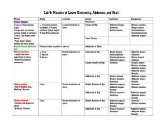
1. Lab 9: Muscles of Lower Extremity, Abdomen, and Skull
Muscle Origin Insertion Action Synergists Antagonists
Pelvic Region Move femur
Iliopsoas: Iliacus/psoas 1. Transverse process Lesser trochanter of Flexion at Hip -Adductor longus -Gluteus maximus
major and bodies of lumbar femur -Sartorius -Biceps femoris
-Can be seen on anterior vertebrae (psoas major) -Rectus femoris -Semitendinosus
surface medial to sartorius 2. Iliac Fossa (iliacus) -Semimembranosus
-Iliacus- fan-shaped, more -Adductor magnus
lateral Torso Flexion
-Psoas major- longer,
thicker and more medial
Gluteal Muscles (posterior Common origin on pelvis or sacrum Abduction of Thigh
hip)
Gluteus maximus 1. Ilium Gluteal tuberosity of Extension at Hip -Biceps femoris -Adductor longus
-Largest and most 2. Sacrum femur -Semitendinosus -Sartorius
superficial of glutes 3. Coccyx -Semimembranosus -Rectus femoris
-Meant for powerful -Adductor magnus -Iliopsoas
movements Lateral rotation at Hip -Sartorius -Gluteus medius
-Gluteus minimus
-Adductor magnus
-Adductor longus
-Gracilis
Abduction at Hip -Gluteus medius -Adductor magnus
-Gluteus minimus -Adductor longus
-Sartorius -Gracilis
Gluteus medius Ilium Greater trochanter of Medial Rotation at Hip -Adductor magnus -Sartorius
-Deep to gluteus max. femur -Adductor longus -Gluteus maximus
Walking” Muscles -Gracilis
Abduction at Hip -Gluteus maximus Adductor magnus
-Sartorius -Adductor longus
-Gracilis
Gluteus minimus Ilium Greater trochanter of Medial Rotation at Hip -Adductor magnus -Sartorius
-Smallest and deepest of femur -Adductor longus -Gluteus maximus
glutes -Gracilis
-Walking” Muscles Abduction at Hip -Gluteus maximus Adductor magnus
-Sartorius -Adductor longus
-Gracilis
2. Piriformis- landmark Anterolateral surface of Greater trochanter of Only need to ID: Follow sciatic nerve up leg, ends in triangular shaped muscle,
-Posterior part of hip joint sacrum femur inferior to the gluteus minimus, pyramidal shaped
Muscle Origin Insertion Action Synergists Antagonists
Thigh (Anterior) Tibial tuberosity of Extension of knee (
tibia
Sartorius (exception) Anterior superior iliac spine Medial proximal tibia Flexion of Knee Biceps femoris -Hamstrings
-Longest muscle, runs -Semimembranosus -Short head biceps
obliquely across anterior -Semitendinosus femoris
surface of thigh -Gracilis -Adductor magnus
-“Indian style” sitting -Gluteus maximus
Abduction at Hip -Gluteal Muscles -Adductor magnus
-Adductor longus
-Gracilis
Lateral rotation at Hip -Gluteus Maximus -Adductors
-Gracilis
Flexion at Hip --Adductor longus --Hamstrings
-Rectus femoris -Adductor magnus
-Iliopsoas
Quadriceps Femoris Tibial tuberosity via Extension of knee - -Biceps femoris
-Rectus femoris patellar tendon -Semimembranosus
-Vastus muscles (3) -Semitendinosus
-Sartorius
-Gracilis
-Gastrocnemius
Rectus Femoris Anterior inferior iliac spine Tibial tuberosity via Extension at Knee Rectus femoris See Quad Antagonists
-Superficial, runs straight patellar tendon Vastus medialis
down anterior surface of Vastus intermedius
thigh Vastus lateralis
Flexion at Hip Semimembranosus -Hamstrings
-Semitendinosus -Adductor magnus
-Sartorius -Gluteus maximus
Vastus lateralis Linea aspera of posterior Tibial tuberosity via Extension at Knee Rectus femoris See Quad Antagonists
-Lateral part of thigh femur patellar tendon Vastus medialis
-Largest head of quads Vastus intermedius
Vastus medialis Linea aspera of posterior Tibial tuberosity via Extension at Knee Rectus femoris See Quad Antagonists
-Inferomedial part of thigh femur patellar tendon Vastus lateralis
3. Vastus intermedius
Vastus intermedius Anterior Femur Tibial tuberosity via Extension at Knee Rectus femoris See Quad Antagonists
-Deep to rectus femoris patellar tendon Vastus medialis
-Between other vastus Vastus lateralis
muscles
Tensor fasciae latae Anterior Iliac crest and IT band ID ONLY- TFL pulls on IT band, which is a thickened fascia that runs down
ASIS lateral side of thigh. TFL overlies rectus femoris and vastus lateralis.
Iliotibial (IT) Band Iliac crest Knee
Muscle Origin Insertion Action Synergists Antagonists
Thigh (Medial) Adductors at Hip
Gracilis Inferior ramus of pubis Medial proximal tibia Flexion of Knee -Biceps femoris -Rectus femoris
-Long, thin superficial -Semimembranosus -Vastus lateralis
muscle of medial thigh -Semitendinosus -Vastus medialis
-Sartorius -Vastus intermedius
-Gastrocnemius
Adduction at Hip -Adductor magnus -Gluteal Muscles
-Adductor longus -Sartorius
Adductor longus Pubis Linea aspera of Flexion at hip -Sartorius -Hamstrings
-Most anterior of adductor posterior femur -Rectus femoris -Adductor magnus
muscles, overlies middle -Iliopsoas -Gluteus maximus
aspect of adductor magnus Adduction at Hip -Adductor Magnus -Gluteal Muscles
-Gracilis -Sartorius
Medial Rotation at Hip -Adductor magnus -Gluteus Maximus
-Gracilis -Sartorius
-Gluteus medius
-Gluteus minimus
Adductor magnus 1. Ischial ramus 1. Linea aspera of Flexion at Hip (ant) -Sartorius -Hamstrings
-Triangular muscle, medial to 2. Inferior pubic ramus posterior femur -Rectus femoris -Adductor magnus
Semimembranosus, lateral to 3. Ischial tuberosity 2. Medial -Iliopsoas -Gluteus maximus
Gracilis supracondylar line of Adduction at Hip (ant) -Adductor longus -Gluteal Muscles
-Anterior part acts like femur -Gracilis -Sartorius
4. adductor 3. Adductor tubercle Medial Rotation (ant) -Adductor longus -Gluteus Maximus
-Posterior part acts like of femur -Gracilis -Sartorius
hamstring -Gluteus medius
-Gluteus minimus
Extension of hip (post) -Hamstrings --Sartorius
-Gluteus maximus -Rectus femoris
-Iliopsoas
-Adductor Longus
Muscle Origin Insertion Action Synergists Antagonists
Thigh (Posterior) Flexion of knee
Hamstrings Ischial tuberosity Extend hip, flex knee
Biceps Femoris 1. Long head- Ischial 1. Head of fibula Flexion at Knee -Gracilis -Rectus femoris
-Long head is medial to short tuberosity 2. Proximal lateral -Semimembranosus -Vastus lateralis
head 2. Short head- Linea aspera tibia -Semitendinosus -Vastus medialis
-Only long head is hamstring of posterior femur -Sartorius -Vastus intermedius
-Most lateral of hamstrings -Gastrocnemius
Extension at Hip (only -Gluteus maximus -Adductor Longus
long head) -Semitendinosus -Iliopsoas
-Semimembranosus -Sartorius
-Adductor magnus -Rectus Femoris
Semitendinosus Ischial tuberosity Proximal medial tibia Flexion at Knee -Gracilis -Rectus femoris
-Medial to biceps femoris (with Gracilis and -Semimembranosus -Vastus lateralis
Sartorius) -Biceps Femoris -Vastus medialis
-Sartorius -Vastus intermedius
-Gastrocnemius
5. Extension at Hip -Gluteus maximus -Adductor Longus
-Long head of biceps -Iliopsoas
femoris -Sartorius
-Semimembranosus -Rectus Femoris
-Adductor magnus
Semimembranosus Ischial tuberosity Proximal medial tibia Flexion at Knee -Gracilis -Rectus femoris
-Deep and medial to (not with Gracilis -Biceps Femoris -Vastus lateralis
Semitendinosus, lateral to and Sartorius) -Semitendinosus -Vastus medialis
adductor magnus -Sartorius -Vastus intermedius
-Gastrocnemius
Extension at Hip -Gluteus maximus -Adductor Longus
-Long head of biceps -Iliopsoas
femoris -Sartorius
-Semitendinosus -Rectus Femoris
-Adductor magnus
Muscle Origin Insertion Action Synergists Antagonists
Leg (Posterior Comp.)
Gastrocnemius 1. Medial condyle of femur Calcaneal tuberosity Flexion of Knee -Gracilis Triceps Brachii
-Part of triceps surae, on 2. Lateral condyle of femur via calcaneal tendon -Semimembranosus
posterior calf -Semitendinosus
-Superficial muscle -Sartorius
-2 heads (lateral and medial) -Biceps femoris
-Forms proximal curve of calf Plantar Flexion at -Tibialis posterior Tibialis anterior
Ankle -Soleus
-Fibularis
6. Soleus 1. Superior tibia Calcaneal tuberosity Plantar Flexion at -Tibialis posterior - Tibialis anterior
-Part of triceps surae, flat / 2. Fibula via calcaneal tendon Ankle -Gastrocnemius
broad 3. Interosseous membrane -Fibularis
-Deep to Gastrocnemius on
posterior surface
Tibialis posterior To locate: originates on superior tibia and fibula and Plantar Flexion at -Soleus -Tibialis anterior
-Thick, flat muscles deep to interosseous membrane, inserts into tarsals and Ankle -Gastrocnemius
soleus metatarsals -Fibularis
-Crossed over by flexor -tendon is directly posterior to tibia, crosses under Inversion at Ankle -Tibialis anterior --Fibularis
digitorum longus on distal flexor digitorum longus
end
-Placed lateral to flexor
digitorum longus
Flexor Digitorum Longus Only Need to ID: long narrow, runs medial to Tibialis posterior and partially overlies it, originates on posterior tibia, inserts into
phalanges
Leg (Anterior Comp.)
Tibialis Anterior To locate: originates on lateral condyle, interosseous Dorsiflexion at Ankle -Extensor digitorum -Gastrocnemius
-Superficial muscles of membrane and inserts into inferior surface of medial longus -Soleus
anterior leg cuneiform and 1st metacarpal -Tibialis posterior
-Laterally parallels anterior -Tendon anterior to tibia -Fibularis
tibia Inversion at Ankle -Tibialis Posterior -Fibularis
Extensor Digitorum Longus Only Need to ID: anterolateral surface of leg, lateral to Tibialis anterior, inserts on phalanges of digits 2-5,
Leg (Lateral Comp.)
Fibularis (longus and brevis) To locate: longus originates on head and upper portion Plantar Flexion at -Tibialis posterior -Tibialis anterior
-Longus: superficial, overlies of lateral side of fibula, same insertion as Tibialis Ankle -Gastrocnemius
fibula anterior-first metatarsal and medial cuneiform -Soleus
-Brevis is deep to longus, Brevis originates on distal fibula shaft and inserts on Eversion at Ankle -Tibialis anterior
smaller and wider proximal end of fifth metatarsal -Tibialis posterior
Muscle Origin Insertion Action Synergist Antagonists
Muscles of Mastication Elevate mandible
7. Temporalis- fan shaped Temporal fossa (lateral Coronoid process of Elevation of Mandible Masseter Digastric
muscles that covers temple of cranium) mandible Pterygoids
skull
Masseter Zygomatic arch (bone + 1. Angle of mandible Elevation of Mandible Temporalis Digastric
-Covers lateral aspect of process of temporal bone) 2. Ramus of mandible Pterygoids
mandibular ramus Lateral Motion of Pterygoids
mandible
Pterygoids Lateral sphenoid Medial ramus of Elevation of Mandible Masseter, Temporalis Digastric
mandible Lateral Motion of Masseter
Mandible
Muscles of Abdominal Wall Direction of Fibers Action Location
External oblique Superolaterally to inferomedially Flex trunk, Most superficial of lateral abs
Rotation of trunk
Lateral flexion of trunk
Internal Oblique Inferolaterally to superomedially Flex trunk Deep to internal oblique
Rotation of trunk
Lateral flexion of trunk
Transversus Abdominis Horizontal Compress viscera Deepest of lateral muscles
Rectus Abdominis (6 pack) Vertical Flexion of trunk Medial muscle group, aponeuroses of
lateral muscles surround, separated
by 3 tendinous intersections
horizontally
Linea alba ID only: tendinous raphe that runs vertically to separate left from right rectus abdominis