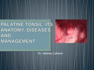
The document provides information on the anatomy, physiology, and diseases of the palatine tonsils. It discusses the embryology, blood supply, innervation, and lymphatic drainage of the tonsils. Common diseases covered include acute and chronic tonsillitis, peritonsillar abscess, and tonsilloliths. The tonsils play an important role in the immune system as part of Waldeyer's ring.
Introduction
Functions
Development
Structure
Nasal cavity
Nasal septum
Lateral wall
Applied anatomy and pathology –
- danger area of nose
- nose bleeding
- foreign body in nose
- developmental nasal deformities
- nasal polyps
- mouth breathing
- rhinitis
Introduction
Functions
Development
Structure
Nasal cavity
Nasal septum
Lateral wall
Applied anatomy and pathology –
- danger area of nose
- nose bleeding
- foreign body in nose
- developmental nasal deformities
- nasal polyps
- mouth breathing
- rhinitis
Imaging of paranasal sinuses (including anatomy and varaints)pk1 pdf pptDr pradeep Kumar
This is very good powerpoint presentation of imaging anatomy and variants of paranasal sinuses and imaging pathology as well as multiple pathological imaging findings and images.it will helps for radiologist and radiology resident and even ent resident. our references is CT and mri whole body by Haaga and various internet sources. THANKS.
263778731218 Abortion Clinic /Pills In Harare ,sisternakatoto
263778731218 Abortion Clinic /Pills In Harare ,ABORTION WOMEN’S CLINIC +27730423979 IN women clinic we believe that every woman should be able to make choices in her pregnancy. Our job is to provide compassionate care, safety,affordable and confidential services. That’s why we have won the trust from all generations of women all over the world. we use non surgical method(Abortion pills) to terminate…Dr.LISA +27730423979women Clinic is committed to providing the highest quality of obstetrical and gynecological care to women of all ages. Our dedicated staff aim to treat each patient and her health concerns with compassion and respect.Our dedicated group ABORTION WOMEN’S CLINIC +27730423979 IN women clinic we believe that every woman should be able to make choices in her pregnancy. Our job is to provide compassionate care, safety,affordable and confidential services. That’s why we have won the trust from all generations of women all over the world. we use non surgical method(Abortion pills) to terminate…Dr.LISA +27730423979women Clinic is committed to providing the highest quality of obstetrical and gynecological care to women of all ages. Our dedicated staff aim to treat each patient and her health concerns with compassion and respect.Our dedicated group of receptionists, nurses, and physicians have worked together as a teamof receptionists, nurses, and physicians have worked together as a team wwww.lisywomensclinic.co.za/
Pulmonary Thromboembolism - etilogy, types, medical- Surgical and nursing man...VarunMahajani
Disruption of blood supply to lung alveoli due to blockage of one or more pulmonary blood vessels is called as Pulmonary thromboembolism. In this presentation we will discuss its causes, types and its management in depth.
Flu Vaccine Alert in Bangalore Karnatakaaddon Scans
As flu season approaches, health officials in Bangalore, Karnataka, are urging residents to get their flu vaccinations. The seasonal flu, while common, can lead to severe health complications, particularly for vulnerable populations such as young children, the elderly, and those with underlying health conditions.
Dr. Vidisha Kumari, a leading epidemiologist in Bangalore, emphasizes the importance of getting vaccinated. "The flu vaccine is our best defense against the influenza virus. It not only protects individuals but also helps prevent the spread of the virus in our communities," he says.
This year, the flu season is expected to coincide with a potential increase in other respiratory illnesses. The Karnataka Health Department has launched an awareness campaign highlighting the significance of flu vaccinations. They have set up multiple vaccination centers across Bangalore, making it convenient for residents to receive their shots.
To encourage widespread vaccination, the government is also collaborating with local schools, workplaces, and community centers to facilitate vaccination drives. Special attention is being given to ensuring that the vaccine is accessible to all, including marginalized communities who may have limited access to healthcare.
Residents are reminded that the flu vaccine is safe and effective. Common side effects are mild and may include soreness at the injection site, mild fever, or muscle aches. These side effects are generally short-lived and far less severe than the flu itself.
Healthcare providers are also stressing the importance of continuing COVID-19 precautions. Wearing masks, practicing good hand hygiene, and maintaining social distancing are still crucial, especially in crowded places.
Protect yourself and your loved ones by getting vaccinated. Together, we can help keep Bangalore healthy and safe this flu season. For more information on vaccination centers and schedules, residents can visit the Karnataka Health Department’s official website or follow their social media pages.
Stay informed, stay safe, and get your flu shot today!
TEST BANK for Operations Management, 14th Edition by William J. Stevenson, Ve...kevinkariuki227
TEST BANK for Operations Management, 14th Edition by William J. Stevenson, Verified Chapters 1 - 19, Complete Newest Version.pdf
TEST BANK for Operations Management, 14th Edition by William J. Stevenson, Verified Chapters 1 - 19, Complete Newest Version.pdf
NVBDCP.pptx Nation vector borne disease control programSapna Thakur
NVBDCP was launched in 2003-2004 . Vector-Borne Disease: Disease that results from an infection transmitted to humans and other animals by blood-feeding arthropods, such as mosquitoes, ticks, and fleas. Examples of vector-borne diseases include Dengue fever, West Nile Virus, Lyme disease, and malaria.
These simplified slides by Dr. Sidra Arshad present an overview of the non-respiratory functions of the respiratory tract.
Learning objectives:
1. Enlist the non-respiratory functions of the respiratory tract
2. Briefly explain how these functions are carried out
3. Discuss the significance of dead space
4. Differentiate between minute ventilation and alveolar ventilation
5. Describe the cough and sneeze reflexes
Study Resources:
1. Chapter 39, Guyton and Hall Textbook of Medical Physiology, 14th edition
2. Chapter 34, Ganong’s Review of Medical Physiology, 26th edition
3. Chapter 17, Human Physiology by Lauralee Sherwood, 9th edition
4. Non-respiratory functions of the lungs https://academic.oup.com/bjaed/article/13/3/98/278874
- Video recording of this lecture in English language: https://youtu.be/lK81BzxMqdo
- Video recording of this lecture in Arabic language: https://youtu.be/Ve4P0COk9OI
- Link to download the book free: https://nephrotube.blogspot.com/p/nephrotube-nephrology-books.html
- Link to NephroTube website: www.NephroTube.com
- Link to NephroTube social media accounts: https://nephrotube.blogspot.com/p/join-nephrotube-on-social-media.html
Tom Selleck Health: A Comprehensive Look at the Iconic Actor’s Wellness Journeygreendigital
Tom Selleck, an enduring figure in Hollywood. has captivated audiences for decades with his rugged charm, iconic moustache. and memorable roles in television and film. From his breakout role as Thomas Magnum in Magnum P.I. to his current portrayal of Frank Reagan in Blue Bloods. Selleck's career has spanned over 50 years. But beyond his professional achievements. fans have often been curious about Tom Selleck Health. especially as he has aged in the public eye.
Follow us on: Pinterest
Introduction
Many have been interested in Tom Selleck health. not only because of his enduring presence on screen but also because of the challenges. and lifestyle choices he has faced and made over the years. This article delves into the various aspects of Tom Selleck health. exploring his fitness regimen, diet, mental health. and the challenges he has encountered as he ages. We'll look at how he maintains his well-being. the health issues he has faced, and his approach to ageing .
Early Life and Career
Childhood and Athletic Beginnings
Tom Selleck was born on January 29, 1945, in Detroit, Michigan, and grew up in Sherman Oaks, California. From an early age, he was involved in sports, particularly basketball. which played a significant role in his physical development. His athletic pursuits continued into college. where he attended the University of Southern California (USC) on a basketball scholarship. This early involvement in sports laid a strong foundation for his physical health and disciplined lifestyle.
Transition to Acting
Selleck's transition from an athlete to an actor came with its physical demands. His first significant role in "Magnum P.I." required him to perform various stunts and maintain a fit appearance. This role, which he played from 1980 to 1988. necessitated a rigorous fitness routine to meet the show's demands. setting the stage for his long-term commitment to health and wellness.
Fitness Regimen
Workout Routine
Tom Selleck health and fitness regimen has evolved. adapting to his changing roles and age. During his "Magnum, P.I." days. Selleck's workouts were intense and focused on building and maintaining muscle mass. His routine included weightlifting, cardiovascular exercises. and specific training for the stunts he performed on the show.
Selleck adjusted his fitness routine as he aged to suit his body's needs. Today, his workouts focus on maintaining flexibility, strength, and cardiovascular health. He incorporates low-impact exercises such as swimming, walking, and light weightlifting. This balanced approach helps him stay fit without putting undue strain on his joints and muscles.
Importance of Flexibility and Mobility
In recent years, Selleck has emphasized the importance of flexibility and mobility in his fitness regimen. Understanding the natural decline in muscle mass and joint flexibility with age. he includes stretching and yoga in his routine. These practices help prevent injuries, improve posture, and maintain mobilit
New Directions in Targeted Therapeutic Approaches for Older Adults With Mantl...i3 Health
i3 Health is pleased to make the speaker slides from this activity available for use as a non-accredited self-study or teaching resource.
This slide deck presented by Dr. Kami Maddocks, Professor-Clinical in the Division of Hematology and
Associate Division Director for Ambulatory Operations
The Ohio State University Comprehensive Cancer Center, will provide insight into new directions in targeted therapeutic approaches for older adults with mantle cell lymphoma.
STATEMENT OF NEED
Mantle cell lymphoma (MCL) is a rare, aggressive B-cell non-Hodgkin lymphoma (NHL) accounting for 5% to 7% of all lymphomas. Its prognosis ranges from indolent disease that does not require treatment for years to very aggressive disease, which is associated with poor survival (Silkenstedt et al, 2021). Typically, MCL is diagnosed at advanced stage and in older patients who cannot tolerate intensive therapy (NCCN, 2022). Although recent advances have slightly increased remission rates, recurrence and relapse remain very common, leading to a median overall survival between 3 and 6 years (LLS, 2021). Though there are several effective options, progress is still needed towards establishing an accepted frontline approach for MCL (Castellino et al, 2022). Treatment selection and management of MCL are complicated by the heterogeneity of prognosis, advanced age and comorbidities of patients, and lack of an established standard approach for treatment, making it vital that clinicians be familiar with the latest research and advances in this area. In this activity chaired by Michael Wang, MD, Professor in the Department of Lymphoma & Myeloma at MD Anderson Cancer Center, expert faculty will discuss prognostic factors informing treatment, the promising results of recent trials in new therapeutic approaches, and the implications of treatment resistance in therapeutic selection for MCL.
Target Audience
Hematology/oncology fellows, attending faculty, and other health care professionals involved in the treatment of patients with mantle cell lymphoma (MCL).
Learning Objectives
1.) Identify clinical and biological prognostic factors that can guide treatment decision making for older adults with MCL
2.) Evaluate emerging data on targeted therapeutic approaches for treatment-naive and relapsed/refractory MCL and their applicability to older adults
3.) Assess mechanisms of resistance to targeted therapies for MCL and their implications for treatment selection
Prix Galien International 2024 Forum ProgramLevi Shapiro
June 20, 2024, Prix Galien International and Jerusalem Ethics Forum in ROME. Detailed agenda including panels:
- ADVANCES IN CARDIOLOGY: A NEW PARADIGM IS COMING
- WOMEN’S HEALTH: FERTILITY PRESERVATION
- WHAT’S NEW IN THE TREATMENT OF INFECTIOUS,
ONCOLOGICAL AND INFLAMMATORY SKIN DISEASES?
- ARTIFICIAL INTELLIGENCE AND ETHICS
- GENE THERAPY
- BEYOND BORDERS: GLOBAL INITIATIVES FOR DEMOCRATIZING LIFE SCIENCE TECHNOLOGIES AND PROMOTING ACCESS TO HEALTHCARE
- ETHICAL CHALLENGES IN LIFE SCIENCES
- Prix Galien International Awards Ceremony
These lecture slides, by Dr Sidra Arshad, offer a quick overview of physiological basis of a normal electrocardiogram.
Learning objectives:
1. Define an electrocardiogram (ECG) and electrocardiography
2. Describe how dipoles generated by the heart produce the waveforms of the ECG
3. Describe the components of a normal electrocardiogram of a typical bipolar leads (limb II)
4. Differentiate between intervals and segments
5. Enlist some common indications for obtaining an ECG
Study Resources:
1. Chapter 11, Guyton and Hall Textbook of Medical Physiology, 14th edition
2. Chapter 9, Human Physiology - From Cells to Systems, Lauralee Sherwood, 9th edition
3. Chapter 29, Ganong’s Review of Medical Physiology, 26th edition
4. Electrocardiogram, StatPearls - https://www.ncbi.nlm.nih.gov/books/NBK549803/
5. ECG in Medical Practice by ABM Abdullah, 4th edition
6. ECG Basics, http://www.nataliescasebook.com/tag/e-c-g-basics
Title: Sense of Smell
Presenter: Dr. Faiza, Assistant Professor of Physiology
Qualifications:
MBBS (Best Graduate, AIMC Lahore)
FCPS Physiology
ICMT, CHPE, DHPE (STMU)
MPH (GC University, Faisalabad)
MBA (Virtual University of Pakistan)
Learning Objectives:
Describe the primary categories of smells and the concept of odor blindness.
Explain the structure and location of the olfactory membrane and mucosa, including the types and roles of cells involved in olfaction.
Describe the pathway and mechanisms of olfactory signal transmission from the olfactory receptors to the brain.
Illustrate the biochemical cascade triggered by odorant binding to olfactory receptors, including the role of G-proteins and second messengers in generating an action potential.
Identify different types of olfactory disorders such as anosmia, hyposmia, hyperosmia, and dysosmia, including their potential causes.
Key Topics:
Olfactory Genes:
3% of the human genome accounts for olfactory genes.
400 genes for odorant receptors.
Olfactory Membrane:
Located in the superior part of the nasal cavity.
Medially: Folds downward along the superior septum.
Laterally: Folds over the superior turbinate and upper surface of the middle turbinate.
Total surface area: 5-10 square centimeters.
Olfactory Mucosa:
Olfactory Cells: Bipolar nerve cells derived from the CNS (100 million), with 4-25 olfactory cilia per cell.
Sustentacular Cells: Produce mucus and maintain ionic and molecular environment.
Basal Cells: Replace worn-out olfactory cells with an average lifespan of 1-2 months.
Bowman’s Gland: Secretes mucus.
Stimulation of Olfactory Cells:
Odorant dissolves in mucus and attaches to receptors on olfactory cilia.
Involves a cascade effect through G-proteins and second messengers, leading to depolarization and action potential generation in the olfactory nerve.
Quality of a Good Odorant:
Small (3-20 Carbon atoms), volatile, water-soluble, and lipid-soluble.
Facilitated by odorant-binding proteins in mucus.
Membrane Potential and Action Potential:
Resting membrane potential: -55mV.
Action potential frequency in the olfactory nerve increases with odorant strength.
Adaptation Towards the Sense of Smell:
Rapid adaptation within the first second, with further slow adaptation.
Psychological adaptation greater than receptor adaptation, involving feedback inhibition from the central nervous system.
Primary Sensations of Smell:
Camphoraceous, Musky, Floral, Pepperminty, Ethereal, Pungent, Putrid.
Odor Detection Threshold:
Examples: Hydrogen sulfide (0.0005 ppm), Methyl-mercaptan (0.002 ppm).
Some toxic substances are odorless at lethal concentrations.
Characteristics of Smell:
Odor blindness for single substances due to lack of appropriate receptor protein.
Behavioral and emotional influences of smell.
Transmission of Olfactory Signals:
From olfactory cells to glomeruli in the olfactory bulb, involving lateral inhibition.
Primitive, less old, and new olfactory systems with different path
3. The palatine tonsils are dense
compact bodies of lymphoid tissue
that are located in the lateral wall
of the oropharynx.
The palatine tonsil represent the
largest accumulation of lymphoid
tissue in Waldeyer's ring.
The Waldeyer ring is involved in
the production of immunoglobulins
and the development of both B-cell
and T-cell lymphocytes
4. Development begin in early in the third month of fetal
life.
Arise from
• The endoderm lining of the second pharyngeal pouch,
• The mesoderm of the second pharyngeal membrane ,
• Adjacent regions of the first and second arches.
• At fourth month, Epithelium of the second pouch
proliferates to form solid endodermal buds, growing into
the underlying mesoderm; these buds give rise to
tonsillar stroma.
• Central cells of the buds later die and slough, converting
the solid buds into hollow tonsillar crypts, which are
infiltrated by lymphoid tissue (1).
• Follicles of lymphoid tissue - begin to collect around buds
in the 5th month of foetal life.
5. Theories regarding origin of lymphoid tissue in tonsils
1. Gulland's theory -
Most recent and accepted theory.
Epithelial endodermal cells, which form the glandular buds of the tonsil, give
rise to broods of lymphoid cells.
2. Older theory –
These lymphoid cells arise from the blood or surrounding connective tissue,
creep in and form follicles round the glandular endodermal buds.
6. Size of the tonsil
The size of the tonsil varies according to the age, individuality, and
pathologic status.
Actual size of the tonsil is bigger than the one that appears from its surface .
At the fifth or sixth year of life, the tonsils rapidly increase in size, reaching
their maximum size at puberty.
At puberty, the tonsils measure 20-25 mm in vertical and 10-15 mm in
transverse diameters (2).
7. Palatine tonsils are two in number and ovoid in
shape.
Situated in Tonsilar fossa in lateral wall of
oropharynx.
Tonsilar fossa - composed of three muscles.
• Palatoglossus muscle - anterior pillar.
• Palatopharyngeal muscle - posterior pillar
• Superior constrictor muscle – laterally – forms
larger part of the tonsillar bed.
8. Tonsil has -
Two surfaces- a medial and a lateral
Two poles - an upper and a lower.
Medial surface
• Covered by non-keratinising
stratified squamous epithelium
• Crypts – epithelium dips in tonsil
stroma to form crypts
• 12-15 crypts
• Crypta magna – largest, a/k/a
intratonsillar cleft, represents the
ventral part of second pharyngeal
pouch.
• From the main crypts arise the
secondary crypts.
9. Lateral surface
Presents a well-defined fibrous capsule.
The tonsillar capsule is a specialized portion of the pharyngobasilar fascia and
extends into it to form septa that conduct the nerves and vessels.
Bed of Tonsil
Lateral surface of tonsil lies over many structures which form bed of tonsil,
1. Capsule
2. Loose areolar tissue
3. Paratonsillar vein
4. Pharyngobasilar fascia
5. Superior constrictor muscle
6. Buccopharyngeal fascia
7. Styloglossus
8. Glossopharyngeal nerve
9. facial artery
10.Medial pterygoid muscle
11.Angle of mandible
12.Submandibular salivary gland
10. Importance of Tonsilar Bed
Capsule - Because of the septa, tonsil is not easily separated from its capsule.
Loose areolar tissue - One can easily dissect the tonsil by separating the
capsule from the muscle through this loose connective tissue.
Glossopharyngeal nerve –
• This nerve can be easily injured if the tonsillar bed is violated
• Commonly affected temporarily by edema after tonsillectomy, which produces
both a transitory loss of taste over the posterior third of the tongue and referred
otalgia.
• Can be addressed surgically through tonsilar bed for its neuralgia.
Styloid process -
Can be addressed surgically through tonsilar bed for Eagle syndrome.
11. Upper Pole
• Extends into soft palate.
• Supratonsillar fossa – potential space
enclosed in a semilunar fold, extending
between anterior and posterior pillars.
• Weber's glands are tubular mucous glands
located at superior pole of the tonsil. The
glands send a common duct to the tonsil and
secrete saliva on to the surface of the tonsillar
crypts.The glands may be left behind following
a tonsillectomy and are therefore a potential
source of quinsy after tonsillectomy(3).
12. Lower Pole
Attached to the tongue.
A triangular fold of mucous membrane
extends from anterior pillar to the
anteroinferior part of tonsil.
Anterior tonsillar space – Space enclosed by
Triangular fold of mucous membrane.
Tonsillolingual sulcus - Sulcus separating
tonsil from base of tongue, may be the seat
of carcinoma.
13. Arterial Supply of Tonsil (4)
The arterial blood supply of the tonsil enters primarily at the lower pole, with
branches also at the upper pole.
At the lower pole:
• Tonsillar branch of the dorsal lingual artery Anteriorly
• Ascending palatine artery (a branch of the facial artery) posteriorly
• Tonsillar branch of the facial artery between them that enters the lower aspect
of the tonsillar bed.
At the upper pole:
• Ascending pharyngeal artery enters posteriorly
• Lesser palatine artery enters on the anterior surface.
15. Nerve Supply of Tonsil (4)
Tonsillar branches of the glossopharyngeal
nerve about the lower pole of the tonsil
Descending branches of the lesser palatine
nerves, which course through the
pterygopalatine ganglion.
Applied Anatomy
The cause of referred otalgia with tonsillitis
is through the tympanic branch of the
glossopharyngeal nerve.
16. Lymphatic Drainage (5)
Upper deep cervical lymph nodes,
especially the jugulodigastric or
tonsillar node.
JD lymph nodes belong to
Anterosuperior group of Level II LN
Bounded by;
• IJV
• Facial Vein
• Posterior belly of Diagastric
Other areas draining into JD LN –
• Submandibular gland
• oropharynx
17. Medial aspect – Non-keratininzing stratified squamous epithelium
Crypts greatly increase the contact surface – 295 cm2
4 lymphoid compartments
Reticular cell/crypt epithelium
Extrafollicular area
Mantle zone of lymhoid follicle
Germinal centre of lymphoid follicle - multiplication of
lymphocytes takes place here.
The immunoreactive lymphoid cells
of the tonsils are found in four
distinct area
18.
19. Tonsil acts as a sentinel to guard against foreign introducers by two
mechanism;
1. Providing local immunity
2. Providing surveillance mechanism
The adenoids and tonsils are predominantly B-cell organs;
• B cells - 50% to 65%
• T cells - 40%,
• Mature plasma cells – 3 %.
Conversely, 70% of the lymphocytes in peripheral blood are T cells.
20. Tonsils are particularly designed for direct transport of foreign material from
the exterior to the lymphoid cells.
This is in contrast to lymph nodes, which depend on antigenic delivery
through
afferent lymphatics.
Intratonsillar defense mechanisms eliminate weak antigenic signals.
Low antigen doses effect the differentiation of lymphocytes to plasma cells,
whereas high antigen doses produce B-cell proliferation.
Immunoglobulins (Igs) produced by the adenoid include IgG,IgA, IgM, and
IgD.
IgG appears to pass into the nasopharyngeal lumen by passive diffusion.
The tonsil produces antibodies locally as well as B cells, which migrate to
other sites around the pharynx and periglandular lymphoid tissues to
produce antibodies.
21. The human tonsils are immunologically most active between ages 4 and 10
years.
Involution of the tonsils begins after puberty, resulting in a decrease of the B-
cell population and a relative increase in the ratio of T to B cells.
Considerable B-cell activity is still seen in clinically healthy tonsils even at age
80 years.
22. Immunology is different in diseased and normal condition (4)
Inflammation of the reticular crypt epithelium results in shedding of
immunologically active cells and decreasing antigen transport function
with subsequent replacement by stratified squamous epithelium.
These changes lead to reduced activation of the local B-cell system,
decreased antibody production, and an overall reduction in density of the
B-cell and germinal centers in extrafollicular areas.
In contrast to recurrent tonsillitis, in adenoid hyperplasia the
immunoregulatory conditions are well preserved.
The reason is most likely that the reticular epithelium is less affected in
inflammation of adenoids than of tonsils.
23.
24. Depending upon the component involved, Acute infections of tonsil classified
as:
1.Acute catarrhal or superficial tonsillitis - tonsillitis is a part of generalized
pharyngitis and is mostly seen in viral infections.
2. Acute follicular tonsillitis. Infection spreads into the crypts which become
filled with purulent material, presenting at the openings of crypts as yellowish
spots.
3. Acute parenchymatous tonsillitis. Here tonsil substance is affected. Tonsil is
uniformly enlarged and red.
4. Acute membranous tonsillitis. It is a stage ahead of acute follicular tonsillitis
when exudation from the crypts coalesces to form a membrane on the surface
of tonsil.
25. Epidemiology
Both sexes equally affected.
All age groups
More common in children: 5-15 years of age.
Peak incidence: 5-6 years of age.
Season: winter months.
Organism Involved
Bacteria - Haemolytic streptococcus is the most commonly infecting organism.
Other causes of infection may be staphylococci, pneumococci or H. influenzae.
Viruses - Influenza, parainfluenza, herpes simplex, coxsackievirus, echovirus,
rhinovirus, RSV
26. Symptomatology
1. Sore throat.
2. Dysphagia / odynophagia.
3. Fever - Associated with chills and rigors.
4. Earache - Referred pain from the tonsil or the result of acute otitis media.
5. Constitutional symptoms - Headache, general body aches, malaise and
constipation. There may be abdominal pain due to mesenteric lymphadenitis
simulating a clinical picture of acute appendicitis.
27. Signs
1. Breath is foetid and tongue is coasted.
2. There is hyperaemia of pillars, soft palate and uvula.
3. Depending upon type of Acute Tonsillitis -
• Acute follicular tonsillitis - Tonsils are red and swollen with yellowish spots
of purulent material presenting at the opening of crypts.
• Acute membranous tonsillitis - There may be a whitish membrane on the
medial surface of tonsil which can be easily wiped away with a swab.
• Acute parenchymatous tonsillitis - The tonsils may be enlarged and
congested so much so that they almost meet in the midline along with
some oedema of the uvula and soft palate.
4. The jugulodigastric lymph nodes are enlarged and tender.
29. Dignosis
Mainly on clinical grounds.
Throat culture - group A -hemolytic streptococcal (GABHS)
• Simple and extremely useful test
• One of the major problems - delay in obtaining results (18 to 48 hours).
Rapid antigen detection test –
• To overcome the problem of delay in throat culture.
• Latex agglutination or enzyme-linked immunosorbent assay (ELISA) methods
to extract the antigen from a swab.
• Need throat culture in case of negative tests.
30. Management
1. Warm saline or Betadine gargles
2. Analgesics – Paracetamol or Ibuprofen.
3. Antibiotics - Penicillin is still the agent of choice in most cases.
• In adults - Penicillin V 500 mg PO BID for 10d or 250 mg PO QID for 10d or
Benzathine penicillin 1.2 million U IM once.
• In pediatrics - Penicillin V 25-50 mg/kg/day divided q6h for 10d or
Benzathine penicillin G 25,000 U/kg IM once (maximum 1.2 million U)
If penicillin is not used;
• Amoxicillin 50 mg/kg/day PO in 2 or 3 divided doses for 10d or
• Amoxicillin-clavulanate 500-875 mg PO q12h for 10d.
• Clindamycin
• Erthyomycin + metronidazole
Antibiotic therapy should be given for 10 days. (Schwartz et al)
31. Chronic tonsillitis – sequel
Recurrent episodes of acute tonsillitis for more than 12 weeks.
Due to incomplete resolution.
Infection may persist in lymphoid follicles of the tonsil in the form of micro
abscesses.
Complications of acute tonsillitis are divided into 2 types –
1. Non Suppurative 2. Suppurative
• Scarlet fever
• Acute rheumatic fever
• Poststreptococcal
glomerulonephritis.
• Peritonsillar abscess.
• Parapharyngeal
abscess.
• Cervical abscess
32. Scarlet fever
Secondary to acute streptococcal tonsillitis or pharyngitis with production of
endotoxins
Manifestations include
• Erythematous rash
• Severe lymphadenopathy with a sore throat
• Vomiting, headache; fever;
• Erythematous tonsils and pharynx;
• Yellow exudate over the tonsils, pharynx, and nasopharynx.
The membrane that is present over the tonsils is usually more friable than that
seen with diphtheria.
A strawberry tongue with a rash and large glossal papillae is a good diagnostic
sign.
Diagnosis –
• Throat Culture
• Dick test - an intradermal injection of dilute streptococcal toxin.
Management - Intravenous administration of penicillin G.
33. Peritonsillar abscess or Quinsy
Collection of pus in Peritonsillar space (between capsule and superior
constrictor muscle).
Aetiology – recurrent tonsillitis sealed off infection in crypta magna
intratonsillar abscess burst to form Peritonsillar abscess.
Organisms - Strept. pyogenes, Staph. aureus or anaerobic organisms
Clinical features –
Mostly affects adults and rarely the children, Unilateral
1. General - fever (up to 104°F), chills and rigors, general malaise, body aches,
headache
2. Local
(i) Severe pain in throat.
(ii) Odynophagia and drooling
(iii) Muffled and thick speech, often called "Hot potato voice".
(iv) Foul breath.
(v) Ipsilateral earache – referred
(vi) Trismus due to spasm of pterygoid muscles.
34. Examination –
1. The tonsil, pillars and soft palate on the involved side are congested and
swollen.
Tonsil itself may not appear enlarged as it gets buried in the oedematous pillars.
2. Uvula is swollen and oedematous and pushed to the opposite side.
3. Bulging of the soft palate and anterior pillar above the tonsil.
4. Mucopus may be seen covering the tonsillar region.
5. Cervical lymphadenopathy - jugulodigastric lymph nodes.
6. Torticollis - to the side of abscess.
Diagnosis of peritonsillar abscess is CLINICAL.
Cellulitis must be differentiated from abscess.
CT neck is required to see the extension of abscess in other spaces.
35. Treatment
Conservative management
1. Hospitalisation.
2. Intravenous fluids to combat dehydration.
3. Antibiotics.
4. Analgesics.
5. Oral hygiene should be maintained by betadine or
saline mouth washes.
Surgical management
1. Needle aspiration –
2. Incision and drainage of abscess - LA
- A peritonsillar abscess is opened at the point of
maximum bulge above the upper pole of tonsil.
3. Interval tonsillectomy -
Tonsils are removed four to six weeks following an attack
of quinsy.
4. Quinsy or hot tonsillectomy - video
36. Controversies in management of peritonsillar abscess (4)
Traditional management has consisted of incision and drainage, with
tonsillectomy 4 to 12 weeks later.
versus
Some surgeons advocate immediate tonsillectomy or Quinsy tonsillectomy as
definitive management to ensure complete drainage of the abscess and to
alleviate the need for a
second hospitalization for an interval tonsillectomy.
Indications of quinsy tonsillectomy –
• If incision and drainage or needle aspiration fails to drain an abscess
adequately.
• A prior history of recurrent peritonsillar abscess or recurrent tonsillitis severe
enough to warrant tonsillectomy
• Favored in children because they are likely to experience further episodes of
tonsillitis, and needle aspiration or incision and drainage with a child under
local anesthesia is often difficult or impossible.
37. Complications of quinsy
Rare with modern therapy.
1. Parapharyngeal abscess (a peritonsillar abscess is a potential
parapharyngeal abscess).
2. Oedema of larynx. Tracheostomy may be required.
3. Septicaemia. Other complications like endocarditis, nephritis, brain abscess
may occur.
4. Pneumonitis or lung abscess.
5. Jugular vein thrombosis – Lemierr’s syndrome
6. Spontaneous hemorrhage from carotid artery or jugular vein.
38.
39. 1. Membranous tonsillitis – abrupt in onset.
• It occurs due to pyogenic organisms.
• An exudative membrane forms over the medial surface
of the tonsils, along with the features of acute tonsillitis.
2. Diphtheria -slower in onset with less local discomfort.
• The membrane in diphtheria extends beyond the
tonsils, on to the soft palate and is dirty grey in colour.
• It is adherent and its removal leaves a bleeding
surface.
• “Bull -neck“ appearance.
• Smear and culture of throat swab will reveal
C.diphtheriae.
• Treatment –
Antidiphtheric antitoxin – given by iv saline infusion over
60 mins after sensitivity test.
Dose depends upon duration and severity of disease;
Within 48 hrs or membrane is limited to tonsils – 20000 to
40000 unit
More than 48 hrs or membrane beyond tonsil – 60000 to
120000 unit.
Antibiotics - benzyl penicillin 600 mg 6-hourly for 7 days.
Erythromycin (500 mg 6 hourly orally)
40. 3. Vincent's angina - Insidious in onset with less fever,
less discomfort
• Membrane, which usually forms over one tonsil,
can be easily removed revealing an irregular ulcer
on the tonsil.
• Throat swab will show fusiform bacilli and
spirochaetes.
• Treatment – antibiotics and irrigation + removal of
necrotic debris.
4. Infectious mononucleosis - affects young adults.
• Both tonsils are very much enlarged, congested
and covered with membrane.
• Local discomfort is marked.
• Lymph nodes are enlarged in the posterior triangle
of neck along with splenomegaly.
• Blood smear may show more than 50%
lymphocytes, of which about 10% are atypical.
• Paul-Bunnell test (mono test) will show high titre of
heterophil antibody.
• Management –
Symptomatic, resolves in few weeks
Antibiotics have no role
41. 5. Agranulocytosis –
• presents with ulcerative necrotic lesions over tonsil
and oropharynx.
• Patient is severely ill.
• In acute fulminant form, total leucocytic count is
decreased to < 2000/cu mm or even as low as
50/cu mm and polymorph neutrophils may be
reduced to 5% or less.
• In chronic or recurrent form, total count is reduced
to 2000/cu mm with less marked granulocytopenia.
6. Leukaemia –
• In children, 75% of leukaemias are acute
lymphoblastic and 25% acute myelogenous or
chronic.
• In adults 20% of acute leukaemias are lymphocytic
and 80% non-lymphocytic.
• Peripheral blood shows TLC > 100,000/cu mm.
• Anaemia is always present and may be
progressive.
• Blasts cells are seen on examination of the bone
marrow.
42. 7. Aphthous ulcers -
• They may involve any part of oral cavity or oropharynx.
Sometimes, it is solitary and may involve the tonsil and
pillars.
• It may be small or quite large and alarming.
• It is very painful.
8. Malignancy tonsil –
9. Candidiasis –
10. Trauma -
43. Investigations for diagnosis of Membrane over tonsil
1. History.
2. Physical examination.
3. Total and differential counts (for agranulocytosis, leukaemia,
neutropenia, infectious mononucleosis).
4. Blood smear (for atypical cells).
5. Throat swab and culture (for pyogenic bacteria), Vincent's angina,
diphtheria candidal infection.
6. Bone marrow aspiration or needle biopsy.
7. Other tests. Paul-Bunnell or mono spot test and biopsy of the lesion.
44. Recurrent tonsillitis for more than 12 weeks.
Aetiology –
1. It may be a complication of acute tonsillitis.
2. Subclinical infections of tonsils without an acute attack.
3. Predisposing factor - Chronic infection in sinuses or teeth.
Mostly affects children and young adults.
45. Types of Chronic tonsillitis
1. Chronic follicular tonsillitis –
• Here tonsillar crypts are full of infected
cheesy material which shows on the
surface as yellowish spots.
2. Chronic parenchymatous tonsillitis –
• There is hyperplasia of lymphoid tissue.
• Tonsils are very much enlarged
• May interfere with speech, deglutition and
respiration.
• Attacks of sleep apnoea may occur.
• Long-standing cases develop features of
cor pulmonale.
3. Chronic fibroid tonsillitis.
• Tonsils are small but infected, with history
of repeated sore throats.
46. Clinical Features
1. Recurrent attacks of sore throat or acute tonsillitis.
2. Chronic irritation in throat with cough.
3. Bad taste in mouth and foul breath (halitosis) due to pus in crypts.
4. Thick speech.
5. Difficulty in swallowing
6. Choking spells at night - obstructive sleep apnea (adenotonsillar
hypertrophy)
47. Signs and Examination
1. Chronic parenchymatous type - Tonsils may show varying degree of
enlargement.
2. Chronic follicular type - There may be yellowish beads of pus on the medial
surface of tonsil.
3. Chronic fibroid type - Tonsils are small but pressure on the anterior pillar
expresses frank pus or cheesy material.
4. Flushing of anterior pillars.
5. Enlargement of jugulodigastric lymph nodes. During acute attacks, become
tender.
Cardinal signs of chronic tonsillitis
1. Hypertrophied non-congested tonsils
2. Flushing of ant pillars
3. Irwin Moore sign
4. Jugulodiagastric LN pathy b/l & nontender
48. Grading of Tonsils (4)
Brodsky and coworkers described an assessment scale for tonsillar
hypertrophy.
• 0 indicates that the tonsils do not impinge on the airway;
• 1+ indicates less than 25% airway obstruction;
• 2+ indicates 25% to 50% airway obstruction;
• 3+ indicates 50% to 75% airway obstruction;
• 4+ indicates more than 75% airway obstruction.
49. Management of Chronic tonsillitis
1. Conservative treatment consists of attention to general health, diet, treatment
of co-existent infection of teeth, nose and sinuses.
2. Tonsillectomy is indicated when tonsils interfere with speech, deglutition and
respiration or cause recurrent attacks
Complications of Chronic tonsillitis
1. Peritonsillar abscess.
2. Parapharyngeal abscess.
3. Intratonsillar abscess.
4. Tonsilloliths.
5. Tonsillar cyst.
6. Focus of infection in rheumatic fever, acute glomerulonephritis, eye and skin
disorders
50. Calculus or stone in the tonsil.
Aetiology – seen in chronic tonsillitis
The blocked tonsilar crypt causes retention of debris,
which consists of inorganic salts of calcium and
magnesium (formation of stone).
Clinical Features
• Usually seen in adults
• „The affected crypt gradually enlarges, and may
ulcerate on medial surface of tonsil.
• „Halitosis and sore throat – d/t sec infection
• „Whitish foul-tasting and foul-smelling cheesy
material can be expressed from tonsils.
• „Local discomfort or foreign body sensation.
Diagnosis – Clinical examination by palpation or
probing.
51. Treatment –
• „„Conservative: Expression of concretions/cheesy material and chemical
cauterization of crypts with topical silver nitrate application.
• „„Tonsillectomy: In cases of persistent pain, halitosis, or foreign body
sensation.
52. • Due to blockage of a tonsillar crypt.
• Appears as a yellowish swelling over the tonsil.
• Symptomless.
• It can be easily drained.
Case of epidermoid cyst in tonsil is reported in
literature.
Treated by tonsillectomy. (6)
53. • Accumulation of pus within the blocked tonsillar crypt can
occur in cases of acute follicular tonsillitis.
• Predisposing factors –
Dehydration
Inflammatory swelling of tonsillar follicles
Previous h/o peritonsillar abscess.
• „Clinical Features -
Marked local pain and dysphagia.
Tonsil swollen and red.
54. • „D/D’s -
Tonsillar cyst
Lymphoma
Malignancy
• Needle aspiration confirms the
diagnosis.
• CT scan
• Treatment
Antibiotics
Drainage of the abscess
Tonsillectomy
Two important clinical
features distinguish it
from peritonsillar
abscess:
1. enlargement of
tonsils with no
significant swelling
and
2. absence of muffled
voice
55. • White or yellowish dots or horny excrescences on the
surface of tonsils, pharyngeal wall or lingual tonsils
characterize this benign condition.
• These excrescences are firmly adherent and cannot
be wiped off.
• They are the result of hypertrophy and keratinization of
epithelium.
• Patient does not have features of acute follicular
tonsillitis.
• Treatment -
The spontaneous regression does occur so, no specific
treatment is required.
The concerned patients need reassurance.
58. A. Infection
1. Recurrent acute tonsillitis (more than 6 episodes per year or 3 episodes
per year for 2 years or longer)
2. Recurrent acute tonsillitis associated with other conditions:
• Cardiac valvular disease associated with recurrent
• streptococcal tonsillitis
• Recurrent febrile seizures
3. Chronic tonsillitis that is unresponsive to medical therapy and is associated
with:
• Halitosis
• Persistent sore throat
• Tender cervical adenitis
• Streptococcal carrier state unresponsive to medical therapy
• Peritonsillar abscess
• Tonsillitis associated with abscessed cervical nodes
• Mononucleosis with severely obstructing tonsils that is unresponsive to
medical therapy
59. B. Obstruction
1. Excessive snoring and chronic mouth-breathing
2. Obstructive sleep apnea or sleep disturbances
3. Adenotonsillar hypertrophy associated with:
• Cor pulmonale
• Failure to thrive
• Dysphagia
• Speech abnormalities
• Craniofacial growth abnormalities
• Occlusion abnormalities
C. Suspected neoplasia
D. As a Part of Another Operation
1. Palatopharyngoplasty.
2. Glossopharyngeal neurectomy.
3. Removal of styloid process.
60. 1. Hemoglobin level less than 10 g%.
2. Presence of acute infection.
3. Children under 3 years of age.
4. Overt or submucous cleft palate.
5. Bleeding disorders, e.g. leukaemia, purpura, aplastic anaemia, haemophilia.
6. At the time of epidemic of polio.
7. Uncontrolled systemic disease, e.g. diabetes, cardiac disease, hypertension
or asthma.
8. Tonsillectomy is avoided during the period of menses.
61. • Hemoglobin, platelet count and TLC
• Bleeding time and clotting time
• Prothrombin time
• Sickling cell test
• Blood group
62. Cold Methods
I -Dissection and snare (most
common)
II -Guillotine method
III -Intracapsular (capsule
preserving) tonsillectomy with
debrider
IV -Harmonic scalpel (ultrasound)
V -Plasma-mediated ablation
technique
VI -Cryosurgical technique
Hot methods
I -Electrocautery
II -Laser tonsillectomy or
tonsillotomy (CO2 or KTP)
III -Coblation tonsillectomy
IV -Radio frequency
63. Anesthesia – GA with Oro/naso tracheal intubation and throat pack.
Position – ROSE position
Patient lies supine with head extended by placing a pillow under the shoulders.
A rubber ring is placed under the head to stabilise it.
Hyperextension should always be avoided.
Advantages of Rose position:
1. There is virtually no aspiration – larynx lies at higher level than oral cavity.
2. Both hands of the surgeon are free.
3. This position helps in proper application of the Boyles Davis mouth gag.
4. The surgeon can be comfortably seated at the head end of the patient
64. • Most common method.
• The tonsil is dissected along with its capsule and lifted out of its bed.
• It is ultimately removed using a tonsilar snare.
• Safe, bleeding is less and the tonsil can be removed in toto.
• Cost effective.
Video
65. • Used during olden days.
• Abandoned because of the risks of bleeding.
• In this method a guillotine is used to simply chop off the tonsil.
• Term guillotine - literally means chop off the head
Recently there are many studies
concluding;
in carefully selected children guillotine
tonsillectomy is a safe, time saving with
less bleeding and cost-effective
procedure.
66. a.k.a Subtotal tonsillectomy.
WHAT IS DONE –
• The only tissue manipulated and dissected is the tonsil itself.
• No mucosal cuts are made.
• The peritonsillar capsule is not dissected
• There should not be any direct cauterization of the peritonsillar
fascia and underlying pharyngeal musculature.
• Tonsils are shaved behind the levels of the anterior and posterior
tonsillar pillars. Exposure is obtained by retraction of the anterior
tonsillar pillar.
• Hemostasis is obtained with suction cautery
Special indication - children without a history of tonsillitis.
67. Advantages –
• Less postoperative pain.
• Less bleeding.
Disadvantages –
• Cost of disposable microdebrider blade
• Residual tonsil tissue may regrow (the incidence of regrowth was 3.2%.).
68. • An ultra sound coagulator and dissector that uses
ultra sonic vibrations to cut and coagulate tissues.
• The cutting operation is made possible by a sharp
knife with a vibratory frequency of 55.5 KHz over a
distance of 89 micro meters.
• Coagulation occurs due to transfer of vibratory
energy to tissues. This breaks hydrogen bonds of
proteins in tissues and generates heat from tissue
friction.
• The temperature generated 50 - 100 degrees
centigrade.
• The major disadvantage is the expense of the
equipment and the increased duration of surgery.
69. Principle – PMA energizes protons to break molecular bonds between
tissues.
Leaves less heat in the tissues and hence less thermal energy.
Less painful recovery.
70. Cryoprobe is based on Joule Thomson effect i.e rapid
expansion of compressed gas through a small probe produces
cooling.
Cryoprobe is applied to tonsil and it is removed by the process
of repeated freezing and thawing.
The temperature reached during cryo is dependent on the
medium used :
- 82 degrees centigrade by carbondioxide
- 196 degrees centigrade by liquid nitrogen
The major advantage of this procedure is minimal bleeding.
The major disadvantage of this procedure is the operating time
involved.
This procedure is used only in patients with known bleeding
72. • This method uses unipolar cautery.
• Heat generated by cautery is used for cutting of tissue.
• Temperature – 150 to 400 degree Celsius.
• Precautions to use –
Prevent damage to posterior pillar and pharyngeal mucosa –
nasopharyngeal stenosis.
Avoid contact between metal instrument and electrocautery.
Electrocautery blade should be guarded with a nonconducting
material.
• Advantage – Rapid, safe and simultaneous hemostasis.
• Disadvantage – post op pain.
73.
74. Principle - Radiofrequency bipolar electrical current that passes
through a medium of normal saline, which results in the production of a
plasma field of sodium ions.
These energized ions are able to break down intercellular bonds and
effectively vaporize tissue at a temperature of only 60° C.
This vaporization theoretically results in effective dissection with less
postoperative pain from thermal injury.
• The technique can be utilized for complete tonsillectomy or for
intracapsular tonsillectomy.
• The major advantage of this procedure is reduced bleeding and
reduced post operative pain.
• Disadvantage – expensive setup and maintance of probe.
75. The Coblator consists of;
Hand piece with a suction irrigation tip that transmits the radiofrequency
current and dissects tissue and also has a cautery capability for
hemostasis.
Video
76. • Laser used are;
1. CO2 laser – 10600 nm
2. KTP (potassium titanyl Phosphate) – 512 nm
3. Diode laser – 600-1000
• Major advantage of laser surgery is reduced bleeding. Laser seals
all bleeders efficiently.
• Disadvantage –
Increased operating time.
Cost of laser equipment.
Maintenance of device. VIDEO
77. 1. Immediate general care
(a) Keep the patient in coma position.
(b) Keep a watch on bleeding from the nose and
mouth.
(c) Keep check on vital signs.
2. Diet -
When patient is fully recovered he is permitted to take
liquids, e.g. cold milk or ice cream.
Sucking of ice cubes gives relief from pain.
Diet is gradually built from soft-liquid to solid food.
3. Oral hygiene –
H2O2 + water gargles for 2 days post op.
Condy's or salt water gargles after every feed helps to
keep the mouth clean.
78. 4. Analgesics - like paracetamol.
5. Antibiotics - Injectable for first 2 days followed by oral antibiotics
for 7 days.
Patients or their parents are instructed to return immediately to the
emergency room if there is any evidence of bright red bleeding from
the nose or oral cavity.
79.
80. A. Immediate
1. Primary haemorrhage - Occurs at the time of operation.
• It can be controlled by pressure, ligation or electrocoagulation.
2. Reactionary haemorrhage - Occurs within a period of 24 hours.
• controlled by removal of the clot, application of pressure or vasoconstrictor.
• If above measures fail, ligation or electrocoagulation.
3. Injury to tonsillar pillars, uvula, soft palate, tongue or superior constrictor muscle
due to bad surgical technique.
4. Injury to teeth.
5. Aspiration of blood or Corner’s clot.
6. Facial oedema.
7. Surgical emphysema. Rarely occurs due to injury to superior constrictor
muscle.
8. Airway obstruction d/t edema of tongue and soft palate.
9. Pulmonary edema
81. B. Delayed
1. Secondary haemorrhage - Between the 5th to 10th post-operative day.
It is the result of sepsis and premature separation of the membrane.
Usually, it is heralded by bloodstained sputum but may be profuse.
M/n –
• Removal of clot, topical application of dilute adrenaline or hydrogen peroxide
with pressure usually suffice.
• For profuse bleeding, bleeding vessel is electrocoagulated or ligated.
• Approximation of pillars with mattress sutures may be required.
• External carotid ligation may also be required.
• Transfusion of blood or plasma, depending on blood loss, is given.
• Systemic antibiotics are given for control of infection.
82. 2. Infection - Infection of tonsillar fossa may lead to parapharyngeal abscess or
otitis media.
3. Lung complications - Aspiration of blood, mucus or tissue fragments may
cause atelectasis or lung abscess.
4. Scarring in soft palate and pillars.
5. Tonsillar remnants - Tonsil tags or tissue, left due to inadequate surgery,
may get repeatedly infected.
6. Hypertrophy of lingual tonsil - compensatory to loss of palatine tonsils.
Lymphoid tissue is left in the plica triangularis near the lower
pole of tonsil, which later gets hypertrophied, therefore PT
should be removed during tonsillectomy.
83.
84. 1.William JL, Lawrence SS, Steven P, William JS. Human Embryology. 3rd ed.
Philadelphia: Elsevier; 2001. 375-376
2.Susan S, Harold E, Jermiah CH, David J, Andrew W. Pharynx (chapter
35). Gray’s Anatomy: The Anatomical Basis of Clinical Practice. 39th ed.
Philadelphia: Elsevier; 2005. 619-631.
3.Al-Kindy S. Post tonsillectomy quinsy. Saudi Med J. 2002;23:240–241.
4. Cummings Otolaryngology Head & Neck Surgery FIFTH EDITION. Chapter
199 pg no 2822.
5. Stell and Maran’sTextbook of Head and Neck Surgery and Oncology. 5th
edition. Anantomy of neck.
6. Epidermoid cyst localized in the palatine tonsil Keles Erol, Kaplama
Mehmet Erkan, Dolen Tolga, and Cobanoglu Bengu. J Oral Maxillofac Pathol.
2013 Jan-Apr; 17(1): 148.
Editor's Notes
Actual size of the tonsil is bigger than the one that appears from its surface as parts of tonsil extend upwards into the soft
palate, downwards into the base of tongue and anteriorly into palatoglossal arch
Crypts may be filled with cheesy material consisting of epithelial cells, bacteria and food debris
Eagle's syndrome is defined as secondary glossopharyngeal neuralgia due to elongated styloid process, resulting from abnormal stylohyoid chain ossification.
The tonsillar branch of the facial artery is the largest.
Primarily, the tonsil consists of (a) surface epithelium which is continuous with the oropharyngeal lining; (b) crypts which are tube-like
invaginations from the surface epithelium; and (c) the lymphoid tissue.
The use of antibiotics also minimizes the chance of suppurative complications and diminishes the likelihood of acute rheumatic
fever
Cervical ab scess due to suppuration of jugulodigastric lymph nodes
rapid lymphatic transport from palatine tonsils and the absence of lymphatic valves prior to the capsule may not allow aggregation of bacteria within the tonsillar parenchyma. This may account for minimal cases of ITA (and more peritonsillar abscess)
owing to the concern
children without a history of tonsillitis about persistent tonsillitis in the residual tonsil tissue
If no active bleeding is apparent and a blood clot is evident in the tonsillar fossa, it should not be disturbed. However, if the surgeon cannot determine whether active bleeding is taking place, the clot should be suctioned to allow better examination.
Blood clots often hide bleeding vessels and may prevent appropriate coagulation as a result of fibrinolysis.