A malunited fractureis one that has healed with the fragments
in a nonanatomic position.
Definition
3.
CLINICAL FEATURES
Malunited fracturescan impair function in several ways:
• Reduced Range of movements
An abnormal joint surface can cause irregular weight transfer and arthritis of
the joint, especially in the lower extremities
• Gait abnormalities
Rotation or angulation of the fragments can interfere with
proper balance or gait in the lower extremities or positioning of the upper
extremities
• Limb length discrepancy
Overriding of fragments or bone loss can result in perceptible shortening
• Deformity
4.
CLASSIFICATION
Based on location
1.Intra articular
2. Extra articular- Metaphyseal, Diaphyseal
3. Combined
Plane malalignment
4. Simple- one plane
5. Complex- several plane and translation
NON UNION VSMAL UNION
• On inspection, look for signs of active discharging sinus, skin
discolouration due to infection.
• On palpation check for raised local temperature due to infection
• The hallmark of nonunion is presence of abnormal painless mobility
at the nonunion that is present in two perpendicular planes and also
on axial rotation.
• Also with axial rotation, there is loss of transmitted movements
proximally.
• Painless crepitus (differentiating from acute fracture) is also a
characteristic
7.
INVESTIGATIONS
• Plain radiographs
•CT scan with 3D reconstruction
To evaluate rotational and angular deformities
• MRI
To evaluate associated ligamentous injuries
• Xray Scannogram
To estimate limb length discrepancies and determine deformity
correction
8.
Indications for surgicalmanagement
• Malunion with functional instability
• Intra articular malunion
• Mechanical overload
• Demand of the patient
• Cosmesis
9.
When treating malunions,the following facts must be
considered.
• Alignment
• Rotation
• Restoration of normal length
• Actual position of fragments
MALUNION TREATMENT PRINCIPLES
10.
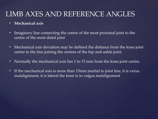
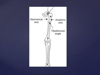
LIMB AXES ANDREFERENCE ANGLES
• Mechanical axis
• Imaginary line connecting the centre of the most proximal joint to the
centre of the most distal joint
• Mechanical axis deviation may be defined the distance from the knee joint
centre to the line joining the centres of the hip and ankle joint.
• Normally the mechanical axis lies 1 to 15 mm from the knee joint centre.
• If the mechanical axis is more than 15mm medial to joint line, it is varus
malalignment, it is lateral the knee is in valgus malalignment
11.
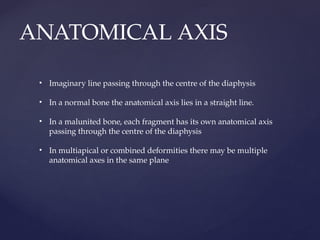
ANATOMICAL AXIS
• Imaginaryline passing through the centre of the diaphysis
• In a normal bone the anatomical axis lies in a straight line.
• In a malunited bone, each fragment has its own anatomical axis
passing through the centre of the diaphysis
• In multiapical or combined deformities there may be multiple
anatomical axes in the same plane
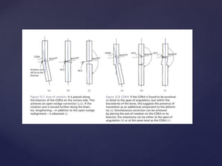
13.
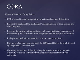
CORA
Centre of Rotationof Angulation
• CORA is used to plan the operative corrections of angular deformities
• It is the intersection of the mechanical/ anatomical axes of the proximal and
distal fragments.
• It reveals the presence of translation as well as angulation as components of
the deformity and can also indicate the presence of multi-apical deformities
• In diaphyseal malunions anatomical axis are more convenient.
• Bisector is a line that passes through the CORA and bisects the angle formed
by the proximal and distal axes.
• Correcting the angular deformity along the bisector results in complete
deformity correction without introducing any iatrogenic translational
deformity.
14.
• When theCORA lies within the boundaries of the bone involved but is at
a different level to that of the apex of deformity, it indicates the presence of
translation and angulation within the deformity.
16.
OSTEOTOMY
• An osteotomyis used to separate deformed bones to align the mechanical
and anatomical axis.
• The ability of an osteotomy to correct a deformity depends upon the
location of the CORA, the axis long which the correction is performed
and the location of the osteotomy.
• Classification
Based on type- open,closed and neutral
Based on cut- straight, dome
17.
WEDGE OSTEOTOMY
• Thetype of wedge osteotomy is determined by the location of the osteotomy
relative to the locations of the CORA and the correction axis.
• When the CORA and correction axis are in the same location (to avoid
translational deformity), they may lie on the cortex on the convex side of the
deformity, on the cortex on the concave side of the deformity, or in the
middle of the bone
• When the CORA and correction axis lie on the convex cortex of the
deformity, the correction will result in an opening wedge osteotomy.
• In an opening wedge osteotomy, the cortex on the concave side of the
deformity is distracted to restore alignment, opening an empty wedge that
traverses the diameter of the bone.
• An opening wedge osteotomy also increases bone length.
18.
• When theCORA and correction axis lie in the middle of the bone, the
correction distracts the concave side cortex and compresses the convex side
cortex.
• A bone wedge is removed from only the convex side to allow realignment.
• This neutral wedge osteotomy has no effect on bone length.
• When the CORA and correction axis lie on the concave cortex of the
deformity, the correction will result in a closing wedge
• In a closing wedge osteotomy, the cortex on the convex side of the deformity
is compressed to restore alignment; this requires removal of a bone wedge
across the entire bone diameter.
• A closing wedge osteotomy also decreases bone length
20.
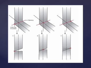
DOME OSTEOTOMY
In contrastto a wedge osteotomy, however, the osteotomy site can never
pass through the mutual CORA-correction axis
Thus, translation will always occur with deformity correction using a
dome osteotomy.
Ideally, the CORA and correction axis are mutually located such that the
angulation and obligatory translation that occurs at the osteotomy site
results in realignment.
Attempts at realignment when the CORA and correction axis are not
mutually located results in a translational deformity
21.
• The principlesguiding wedge osteotomies are also true for dome osteotomies.
• When the CORA and correction axis lie on the convex cortex of the deformity,
the correction will result in an opening dome osteotomy
• The translation that occurs in an opening dome osteotomy increases final bone
length.
• When the CORA and correction axis lie in the middle of the bone, the
correction will result in a neutral dome osteotomy.
• A neutral dome osteotomy has no effect on bone length. When the CORA and
correction axis lie on the concave cortex of the deformity, the correction will
result in a closing dome osteotomy.
• The translation that occurs in a closing dome osteotomy decreases final bone
length.
• Unlike wedge osteotomies, the movement of one bone segment on the other is
rarely impeded, so removal of bone is not typically required unless the final
22.
Limb Length restoration
•Acute distraction or compression methods obtain immediate correction of limb
length by acute lengthening with bone grafting or acute shortening, respectively.
• The extent of acute lengthening or shortening that is possible is limited by the
soft tissues (soft tissue compliance, surgical and open wounds, and
neurovascular structures).
• Acute distraction treatment methods involve distracting the bone ends to the
appropriate length, applying a bone graft, and stabilizing the construct to allow
incorporation of the graft.
• Options for treating length deformities include the use of:
(i) autogenous cancellous or cortical bone grafts
(ii) vascularized autografts
(iii) bulk or strut cortical allografts
(iv) mesh cagebone graft constructs
23.
• The amountof shortening that requires lengthening correction is
uncertain.
• In the upper extremity, up to 3 to 4 cm of shortening is generally well
tolerated, and restoring length when shortening exceeds this value have
been reported to improve
• In the lower extremity, up to 2 cm of shortening may be treated with a
shoe lift; tolerance for a 2 to 4 cm shoe lift is poor for most patients, and
most patients with shortening of greater than 4 cm will benefit from
restoration of length
• Acute compression methods are used to correct overdistraction by first
resecting the appropriate length of bone and then stabilizing the
approximated bone ends under compression.
• For the paired bones of the forearm and leg, the unaffected bone
requires partial excision to allow shortening and compression of the
affected bone.
24.
• Gradual correctiontechniques for length deformities typically use tensioned-
wire (Ilizarov) external fixation
• The most common form of gradual correction is gradual distraction to correct
limb shortening.
• Gradual correction methods for length deformities can also be used to correct
associated angular, translational, or rotational deformities simultaneously while
restoring length.
• Gradual distraction involves the creation of a corticotomy (usually metaphyseal)
and distraction of the bone segments at a rate of 1 mm per day using a rhythm of
0.25 mm of distraction repeated four times per day.
• The bone formed at the distraction site is formed through the process of
distraction osteogenesis