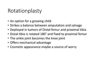
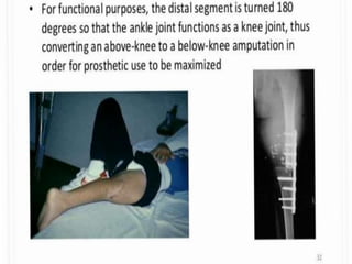
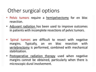
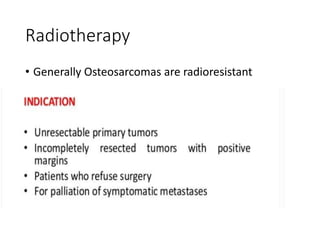
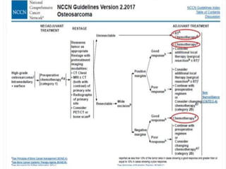
Pathology and
management of
Osteosarcoma
Dr Okonkwo chukwuebuka Augustine
Outline
• Introduction
• Statement of surgical importance
• Anatomy of long bones
• Pathology
1. Epidemiology
2. Aetiology
3. Risk factors
4. Genetic predisposition
5. Pathological types
6. Pathogenesis
7. Staging
• Clinical presentation
1. History
2. Physical examination
• Investigations
• Treatment
Outline contd
• NCCN guidelines
• Future considerations
• Complications
• Prognosis
• Prevention
• Conclusion
• References
Introduction
• Osteosarcoma/osteogenic sarcoma is a malignant
tumor of the bone
• Usually common in fast growing bones - first and 2nd
decades of life
• Histological Hallmark is the production of malignant
osteoid
• Pain is commonest presenting feature as
• Neoadjuvant chemotherapy has improved the long
term survival
• Limb salvage is currently advocated
Statement of surgical importance
• Highly malignant tumor
• > 20% at Presentation already have pulmonary
metastasis
• chemotherapy has improved prognosis
• Limb salvage including metastatectomy is now the
standard of care as against amputation
Long bone Anatomy
Epidemiology
• 80.1% of all primary malignant bone tumors, M:F - 1.35:1
(Lasebikan et al, 2014)
• Peaks in the 2nd decade, (13-16 years of age)
• Prevalence: 2.13% of all childhood malignancies with a male
preponderance (A.Mohammed and Halima Aliyu,2009)
• 3.1/million in the USA
• More in blacks, 5.2/million as against 4.6/million (Michael
Gibson et al,)
• Affects appendicular skeleton ( Distal femur, proximal tibia,
proximal humerus)
• Jaws , axial skeleton
Contd
Aetiology
• Exact aetiology - unknown
• Predispositions
1. Age, sex, race
2. Bone dysplasia, Paget's disease of bone,
enchondromatosis, hereditary multiple exostosis
3. Mutations in germline - Retinoblastoma Gene, p53
gene- Li Fraumeni syndrome
4. Autosomal recessive association -Rothmund
Thomson's syndrome
5. Exposure to radiation
Pathological types
• Primary
1. Intramedullary
• -Conventional/ classic variant
• -Telangiectatic
• -Low grade intramedullary
• -small cell
• 2 Surface Types
• -Parosteal
• -Periosteal
• -high grade surface
Contd
• Secondary : occurs at the site of another disease, > 50
years
1. Paget's disease
2. Previous irradiation
3. Fibrous dysplasia
4. Bone infarcts
5. Osteochondromas
6. Chronic osteomyelitis
7. De-differentiated chondrosarcoma
8. Osteogenesis imperfecta
Conventional/classic variant
• Most prevalent,80% of osteosarcomas,
metaphyseal origin
• High grade
• Mixed osteolytic/ blastic lesion.
• Based on predominant EC matrix ,(osteoblastic,
chondroblastic, fibroblastic)
Telangiectatic
• <4%
• 25% presents with pathological fracture
• Radiography- osteolytic-eccentric lesion looking like
aneurysmal bone cyst, however on histology septa
has high grade sarcoma cells
Low grade intramedullary
• 1-2%, 3rd-4th decades
• Mostly in close proximity to the knee
• Could be lytic, mixed lytic/blastic,or blastic
• Resembles fibrous dysplasia but cortical violation
on ct/MRI gives it out.
Small cell type
Rare,1.5%
Similar to classic variant but for the cells which are
round in shape
Positive for CD 99
Parosteal
• 1-6%, arises from surface of long bones
• Medullary canal is spared
• Affects more females, slow growing
• Consists of a well differentiated fibrous stroma with
osseous components
• Bony trabeculae has a parallel Orientation and a
• "pulled steel wool pattern " on H and E
Periosteal
• 1-2%, juxtacortical, more aggressive,
• Demonstrates the typical sunburst appearance and
codmans triangle
• Matrix is usually cartilaginous with areas of
calcification
High grade
• <1%
• Radiographically has partial mineralisation
• Disruption of cortex
• High grade spindle cell with atypia
Pathogenesis
• Develops at sites of rapid bone turnover
• TUMOR SUPPRESSOR GENES
• Familial syndromes associated with osteosarcoma
1. Retinoblastoma (RB1 gene, 13q14)
2. Li Fraumeni (p53)
3. Rothmund Thomson
4. Werner
5. Bloom syndromes
Contd
• Mutations could be from
• For RB1
1. Loss of heterozygosity
2. Structural rearrangements
3. Point mutations
• Which causes phosphorylation of pRb , transition G1-S is blocked
• For p53
1. Allelic loss
2. Point mutations
3. gene rearrangements
• Causes defective repair and apoptosis
Contd
Contd
• For REcQ helicases
1. Increased mitiotic recombination
2. Elevated chromosome mis-segregation
3. Hypersensitivity to DNA damaging agents
4. Defect in meiosis
• RANKL - Protein
Pathogenesis 2
• PROTO-ONCOGENES
1. MYC
2. FOS
3. GL11
• GROWTH FACTORS AND RECEPTORS
1. FGFR2 (10q11)
2. IGF-1R
3. BMP and BMPR
4. TGF-B
5. VEGF
Contd
• FACTORS FOR METASTASIS
1. NF-2 which codes Merlin (mediates contact
inhibition) and Ezrin
Contd
• Excessive cell proliferation
• Cellular atypia
• Demand necrosis
• Pain
• Attempt at healing
• Loss of inhibition by EC matrix
• Access to medullary and periosteal vessels
• Distant metastasis
Staging
• Enneking staging is the commonest
Default extra compartment sites
• Antecubital fossa
• Inguinal region
• Popliteal fossa
• Intra Pelvic space
• Paraspinal space
Clinical presentation
• History
• Age, sex,
• Pain usually prevents sleep.
• Swelling- distal femur, prox tibia, prox humerus
• Multiple bone lumps( within 6 months synchronous, 6 months
apart - metachronous)
• Ulcers that may fungate
• Weight loss, fever
• Pathologic fractures
• Hemoptysis/ persistent cough
• Low back pain
Examination
• Chronically ill looking
• Painful distress
• Pallor, icterus, febrile
• Lymphadenopathy
• Mass
• Decreased ROM
• Ulcers
• Differential warmth
• Deformities/abnormal movement
• Respiratory signs in late cases (decreased air entry, tachypnea, coarse
crepitation)
• Performance status (Karnorfsky,EcOG)
Investigations
• To confirm diagnosis
• To stage disease
• To work up for surgery
• To monitor treatment
• Could be
1. Radiological
2. Tissue diagnosis
3. Laboratory
Radiological
• X ray (uses the rule of 2s)
• Shows bone destruction
• Soft tissue extension
• Classical sun burst appearance and Codman
triangle
• Cystic lesion
• Fractures
Contd
• CT scan
1. Best modality for excluding distant metastasis
(lungs)
2. Primary tumor site CT can be used to predict the
risk of pathologic fracture
3. Can be used to guide biopsy
Contd
• MRI
• Single most important study for accurate clinical
staging using Enneking
• Can help demonstrate skip lesions
• Gives extent of disease and extracompartmental
involvement
• Axial view shows presence of neurovascular
involvement
Skip lesions
Bone scanning
• Radionuclide bone scanning with technetium-99
(99mTc)-methylene diphosphonate (MDP/MDI) is
important in evaluating for the presence of
metastatic or multifocal disease
• After the bone scan, an image of abnormal areas
should be obtained with computed tomography
(CT) or magnetic resonance imaging (MRI).
Laboratory Studies
• Full blood count- Anemia, neutrophilia
• Esr
• SEUCR
• Urinalysis
• Liver function test
• Prognostic markers ALP, LDH
Biopsy
• Process of obtaining tissue for histological diagnosis
• Should be preferably performed by the definitive surgeon
• Scar of biospy should be incorporated into the enbloc tissue
to be removed
• Frozen section usually done to be sure of tissue being
sampled
• Drain(vacuum) should pass through short incision
• Open biopsy (preferred to avoid sampling error and to
provide adequate tissue for biologic studies)
• Trephine biopsy or core-needle biopsy (preferred for
vertebral bodies and many pelvic lesions)
• Fine-needle aspiration (FNA; not recommended)
Contd
• Craig needle used for bone core needle biopsy
Other investigations
• 2D -ECHO
• Multi gated acquisition scanning (MUGA)
• Audiography
Treatment
Multidisciplinary
• Algorithm
1. Radiological staging
2. Biopsy
3. Pre - op chemotherapy
4. Repeat radiology (to assess and redefine)
5. Surgical resection
6. Reconstruction
7. Adjuvant care
Neoadjuvant Chemotherapy
• Facilitates tumor shrinkage
• Reduces tumor spread at time of surgery
• Takes care of micrometastasis
• Ascertains response
• chemotherapy regimen comprising both a cell
cycle–specific drug and a cell cycle–nonspecific
drug could increase response rates.(Xiao et al)
• Given 3-4 weeks before surgery
Contd
• Response to neoadjuvant Chemotherapy Is
ascertained objectively after surgery
• Based on histological disapperance of neoplastic
cells and preservation of stromal and matrix cells
• Huvos classification
1. Poor responders < 95% kill
2. Good responders > 95 % kill
Surgery
• Mainstay
• Limb salvage currently advocated when possible
• All clinically detectable tumor, including metastases,
should be excised
• The two primary surgical options are
• tumor excision with limb salvage, and
• amputation.
• Surgical margins in excision should encompass
resection of tumor, pseudocapsule, and a cuff of
normal tissue en bloc
• As many detectable metastatic lesion as is technically
feasible should be excised (5 fold increase in survival)
Contd
• When limb-salvage reconstruction is possible, the
following options exist, which must be chosen on the
basis of individual considerations:
• Arthrodesis
• Autologous bone graft
• Allograft
• Allograft prosthesis construct
• Prosthesis
• Rotationplasty
Prosthesis/Endoprosthesis
• Used to reconstruct articular joints
• Provides immediate skeletal and joint stability
• Can allow interval lengthening in growing bones
• Life span of about 15 years-mechanical failure
Intraoperative photography of
salvage surgery
Neurovascular bundle identified
and tagged
En Bloc excision
Further sizing of endoprosthesis
Implantation
Rotationplasty
• An option for a growing child
• Strikes a balance between amputation and salvage
• Deployed in tumors of Distal femur and proximal tibia
• Distal tibia is rotated 180° and fixed to proximal femur
• The ankle joint becomes the knee joint
• Offers mechanical advantage
• Cosmetic appearance maybe a source of worry
Other surgical options
Post op chemotherapy
• Essentially same as pre op chemotherapy
• Usually commenced 2 weeks after surgery
• Poor responders based on pre op gets salvage
therapeutic regimen
1. Increased dose
2. Increased duration
3. Change of regimen
Contd
• For second-line therapy (relapsed/refractory or
metastatic disease)
• Ifosfamide (high dose) ± etoposide
• Regorafenib
• Sorafenib
• Sorafenib ± everolimus
• Cyclophosphamide and topotecan
• Docetaxel and gemcitabine
• Gemcitabine
Radiotherapy
• Generally Osteosarcomas are radioresistant
Radiotherapy contd
National comprehensive cancer
network guidelines NCCN
• Recommends that in all patients with
Osteosarcoma, enrolment into clinical trial should
be done where possible
Future direction in care
• Tyrosine kinase inhibitors- shut down signal pathway
that are necessary for tumor survival
• Liposomal muramyl
tripeptidephosphatidylethanolamine (L-MTP-PE) is a
promising drug in clinical trial, stimulates the formation
of tumoricidal macrophages.
• Intra arterial cisplatin
• Intra- arterial embolisation
• Samarium-153 ethylene diamine tetramethylene
phosphonate (SM-EDTMP) for relapsed or refractory
disease beyond second-line therapy
Recurrence
• Same frequency for appropriately done limb
salvage and amputation
• Primary site recurrence 4-6%
• 30-40% relapse in 3 years
• Lungs is the commonest site, prognosis is poor
• Re-excision offers better outcome
Prognosis
• Depends
• Age at Presentation
• Histologic type
• Tumor biology
• Presence of metastasis at Presentation
• Location of primary tumor
• Size of tumor
• Response to pre-op chemotherapy
• Lymph node involvement
• Elevated ALP, LDH
• Interval before recurrence
Tumor markers
• Wingless-Type MMTV Integration Site Family,
Member 6 (WNT6), located in chromosome 2q35,
belongs to the WNT gene family showing diverse
functions on cell fate, proliferation, migration,
polarity, and death (Jiang K. et al, 2018)
• Ancillary assays- LDH, ALP
Complications
• From tumor itself
1. Pain, fractures, psychology
• From chemotherapy
1. Hearing loss
2. Cardiac abnormality
3. Mucositis
4. Bone marrow failure
• From surgery
1. Wound infection, breakdown, sepsis
2. Hemorrhage
3. Phantom pain
• From radiotherapy
1. Diarrhoea
2. Radiation necrosis
Prevention
• Primary and secondary prevention may not be
maximally feasible
• Early presentation to hospital through health
education may boost secondary and tertiary
prevention
Follow up
• No reliable tumor markers
• History, physical sign, blood work and radiology are
used
• Generally, these visits occur every 3 months for the first
2 years; every 4 months for year 4 and every 6 months
for years 4 and 5 and yearly thereafter.
• When patients have been without therapy for 5 or
more years, they are considered long-term survivors.
These individuals should be seen annually
Conclusion
• Osteosarcoma is an aggressive malignant tumor of
bone mostly affecting skeletally immature bones
• Care is multidisciplinary
• A combination of chemotherapy and wide excision is
currently favored and has improved survival and quality
of life
• Advances in molecular biology may produce a more
reliable target therapy
• Lack of biomarkers makes follow up and prevention
tasky
Thank you
For your Time
References
• Meyers PA, Schwartz CL, Krailo M,et al: Osteosarcoma: The
addition ofmuramyl tripeptide to chemotherapy improves
overall survival: A report fromthe Children’s Oncology Group. J
ClinOncol 2008;26:633-638
• Gorlick R, Janeway K, Marina N. Osteosarcoma. Pizzo PA,
Poplack DG, eds. Principles and Practice of Pediatric Oncology.
7th ed. Philadelphia: Wolters Kluwer; 2016. 876-97
• Orthobullets
• Kim SY, Helman LJ. Strategies to Explore New Approaches in
the Investigation and Treatment of Osteosarcoma. Cancer
Treat Res. 2010. 152:517-528. [QxMD MEDLINE Link].

Pathology and Osteosarcoma.pptx

  • 1.
    Pathology and management of Osteosarcoma DrOkonkwo chukwuebuka Augustine
  • 2.
    Outline • Introduction • Statementof surgical importance • Anatomy of long bones • Pathology 1. Epidemiology 2. Aetiology 3. Risk factors 4. Genetic predisposition 5. Pathological types 6. Pathogenesis 7. Staging • Clinical presentation 1. History 2. Physical examination • Investigations • Treatment
  • 3.
    Outline contd • NCCNguidelines • Future considerations • Complications • Prognosis • Prevention • Conclusion • References
  • 4.
    Introduction • Osteosarcoma/osteogenic sarcomais a malignant tumor of the bone • Usually common in fast growing bones - first and 2nd decades of life • Histological Hallmark is the production of malignant osteoid • Pain is commonest presenting feature as • Neoadjuvant chemotherapy has improved the long term survival • Limb salvage is currently advocated
  • 5.
    Statement of surgicalimportance • Highly malignant tumor • > 20% at Presentation already have pulmonary metastasis • chemotherapy has improved prognosis • Limb salvage including metastatectomy is now the standard of care as against amputation
  • 6.
  • 7.
    Epidemiology • 80.1% ofall primary malignant bone tumors, M:F - 1.35:1 (Lasebikan et al, 2014) • Peaks in the 2nd decade, (13-16 years of age) • Prevalence: 2.13% of all childhood malignancies with a male preponderance (A.Mohammed and Halima Aliyu,2009) • 3.1/million in the USA • More in blacks, 5.2/million as against 4.6/million (Michael Gibson et al,) • Affects appendicular skeleton ( Distal femur, proximal tibia, proximal humerus) • Jaws , axial skeleton
  • 8.
  • 9.
    Aetiology • Exact aetiology- unknown • Predispositions 1. Age, sex, race 2. Bone dysplasia, Paget's disease of bone, enchondromatosis, hereditary multiple exostosis 3. Mutations in germline - Retinoblastoma Gene, p53 gene- Li Fraumeni syndrome 4. Autosomal recessive association -Rothmund Thomson's syndrome 5. Exposure to radiation
  • 10.
    Pathological types • Primary 1.Intramedullary • -Conventional/ classic variant • -Telangiectatic • -Low grade intramedullary • -small cell • 2 Surface Types • -Parosteal • -Periosteal • -high grade surface
  • 11.
    Contd • Secondary :occurs at the site of another disease, > 50 years 1. Paget's disease 2. Previous irradiation 3. Fibrous dysplasia 4. Bone infarcts 5. Osteochondromas 6. Chronic osteomyelitis 7. De-differentiated chondrosarcoma 8. Osteogenesis imperfecta
  • 12.
    Conventional/classic variant • Mostprevalent,80% of osteosarcomas, metaphyseal origin • High grade • Mixed osteolytic/ blastic lesion. • Based on predominant EC matrix ,(osteoblastic, chondroblastic, fibroblastic)
  • 14.
    Telangiectatic • <4% • 25%presents with pathological fracture • Radiography- osteolytic-eccentric lesion looking like aneurysmal bone cyst, however on histology septa has high grade sarcoma cells
  • 16.
    Low grade intramedullary •1-2%, 3rd-4th decades • Mostly in close proximity to the knee • Could be lytic, mixed lytic/blastic,or blastic • Resembles fibrous dysplasia but cortical violation on ct/MRI gives it out.
  • 18.
    Small cell type Rare,1.5% Similarto classic variant but for the cells which are round in shape Positive for CD 99
  • 19.
    Parosteal • 1-6%, arisesfrom surface of long bones • Medullary canal is spared • Affects more females, slow growing • Consists of a well differentiated fibrous stroma with osseous components • Bony trabeculae has a parallel Orientation and a • "pulled steel wool pattern " on H and E
  • 21.
    Periosteal • 1-2%, juxtacortical,more aggressive, • Demonstrates the typical sunburst appearance and codmans triangle • Matrix is usually cartilaginous with areas of calcification
  • 22.
    High grade • <1% •Radiographically has partial mineralisation • Disruption of cortex • High grade spindle cell with atypia
  • 23.
    Pathogenesis • Develops atsites of rapid bone turnover • TUMOR SUPPRESSOR GENES • Familial syndromes associated with osteosarcoma 1. Retinoblastoma (RB1 gene, 13q14) 2. Li Fraumeni (p53) 3. Rothmund Thomson 4. Werner 5. Bloom syndromes
  • 25.
    Contd • Mutations couldbe from • For RB1 1. Loss of heterozygosity 2. Structural rearrangements 3. Point mutations • Which causes phosphorylation of pRb , transition G1-S is blocked • For p53 1. Allelic loss 2. Point mutations 3. gene rearrangements • Causes defective repair and apoptosis
  • 26.
  • 28.
    Contd • For REcQhelicases 1. Increased mitiotic recombination 2. Elevated chromosome mis-segregation 3. Hypersensitivity to DNA damaging agents 4. Defect in meiosis • RANKL - Protein
  • 29.
    Pathogenesis 2 • PROTO-ONCOGENES 1.MYC 2. FOS 3. GL11 • GROWTH FACTORS AND RECEPTORS 1. FGFR2 (10q11) 2. IGF-1R 3. BMP and BMPR 4. TGF-B 5. VEGF
  • 30.
    Contd • FACTORS FORMETASTASIS 1. NF-2 which codes Merlin (mediates contact inhibition) and Ezrin
  • 31.
    Contd • Excessive cellproliferation • Cellular atypia • Demand necrosis • Pain • Attempt at healing • Loss of inhibition by EC matrix • Access to medullary and periosteal vessels • Distant metastasis
  • 32.
  • 33.
    Default extra compartmentsites • Antecubital fossa • Inguinal region • Popliteal fossa • Intra Pelvic space • Paraspinal space
  • 34.
    Clinical presentation • History •Age, sex, • Pain usually prevents sleep. • Swelling- distal femur, prox tibia, prox humerus • Multiple bone lumps( within 6 months synchronous, 6 months apart - metachronous) • Ulcers that may fungate • Weight loss, fever • Pathologic fractures • Hemoptysis/ persistent cough • Low back pain
  • 36.
    Examination • Chronically illlooking • Painful distress • Pallor, icterus, febrile • Lymphadenopathy • Mass • Decreased ROM • Ulcers • Differential warmth • Deformities/abnormal movement • Respiratory signs in late cases (decreased air entry, tachypnea, coarse crepitation) • Performance status (Karnorfsky,EcOG)
  • 37.
    Investigations • To confirmdiagnosis • To stage disease • To work up for surgery • To monitor treatment • Could be 1. Radiological 2. Tissue diagnosis 3. Laboratory
  • 38.
    Radiological • X ray(uses the rule of 2s) • Shows bone destruction • Soft tissue extension • Classical sun burst appearance and Codman triangle • Cystic lesion • Fractures
  • 40.
    Contd • CT scan 1.Best modality for excluding distant metastasis (lungs) 2. Primary tumor site CT can be used to predict the risk of pathologic fracture 3. Can be used to guide biopsy
  • 42.
    Contd • MRI • Singlemost important study for accurate clinical staging using Enneking • Can help demonstrate skip lesions • Gives extent of disease and extracompartmental involvement • Axial view shows presence of neurovascular involvement
  • 44.
  • 45.
    Bone scanning • Radionuclidebone scanning with technetium-99 (99mTc)-methylene diphosphonate (MDP/MDI) is important in evaluating for the presence of metastatic or multifocal disease • After the bone scan, an image of abnormal areas should be obtained with computed tomography (CT) or magnetic resonance imaging (MRI).
  • 48.
    Laboratory Studies • Fullblood count- Anemia, neutrophilia • Esr • SEUCR • Urinalysis • Liver function test • Prognostic markers ALP, LDH
  • 49.
    Biopsy • Process ofobtaining tissue for histological diagnosis • Should be preferably performed by the definitive surgeon • Scar of biospy should be incorporated into the enbloc tissue to be removed • Frozen section usually done to be sure of tissue being sampled • Drain(vacuum) should pass through short incision • Open biopsy (preferred to avoid sampling error and to provide adequate tissue for biologic studies) • Trephine biopsy or core-needle biopsy (preferred for vertebral bodies and many pelvic lesions) • Fine-needle aspiration (FNA; not recommended)
  • 50.
    Contd • Craig needleused for bone core needle biopsy
  • 51.
    Other investigations • 2D-ECHO • Multi gated acquisition scanning (MUGA) • Audiography
  • 52.
    Treatment Multidisciplinary • Algorithm 1. Radiologicalstaging 2. Biopsy 3. Pre - op chemotherapy 4. Repeat radiology (to assess and redefine) 5. Surgical resection 6. Reconstruction 7. Adjuvant care
  • 53.
    Neoadjuvant Chemotherapy • Facilitatestumor shrinkage • Reduces tumor spread at time of surgery • Takes care of micrometastasis • Ascertains response • chemotherapy regimen comprising both a cell cycle–specific drug and a cell cycle–nonspecific drug could increase response rates.(Xiao et al) • Given 3-4 weeks before surgery
  • 55.
    Contd • Response toneoadjuvant Chemotherapy Is ascertained objectively after surgery • Based on histological disapperance of neoplastic cells and preservation of stromal and matrix cells • Huvos classification 1. Poor responders < 95% kill 2. Good responders > 95 % kill
  • 56.
    Surgery • Mainstay • Limbsalvage currently advocated when possible • All clinically detectable tumor, including metastases, should be excised • The two primary surgical options are • tumor excision with limb salvage, and • amputation. • Surgical margins in excision should encompass resection of tumor, pseudocapsule, and a cuff of normal tissue en bloc • As many detectable metastatic lesion as is technically feasible should be excised (5 fold increase in survival)
  • 59.
    Contd • When limb-salvagereconstruction is possible, the following options exist, which must be chosen on the basis of individual considerations: • Arthrodesis • Autologous bone graft • Allograft • Allograft prosthesis construct • Prosthesis • Rotationplasty
  • 60.
    Prosthesis/Endoprosthesis • Used toreconstruct articular joints • Provides immediate skeletal and joint stability • Can allow interval lengthening in growing bones • Life span of about 15 years-mechanical failure
  • 61.
  • 62.
  • 63.
    En Bloc excision Furthersizing of endoprosthesis
  • 64.
  • 65.
    Rotationplasty • An optionfor a growing child • Strikes a balance between amputation and salvage • Deployed in tumors of Distal femur and proximal tibia • Distal tibia is rotated 180° and fixed to proximal femur • The ankle joint becomes the knee joint • Offers mechanical advantage • Cosmetic appearance maybe a source of worry
  • 70.
  • 71.
    Post op chemotherapy •Essentially same as pre op chemotherapy • Usually commenced 2 weeks after surgery • Poor responders based on pre op gets salvage therapeutic regimen 1. Increased dose 2. Increased duration 3. Change of regimen
  • 72.
    Contd • For second-linetherapy (relapsed/refractory or metastatic disease) • Ifosfamide (high dose) ± etoposide • Regorafenib • Sorafenib • Sorafenib ± everolimus • Cyclophosphamide and topotecan • Docetaxel and gemcitabine • Gemcitabine
  • 73.
  • 75.
  • 76.
    National comprehensive cancer networkguidelines NCCN • Recommends that in all patients with Osteosarcoma, enrolment into clinical trial should be done where possible
  • 79.
    Future direction incare • Tyrosine kinase inhibitors- shut down signal pathway that are necessary for tumor survival • Liposomal muramyl tripeptidephosphatidylethanolamine (L-MTP-PE) is a promising drug in clinical trial, stimulates the formation of tumoricidal macrophages. • Intra arterial cisplatin • Intra- arterial embolisation • Samarium-153 ethylene diamine tetramethylene phosphonate (SM-EDTMP) for relapsed or refractory disease beyond second-line therapy
  • 80.
    Recurrence • Same frequencyfor appropriately done limb salvage and amputation • Primary site recurrence 4-6% • 30-40% relapse in 3 years • Lungs is the commonest site, prognosis is poor • Re-excision offers better outcome
  • 81.
    Prognosis • Depends • Ageat Presentation • Histologic type • Tumor biology • Presence of metastasis at Presentation • Location of primary tumor • Size of tumor • Response to pre-op chemotherapy • Lymph node involvement • Elevated ALP, LDH • Interval before recurrence
  • 82.
    Tumor markers • Wingless-TypeMMTV Integration Site Family, Member 6 (WNT6), located in chromosome 2q35, belongs to the WNT gene family showing diverse functions on cell fate, proliferation, migration, polarity, and death (Jiang K. et al, 2018) • Ancillary assays- LDH, ALP
  • 83.
    Complications • From tumoritself 1. Pain, fractures, psychology • From chemotherapy 1. Hearing loss 2. Cardiac abnormality 3. Mucositis 4. Bone marrow failure • From surgery 1. Wound infection, breakdown, sepsis 2. Hemorrhage 3. Phantom pain • From radiotherapy 1. Diarrhoea 2. Radiation necrosis
  • 84.
    Prevention • Primary andsecondary prevention may not be maximally feasible • Early presentation to hospital through health education may boost secondary and tertiary prevention
  • 85.
    Follow up • Noreliable tumor markers • History, physical sign, blood work and radiology are used • Generally, these visits occur every 3 months for the first 2 years; every 4 months for year 4 and every 6 months for years 4 and 5 and yearly thereafter. • When patients have been without therapy for 5 or more years, they are considered long-term survivors. These individuals should be seen annually
  • 86.
    Conclusion • Osteosarcoma isan aggressive malignant tumor of bone mostly affecting skeletally immature bones • Care is multidisciplinary • A combination of chemotherapy and wide excision is currently favored and has improved survival and quality of life • Advances in molecular biology may produce a more reliable target therapy • Lack of biomarkers makes follow up and prevention tasky
  • 87.
  • 88.
    References • Meyers PA,Schwartz CL, Krailo M,et al: Osteosarcoma: The addition ofmuramyl tripeptide to chemotherapy improves overall survival: A report fromthe Children’s Oncology Group. J ClinOncol 2008;26:633-638 • Gorlick R, Janeway K, Marina N. Osteosarcoma. Pizzo PA, Poplack DG, eds. Principles and Practice of Pediatric Oncology. 7th ed. Philadelphia: Wolters Kluwer; 2016. 876-97 • Orthobullets • Kim SY, Helman LJ. Strategies to Explore New Approaches in the Investigation and Treatment of Osteosarcoma. Cancer Treat Res. 2010. 152:517-528. [QxMD MEDLINE Link].