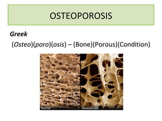
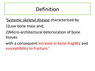
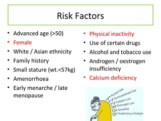
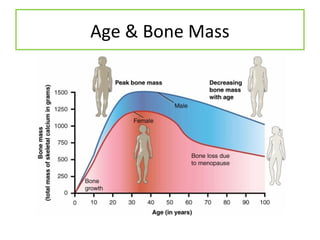
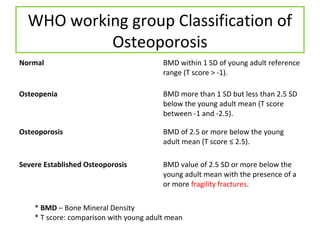
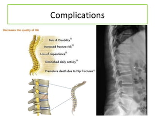
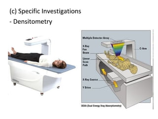
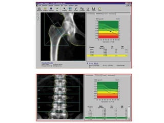
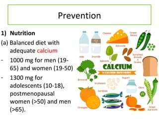
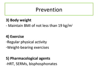
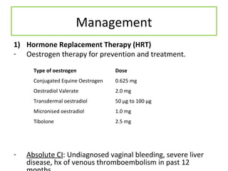
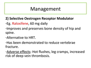
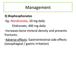
OSTEOPOROSI
S
& OTHER METABOLIC BONE DISEASES
DSL
OSTEOPOROSIS
Greek
(Osteo)(poro)(osis) – (Bone)(Porous)(Condition)
Definition
‘Systemic skeletal disease characterized by
1)Low bone mass and;
2)Micro-architectural deterioration of bone
tissues
with a consequent increase in bone fragility and
susceptibility to fracture.’
Aetiology
Primary osteoporosis
1) Juvenile
2) Post-menopausal (Type I)
3) Senile (Type II)
Secondary osteoporosis
1) Congenital
2) Hypogonadal states
3) Endocrine disorders
4) Deficiency states
5) Inflammatory diseases
6) Haematologic and
neoplastic disorders
7) Medications
8) Miscellaneous
Risk Factors
• Advanced age (>50)
• Female
• White / Asian ethnicity
• Family history
• Small stature (wt.<57kg)
• Amenorrhoea
• Early menarche / late
menopause
• Physical inactivity
• Use of certain drugs
• Alcohol and tobacco use
• Androgen / oestrogen
insufficiency
• Calcium deficiency
Age & Bone Mass
WHO working group Classification of
Osteoporosis
Normal BMD within 1 SD of young adult reference
range (T score > -1).
Osteopenia BMD more than 1 SD but less than 2.5 SD
below the young adult mean (T score
between -1 and -2.5).
Osteoporosis BMD of 2.5 or more below the young
adult mean (T score ≤ 2.5).
Severe Established Osteoporosis BMD value of 2.5 SD or more below the
young adult mean with the presence of a
or more fragility fractures.
* BMD – Bone Mineral Density
* T score: comparison with young adult mean
Complications
Diagnosis
1) Clinical Presentation
Mostly asymptomatic; incidentally found after a
fracture
- Increasing dorsal kyphosis (Dowager’s hump)
- Low-trauma fracture
- Loss of height
- Back pain
2) Exclusion of secondary causes
-Careful history
-Physical examination (Low body weight, loss of
height, kyphosis, tenderness over vertebrae)
3) Investigations
(a)General
FBC, ESR, serum calcium, phosphate and
albumin, alkaline phophatase, RFT, plain X-rays
(osteoporosis apparent only >30% of bone loss
has occurred)
(b) Others – as indicated
FT4, TSH, testosterone, FSH, LH, etc.
(c) Specific Investigations
- Densitometry
Monitoring
• Regular clinical assessments
• DEXA every 1 -2 years
• Dietary counselling
Prevention
1) Nutrition
(a) Balanced diet with
adequate calcium
- 1000 mg for men (19-
65) and women (19-50)
- 1300 mg for
adolescents (10-18),
postmenopausal
women (>50) and men
(>65).
Prevention
(b) Vitamin D
-Exposure to sunlight >15
min / day
-Otherwise 800IU Vit D
supplement daily.
3) Body weight
- Maintain BMI of not less than 19 kg/m2
4) Exercise
-Regular physical activity
-Weight-bearing exercises
5) Pharmacological agents
-HRT, SERMs, bisphosphonates
Prevention
Management
1) Hormone Replacement Therapy (HRT)
- Oestrogen therapy for prevention and treatment.
- Absolute CI: Undiagnosed vaginal bleeding, severe liver
disease, hx of venous thromboembolism in past 12
Type of oestrogen Dose
Conjugated Equine Oestrogen 0.625 mg
Oestradiol Valerate 2.0 mg
Transdermal oestradiol 50 μg to 100 μg
Micronised oestradiol 1.0 mg
Tibolone 2.5 mg
Management
2) Selective Oestrogen Receptor Modulator
-Eg. Raloxifene, 60 mg daily
-Improves and preserves bone density of hip and
spine.
-Alternative to HRT.
-Has been demonstrated to reduce vertebrae
fracture.
-Adverse effects: Hot flushes, leg cramps, increased
risk of deep vein thrombosis.
Management
3) Bisphosphonates
-Eg. Alendronate, 10 mg daily
Etidronate, 400 mg daily
-Increases bone mineral density and prevents
fractures.
-Adverse effects: Gastrointestinal side effects
(oesophageal / gastric irritation)
Management
3) Calcitonin
-Eg. 200IU intranasal daily
-Anti-resorptive agent
-Analgesic effect for acute pain relief in
osteoporotic fractures
-Adverse effects: Nausea, flushing, vomiting,
nasal irritation.
• Excessive parathyroid hormone resulting in
abnormal calcium homeostasis
HYPERPARATHYROIDISM
Effects on bone
• Osteopenia from chronic
excessive bone resorption
• In severe cases – osteitis
fibrosa cystica
- Subperiosteal resorption
of the distal phalanges
- Tapering of the distal
clavicles
- Salt-and-pepper
appearance of the skull
- Brown tumour of long
bones
• Vitamin D is important for calcium
homeostasis
• Increases the efficiency of calcium absorption
from the small intestine
• Enhances absorption of phosphorus from the
distal small bowel
• Both important for proper bone
mineralization
VITAMIN D DEFICIENCY
Effects on bone
• Rickets – in children
before growth plate
fuses – leg bowing
• Osteomalacia – in
adults, presents as
poorly mineralized
skeletal matrix –
periosteal bone pain
• “Brittle/fragile-bone disease”
• Generalized disease of connective tissue due
to qualitative or quantitative reduction in type
1 collagen
OSTEOGENESIS IMPERFECTA
• Localized disorder of bone remodelling
• Begins with excessive bone resorption
followed by an increase in bone formation
• Structurally disorganized bone – susceptible to
fracture
Phases: 1) Lytic
2) Mixed lytic and blastic
3) Sclerotic
PAGET DISEASE
Effects on bone
• May be monostotic /
polyostotic
• Axial skeleton – pelvis
and spine
• Mostly asymptomatic,
if not – bone pain,
manifestations of
complications
(deafness, nerve
palsies)
• “Marble bone disease”
• Failure of osteoclasts to
resorb bone
• Due to various gene
mutations
• Impaired bone modelling
and remodelling
• Skeletal fragility despite
increased bone mass –
thick, dense, sclerotic
bone
OSTEOPETROSIS

Osteoporosis

  • 1.
  • 2.
  • 3.
    Definition ‘Systemic skeletal diseasecharacterized by 1)Low bone mass and; 2)Micro-architectural deterioration of bone tissues with a consequent increase in bone fragility and susceptibility to fracture.’
  • 4.
    Aetiology Primary osteoporosis 1) Juvenile 2)Post-menopausal (Type I) 3) Senile (Type II) Secondary osteoporosis 1) Congenital 2) Hypogonadal states 3) Endocrine disorders 4) Deficiency states 5) Inflammatory diseases 6) Haematologic and neoplastic disorders 7) Medications 8) Miscellaneous
  • 5.
    Risk Factors • Advancedage (>50) • Female • White / Asian ethnicity • Family history • Small stature (wt.<57kg) • Amenorrhoea • Early menarche / late menopause • Physical inactivity • Use of certain drugs • Alcohol and tobacco use • Androgen / oestrogen insufficiency • Calcium deficiency
  • 6.
  • 7.
    WHO working groupClassification of Osteoporosis Normal BMD within 1 SD of young adult reference range (T score > -1). Osteopenia BMD more than 1 SD but less than 2.5 SD below the young adult mean (T score between -1 and -2.5). Osteoporosis BMD of 2.5 or more below the young adult mean (T score ≤ 2.5). Severe Established Osteoporosis BMD value of 2.5 SD or more below the young adult mean with the presence of a or more fragility fractures. * BMD – Bone Mineral Density * T score: comparison with young adult mean
  • 8.
  • 9.
    Diagnosis 1) Clinical Presentation Mostlyasymptomatic; incidentally found after a fracture - Increasing dorsal kyphosis (Dowager’s hump) - Low-trauma fracture - Loss of height - Back pain
  • 11.
    2) Exclusion ofsecondary causes -Careful history -Physical examination (Low body weight, loss of height, kyphosis, tenderness over vertebrae)
  • 12.
    3) Investigations (a)General FBC, ESR,serum calcium, phosphate and albumin, alkaline phophatase, RFT, plain X-rays (osteoporosis apparent only >30% of bone loss has occurred) (b) Others – as indicated FT4, TSH, testosterone, FSH, LH, etc.
  • 13.
  • 15.
    Monitoring • Regular clinicalassessments • DEXA every 1 -2 years • Dietary counselling
  • 16.
    Prevention 1) Nutrition (a) Balanceddiet with adequate calcium - 1000 mg for men (19- 65) and women (19-50) - 1300 mg for adolescents (10-18), postmenopausal women (>50) and men (>65).
  • 17.
    Prevention (b) Vitamin D -Exposureto sunlight >15 min / day -Otherwise 800IU Vit D supplement daily.
  • 18.
    3) Body weight -Maintain BMI of not less than 19 kg/m2 4) Exercise -Regular physical activity -Weight-bearing exercises 5) Pharmacological agents -HRT, SERMs, bisphosphonates Prevention
  • 19.
    Management 1) Hormone ReplacementTherapy (HRT) - Oestrogen therapy for prevention and treatment. - Absolute CI: Undiagnosed vaginal bleeding, severe liver disease, hx of venous thromboembolism in past 12 Type of oestrogen Dose Conjugated Equine Oestrogen 0.625 mg Oestradiol Valerate 2.0 mg Transdermal oestradiol 50 μg to 100 μg Micronised oestradiol 1.0 mg Tibolone 2.5 mg
  • 20.
    Management 2) Selective OestrogenReceptor Modulator -Eg. Raloxifene, 60 mg daily -Improves and preserves bone density of hip and spine. -Alternative to HRT. -Has been demonstrated to reduce vertebrae fracture. -Adverse effects: Hot flushes, leg cramps, increased risk of deep vein thrombosis.
  • 21.
    Management 3) Bisphosphonates -Eg. Alendronate,10 mg daily Etidronate, 400 mg daily -Increases bone mineral density and prevents fractures. -Adverse effects: Gastrointestinal side effects (oesophageal / gastric irritation)
  • 22.
    Management 3) Calcitonin -Eg. 200IUintranasal daily -Anti-resorptive agent -Analgesic effect for acute pain relief in osteoporotic fractures -Adverse effects: Nausea, flushing, vomiting, nasal irritation.
  • 23.
    • Excessive parathyroidhormone resulting in abnormal calcium homeostasis HYPERPARATHYROIDISM
  • 24.
    Effects on bone •Osteopenia from chronic excessive bone resorption • In severe cases – osteitis fibrosa cystica - Subperiosteal resorption of the distal phalanges - Tapering of the distal clavicles - Salt-and-pepper appearance of the skull - Brown tumour of long bones
  • 25.
    • Vitamin Dis important for calcium homeostasis • Increases the efficiency of calcium absorption from the small intestine • Enhances absorption of phosphorus from the distal small bowel • Both important for proper bone mineralization VITAMIN D DEFICIENCY
  • 26.
    Effects on bone •Rickets – in children before growth plate fuses – leg bowing • Osteomalacia – in adults, presents as poorly mineralized skeletal matrix – periosteal bone pain
  • 27.
    • “Brittle/fragile-bone disease” •Generalized disease of connective tissue due to qualitative or quantitative reduction in type 1 collagen OSTEOGENESIS IMPERFECTA
  • 28.
    • Localized disorderof bone remodelling • Begins with excessive bone resorption followed by an increase in bone formation • Structurally disorganized bone – susceptible to fracture Phases: 1) Lytic 2) Mixed lytic and blastic 3) Sclerotic PAGET DISEASE
  • 29.
    Effects on bone •May be monostotic / polyostotic • Axial skeleton – pelvis and spine • Mostly asymptomatic, if not – bone pain, manifestations of complications (deafness, nerve palsies)
  • 30.
    • “Marble bonedisease” • Failure of osteoclasts to resorb bone • Due to various gene mutations • Impaired bone modelling and remodelling • Skeletal fragility despite increased bone mass – thick, dense, sclerotic bone OSTEOPETROSIS