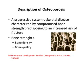
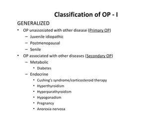
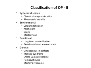
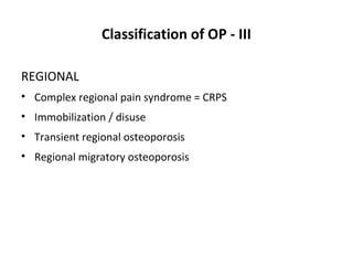
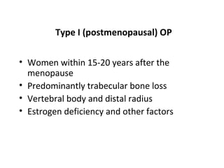
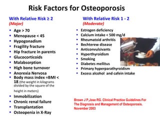
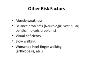
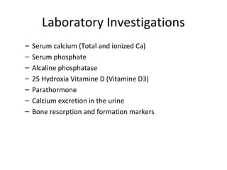
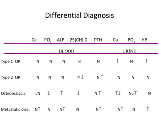
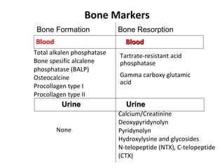
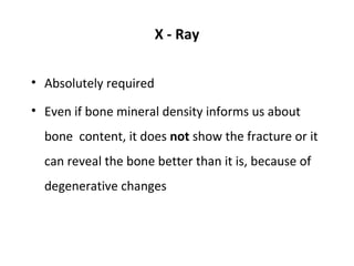
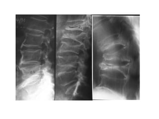
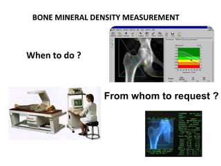
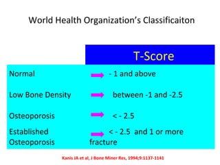
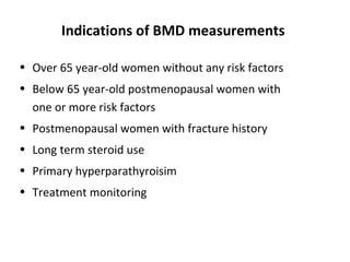
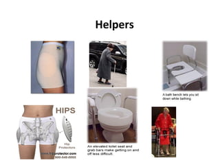
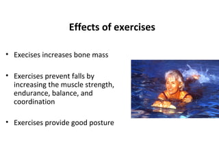
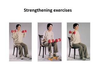
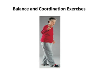
3. Description of Osteoporosis
• A progressive systemic skeletal disease
characterized by compromised bone
strength predisposing to an increased risk of
fracture
• Bone strenght :
– Bone density
– Bone quality
NIH Consensus Development Panel of Osteoporosis JAMA 285 :785
95,2001
4. The New Concept: Osteoporomalacia
matrix matrix
mineral mineral
Normal Osteoporosis
matrix matrix
mineral mineral
Osteomalacia Osteoporosis / malacia
5. Classification of OP - I
GENERALIZED
• OP unassosiated with other disease (Primary OP)
– Juvenile idiopathic
– Postmenopausal
– Senile
• OP associated with other diseases (Secondary OP)
– Metabolic
• Diabetes
– Endocrine
• Cushing’s syndrome/corticosteroid therapy
• Hyperthyroidism
• Hyperparathyroidism
• Hypogonadism
• Pregnancy
• Anorexia nervosa
7. Classification of OP - III
REGIONAL
• Complex regional pain syndrome = CRPS
• Immobilization / disuse
• Transient regional osteoporosis
• Regional migratory osteoporosis
8. Type I (postmenopausal) OP
• Women within 15-20 years after the
menopause
• Predominantly trabecular bone loss
• Vertebral body and distal radius
• Estrogen deficiency and other factors
9. Type II (senile) OP
• Both men and women over age 70
• Proportionate loss of cortical and
trabecular bone
• Hip and vertebral fractures
• Factors related to aging
– Impaired osteoblastic function
– Impairment of renal 1-alpha-hydroxylase
activity
10. TYPE I TYPE II
Postmenopausal Senile
Age (years) 50-70 >70
Sex ratio (F:M) 6:2 2:1
Types of bone Mainly Trabecular and
loss trabecular cortical
Fracture sites Vertebrae and/or Proximal femur
distal radius
Main causes Menopause Aging
11. Pathogenesis of OP - I
• Peak bone mass
• The rate of bone turnover
• Menopause age (early menopause)
• Microarchitectural deterioration of bone
tissue
• Repair disorders of bone
12. Pathogenesis of OP - II
A- The factors affecting peak bone mass
• Genetic
– Vitamine D receptor gene
– Procollagen type I gene
– The receptors of estrogen
• Hormonal and nutritional factors
• Environmental factors
B- The factors affecting the rate of bone turnover
• The differences between trabecular and cortical bone structure
• The differences between men and women
13. Signs and Symptoms of OP - I
• The cardinal symptom of OP is “fracture”
• Vertebral fractures present with acute back pain after
sudden bending, lifting or coughing
• It is associated with progressive kyphosis
• Vertebral fracture can be painless -asymptomatic- and
incidentally discovered in the X-Rays
• Fractures of the distal forearm and proximal femur usually
follow falls
• Rib fractures can be seen
14. Signs and Symptoms of OP - II
• back pain
• loss of height
• increased kyphosis
• immobility
• increased number of bed days
• loss of self-esteem
• distorted body image
• depression
15. Signs and Symptoms of OP - III
• Reduced pulmonary function
• Bone tenderness
• Extreme fatigue
• Brittle or soft fingernails
• Premature grey hair
• Leg cramps at nights
17. Risk Factors for Osteoporosis
With Relative Risk ≥ 2 With Relative Risk 1 - 2
(Major) (Moderate)
• Age > 70 • Estrogen deficiency
• Menopause < 45 • Calcium intake < 500 mg/d
• Hypogonadism • Rheumatoid arthritis
• Bechterew disease
• Fragility fracture
• Anticonvulsivants
• Hip fracture in parents
• Hyperthyroidism
• Glucocorticoids • Smoking
• Malabsorption • Diabetes mellitus
• High bone turnover • Primary hyperparathyroidism
• Anorexia Nervosa • Excess alcohol and cafein intake
• Body mass index =BMI <
18 (the weight in kilograms
divided by the square of the
height in meters)
• Immobilization
• Brown J P,Jose RG. Clinical Practice Guidelines For
Chronic renal failure
The Diagnosis and Management of Osteoporosis.
• Transplantation November 2003
• Osteopenia in X-Ray
20. Laboratory Investigations
– Serum calcium (Total and ionized Ca)
– Serum phosphate
– Alcaline phosphatase
– 25 Hydroxia Vitamine D (Vitamine D3)
– Parathormone
– Calcium excretion in the urine
– Bone resorption and formation markers
21. Differential Diagnosis
Ca PO4 ALP 25(OH) D PTH Ca PO4 HP
BLOOD URINE
Type 1 OP N N N N N ↑ N ↑
Type 2 OP N N N N↓ N↑ N N N
Osteomalacia ↓N ↓ ↑ ↓ N↑ ↑↓ N↓↑ N
Metastatic dise. N↑ N N↑ N N↑ N↑ N ↑
22. Bone Markers
Bone Formation Bone Resorption
Blood Blood
Total alkalen phosphatase Tartrate-resistant acid
Bone spesific alcalene phosphatase
phosphatase (BALP)
Osteocalcine Gamma carboxy glutamic
acid
Procollagen type I
Procollagen type II
Urine Urine
Calcium/Creatinine
Deoxypyridynolyn
None Pyridynolyn
Hydroxylysine and glycosides
N-telopeptide (NTX), C-telopeptide
(CTX)
23. X - Ray
• Absolutely required
• Even if bone mineral density informs us about
bone content, it does not show the fracture or it
can reveal the bone better than it is, because of
degenerative changes
24. Semiquantitative assessment of vertebral
fractures
SQ stage
0 normal
1 mild
2 moderate
3 severe
Genant et al. J BoneGenant et al. 1993: 8;Mineral Res 1993: 8; 1137-48
Mineral Res J Bone 1137-48
27. World Health Organization’s Classificaiton
T-Score
Normal - 1 and above
Low Bone Density between -1 and -2.5
Osteoporosis < - 2.5
Established < - 2.5 and 1 or more
Osteoporosis fracture
Kanis JA et al, J Bone Miner Res, 1994;9:1137-1141
28. Indications of BMD measurements
• Over 65 year-old women without any risk factors
• Below 65 year-old postmenopausal women with
one or more risk factors
• Postmenopausal women with fracture history
• Long term steroid use
• Primary hyperparathyroisim
• Treatment monitoring
29. Advantages of DEXA
• Useful method in patient follow up
• Low radiaton dose (2-4 mRem)
• High precision and accuracy
• Short time for assessment
• Peripheral measurement is available
30. Disadvantages of DEXA
• No differentiation between cortical and trabecular bone
tissues
• Degenerative changes effet the results negatively
• Obesity is a serious problem !
• Expensive
• Reference values change from country to country
• No standardization in the different devices
31. Bone Biopsy
– It is usually preferrred from the iliac crest
– Invasive but definite diagnostic tool
– It must be done in certain conditions such as renal
osteodystrophy and osteomalacia
Normal Bone Osteoporotic Bone
33. Management of OP - I
• The therapeutic approach to OP is a complex
• It means not only taking some medicine but also
– changing a life style
– leaving some harmful habituations (e.g. smoking,
alcohol), and
– increasing physical activity and exercise
34. Management of OP- II
• Early diagnosis is the best to preserve bone mass
• Medical treatment can slow bone loss and
decrease the risk of fracture
• A rehabilitation program designed to reduce pain,
increase mobility, and minimize risk of falling is
necessary
36. Osteoporosis Rehabilitation
• Treatment of pain
• Physical restoration
• Diet, medical treatment, exercises
• Correcting the disability
• Education of the patient and his/her family
• Prevention of falls
37. Who falls and how?
• People at the age of 50s walk fast
and they cast forward themselves
and lean on their hands over the
floor while they are falling
• That is why it is easy for them to
have wrist fractures
• Advanced in years they walk
more slowly and they fall over
their hips
38. Reasons for increase in the risk of falling
• Diminished hearing and vision
• Muscle weakness
• Posture and balance disorder
• Excess alcohol intake
• Hypertension
• Diabetes mellitus
• Parkinson’s disease
39.
40.
41. How to prevent falls ?
• Appropriate clothes and
shoes
• Regularly ophthalmologic
examination
• Use of supportive devices
• Walking on a smooth way
• Moving slowly and safely
43. Effects of exercises
• Execises increases bone mass
• Exercises prevent falls by
increasing the muscle strength,
endurance, balance, and
coordination
• Exercises provide good posture
44. Types of Exercises
There are 5 types of exercises recommended for
osteoporosis:
• Stretching exercises
• Aerobic exercises performed by body mass
• Strengthening exercises
• Exercises with high power
• Balance exercises