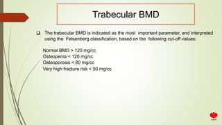
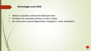
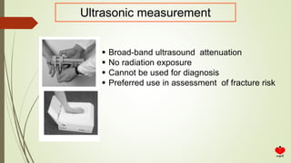
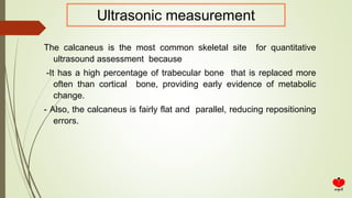
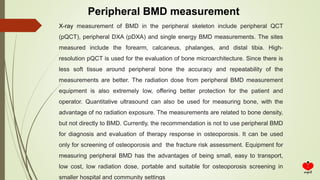
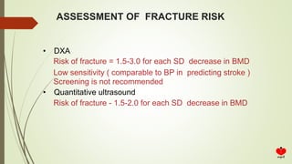
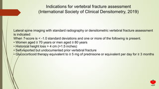
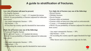
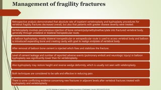
1. IMAGING IN OSTEOPOROSIS
DR. VANDANA BANSAL
MS, D.phil. (Gold Medalist), DGO, FCGP
Infertility & IVF Specialist & Advance Laparoscopic Surgeon
DIRECTOR
Arpit Test Tube Baby Centre, Prayagraj
Jeevan Jyoti Hospital, Prayagraj (U.P.)
2. INTRODUCTION OF OSTEOPOROSIS
1. Osteoporosis and associated fractures are the most common chronic metabolic bone
disease and represent a major global health problem, contributing to 8.9 million fractures
worldwide on an annual basis. Worldwide, there are marked variations in the rates of hip
fracture and major osteoporotic fractures. Fractures associated with osteoporosis cause
not only increased morbidity but also an increased mortality.
2. Osteoporosis may significantly affect life expectancy and quality of life.
3. Osteoporosis is often thought of as an older person’s disease, it can strike at any age.
3. DEFINITION
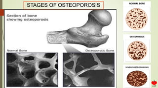
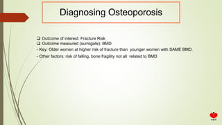
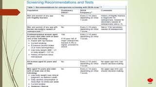
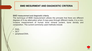
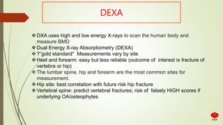
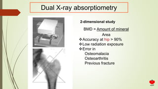
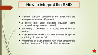
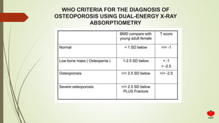
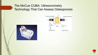
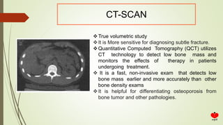
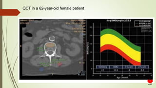
• Osteoporosis is a systemic skeletal disease characterized by low bone mass and deterioration
of bone microarchitecture with a consequent increase in fragility and susceptibility to
insufficiency fracture. It can be classified as primary or secondary. Primary osteoporosis
includes post-menopausal osteoporosis (type I), osteoporosis in the elderly (type II), and
idiopathic osteoporosis (including in adolescents). Secondary osteoporosis refers to patients
with underlying metabolic disease and/or drug induced or from other obvious causes of
osteoporosis. This consensus document mainly applies to the diagnosis of primary
osteoporosis.
• The vertebrae .wrists ,and hips are the
most common sites of fractures
4. Fragility fracture is one caused by a degree of trauma not expected to cause a
fracture; for example, a fall from standing height or lower. Fragility fractures, such as
vertebral compression fractures and distal forearm fractures, are common in the elderly
but can occur at any age. Exclusions: toes, fingers, face, skull, and ribs.
Major osteoporotic fracture is a fracture of the hip, spine (clinical), wrist, or humerus.
Osteoporosis is defined as a history of fragility fracture and/or a T-score of -2.5 or lower
on dual energy X-ray absorptiometry (DEXA).
Osteopenia (or low bone mass) is defined as a T-score between -1.0 and -2.5 on
DEXA.
DEFINITIONS
6. India is home to more than 1.3 billion people, with
approximately 230 million Indians over 50 years.
About 20% of the 230 million Indian women over age 50
have osteoporosis
Prevalence of osteoporosis ranging from 8 to 62% in Indian
women of different age groups has been reported in several
studies
The current Indian population as a whole, the number of hip
fractures every year would be more than 440,000, with a
female to male ratio of about 3:1, with a projected incidence
of more than 600,000 in 2020 and over 1 million in 2050.
Prevalence of Osteoporosis
7. Result of prolonged imbalance of Bone Rent(/deling; •
Mechanisms causing osteoporosis Imbalance between rate of resorption and formation
Failure to complete stages of remodeling
- Bone remodeling occurs throughout an individual’s lifetime.
- In normal adults, the activity of osteoclasts (bone resorption) is balanced by that of osteoblasts
(bone formation).
- normal bone remodeling in the adult result in gradually increase bone mass until the early 30s.
* With ageing the peak bone mass is gradually decrease and
1. Calicitonin which inhibit bone resorption and promote bone formation, (decrease)
2. Estrogen which inhibit bone breakdown, (decrease)
3. PTH increase bone turnover and resorption, (increase)
PATHOPHYSIOLOGY
8. CLASSIFICATION
Primary
Postmenopausal (Type I)
Caused by lack of estrogen
Causes PTH to overstimulate osteoclast
Bone loss - 2-3% per year of total bone mass Most com men fx: vertebral, distal forearm
Age related (Type II)
Bone loss due to increased bone turnover Malabsorption
Mineral and vitamin deficiency
- 3rd decade of life starts slow decline in bone mass at rate of 0.5-1% per year
Most common types of fx: hip and radius
> F>M
IDIOPATHIC (Including adolescence)
Secondary
12. Risk Factors For Osteoporosis And Osteoporotic Fractures
• Women aged > 65
• Caucasian or Asian race
• Low body weight; (< 127 lbs or BMI < 20)
• Family history of Osteoporosis
• Personal history of fragility fracture
and/or fracture as an adult
• History of fragility fracture in a first-
degree relative
• Long-term use of Glucocorticoids or
others
• Current tobacco smoking
• Alcohol in amounts > 2-3 drinks per day
• Estrogen deficiency at an early age (< 45yrs)
• Low calcium intake (lifelong) and/or Vitamin D
deficiency
• Sedentary lifestyle
• Testosterone/Estrogen depletion in men
• Increased risk of falling due to:
o Dementia
o Poor health/frailty
o Recent falls
o Poor vision
14. Diagnosis of Osteoporosis
The aim is to –
1. To diagnose osteoporosis and osteopenia
2. Predicting fracture risk
3. Evaluating management
15. Diagnosing Osteoporosis
Outcome of interest: Fracture Risk
Outcome measured (surrogate): BMD
- Key: Older women at higher risk of fracture than younger women with SAME BMD.
- Other factors: risk of falling, bone fragility not all related to BMD
16. Diagnostic criteria of Osteoporosis
The principle of diagnosing osteoporosis is to combine clinical history, risk
factors, clinical manifestations, imaging findings, BMD measurement and
laboratory results together. Osteoporosis can be diagnosed if BMD
measurements and clinical manifestations indicate osteoporotic status.
Imaging findings and BMD measurements play a critical role here. Once
various imaging studies (such as X-ray, CT, MRI, and nuclear medicine
studies) demonstrate an insufficiency fracture, osteoporosis can be
diagnosed regardless of the findings of a BMD measurement. However,
before the occurrence of insufficiency fracture, the diagnosis is mainly
based on BMD measurement.
It is important to differentiate primary, secondary, or idiopathic osteoporosis,
which should be based on age, sex, history, clinical manifestations,
laboratory results, and imaging findings. Biochemistry laboratory results can
reflect bone formation and bone resorption, aiding classification, differential
diagnose and early evaluation of treatment. However, biochemistry cannot
be used alone for diagnosing osteoporosis.
17. Diagnosis of Osteoporosis
The aim is to –
Physical examination
Measurement of bone mineral content
Dual X-ray absorptiometry (DXA)
Ultrasonic measurement of bone
CT scan
Radiography
Nuclear medicine studies
MRI
QCT measurement of BMD
18. Physical examination
Height loss
Body Weight
Kyphosis
Human back
Tooth loss
Skinfold
Thickness
Grip strength
Osteoporosis Vertebral fracture
Arm span-height difference
Wall- occiput distance
Rib-pelvis distance
20. Physical examination
No single maneuver is sufficient to rule in or
rule out osteoporosis or vertebral fracture
without further testing.
21. Work-up
Screen for secondary causes
Serum calcium, phosphorus, alk phos
PTH if calcium is high
25-hydroxyvitamin D if low ca,
low phos and high alk. phos
Thyroid function tests
SPEP, UPEP
24-hour urinary calcium
Serum testosterone
(hyperparathyroidism)
(osteomalacia)
(thyrotoxicosis)
(multiple myeloma)
(hyper or hypo calciuria)
(hypogonadism)
22. Plan radiography
Low sensitivity
High availability
If there is bone loss on a plain X-ray, further evaluation with a BMD
measurement is warranted.
Subclinical vertebral fracture is a strong risk factor for subsequent
fractures at new vertebral site and other sites
if there is subtle vertebral compression, the recommendation is to
combine an X-ray with CT and/or MRI
23. The main radiographic features of generalized osteoporosis are
cortical thinning and increased radiolucency
24. Singh Index
The Singh index describes the trabecular patterns in the bone at
the top of the thighbone (femur).
X-rays are graded 1 through 6 according to the disappearance of
the normal trabecular pattern.
Studies have shown a link between a Singh index of less than 3
and fractures of the hip, wrist, and spine.
25. When to Measure BMD in Postmenopausal Women
• All women 65 years and older
• Postmenopausal women <65 years of age:
• If result might influence decisions about intervention
• One or more risk factors History of fracture
26. When Measurement of BMD Is Not Appropriate
Healthy premenopausal women
Healthy children and adolescents
Women initiating ET/HT for menopausal symptom relief (other osteoporosis
therapies should not be initiated without BMD measurement)
27.
28. BMD MESURMENT AND DIAGNOSTIC CRITERIA
BMD measurement and diagnostic criteria.
The technique of BMD measurement utilizes the principle that there are different
degrees of X-ray attenuation when X-rays pass through different media. It is a non-
invasive measurement of human bone mineral content, bone density and
composition. The current commonly used methods include
• DXA,
• QCT
• peripheral DXA.
29. DEXA
DXA uses high and low energy X-rays to scan the human body and
measure BMD
Dual Energy X-ray Absorptiometry (DEXA)
?”gold standard" Measurements vary by site
Heel and forearm: easy but less reliable (outcome of interest is fracture of
vertebra or hip)
The lumbar spine, hip and forearm are the most common sites for
measurement.
Hip site: best correlation with future risk hip fracture
Vertebral spine: predict vertebral fractures; risk of falsely HIGH scores if
underlying OA/osteophytes
30. Dual X-ray absorptiometry
2-dimensional study
BMD = Amount of mineral
Area
Accuracy at hip > 90%
Low radiation exposure
Error in
Osteomalacia
Osteoarthritis
Previous fracture
31. How to interpret the BMD
T score: standard deviation of the BMD from the
average sex matched 35-year-old
Z score: less used; standard deviation score
compared to age matched control
For every 1 decrease in T score, double risk of
fracture
1 SD decrease in BMD -14 year increase in age for
predicting hip fracture risk
Regardless of BMD, patients with prior osteoporotic
fracture have up to 5 times risk of future fracture.
32. WHO CRITERIA FOR THE DIAGNOSIS OF
OSTEOPOROSIS USING DUAL-ENERGY X-RAY
ABSORPTIOMETRY
33. The WHO criteria are suitable for use in post-menopausal women and
men over the age of 50. WHO T-scores should never be used to diagnose
osteoporosis in children and adolescents. For children, adolescents, pre-
menopausal women, and men under the age of 50, the Z-score value is
recommended for interpreting BMD measurements. Z-scores are
calculated using BMD reference data derived from healthy subjects of the
same age, race and sex. A Z-score <=−2.0 SD (standard deviation) is
defined as “lower than expected range of the same age group” or low bone
density (2,8). The normal reference data for children and adolescents
should be derived from data for the Chinese population (18). Women with
a history of ovariectomy are regarded as equivalent to “post-menopausal”
and the T-score can be used for diagnosis.
How to interpret the BMD
34. -scanners are readily available and relatively inexpensive.
-The radiation dose is negligible
-The T-score scale, defined by the WHO specifically for DXA, provides a
standardized classification.
Clinicians and researchers favor DXA because
35. LIMITATION OF DEXA
BMD measurements are influenced by osteoarthritis, scoliosis,
osteophytes, vertebral body fracture, vascular calcification and
obesity, and these decrease the accuracy of the measurements and
may lead to misdiagnosis (19,20). When patients are underweight,
overweight or have scoliosis or degenerative changes, QCT is
recommended for BMD measurement to minimize their influence or
search for evidence of insufficiency fracture.
36. The trabecular BMD is indicated as the most important parameter, and interpreted
using the Felsenberg classification, based on the following cut-off values:
Normal BMD > 120 mg/cc
Osteopenia < 120 mg/cc
Osteoporosis < 80 mg/cc
Very high fracture risk < 50 mg/cc
Trabecular BMD
37. Ability to separate cortical and trabecular bone
Provides true volumetric density in units of mg/cc
No errors due to spinal degenerative changes or aortic calcification
Advantages over DXA:
38. Ultrasonic measurement
Broad-band ultrasound attenuation
No radiation exposure
Cannot be used for diagnosis
Preferred use in assessment of fracture risk
39. The calcaneus is the most common skeletal site for quantitative
ultrasound assessment because
-It has a high percentage of trabecular bone that is replaced more
often than cortical bone, providing early evidence of metabolic
change.
- Also, the calcaneus is fairly flat and parallel, reducing repositioning
errors.
Ultrasonic measurement
40. The McCue CUBA: Ultrasonometry
Technology That Can Assess Osteoporosis
41. Heel BUA is Significantly Lower in Subjects
With Future Hip Fracture.
42. CT-SCAN
True volumetric study
It is More sensitive for diagnosing subtle fracture.
Quantitative Computed Tomography (QCT) utilizes
CT technology to detect low bone mass and
monitors the effects of therapy in patients
undergoing treatment.
It is a fast, non-invasive exam that detects low
bone mass earlier and more accurately than other
bone density exams
It is helpful for differentiating osteoporosis from
bone tumor and other pathologies.
44. Peripheral BMD measurement
X-ray measurement of BMD in the peripheral skeleton include peripheral QCT
(pQCT), peripheral DXA (pDXA) and single energy BMD measurements. The sites
measured include the forearm, calcaneus, phalanges, and distal tibia. High-
resolution pQCT is used for the evaluation of bone microarchitecture. Since there is
less soft tissue around peripheral bone the accuracy and repeatability of the
measurements are better. The radiation dose from peripheral BMD measurement
equipment is also extremely low, offering better protection for the patient and
operator. Quantitative ultrasound can also be used for measuring bone, with the
advantage of no radiation exposure. The measurements are related to bone density,
but not directly to BMD. Currently, the recommendation is not to use peripheral BMD
for diagnosis and evaluation of therapy response in osteoporosis. It can be used
only for screening of osteoporosis and the fracture risk assessment. Equipment for
measuring peripheral BMD has the advantages of being small, easy to transport,
low cost, low radiation dose, portable and suitable for osteoporosis screening in
smaller hospital and community settings
45. MRI
• MRI can be used to evaluate the osteoporosis quantitatively
• Currently MRI is not suitable for clinical diagnosis or screening for
osteoporosis, but it is used for the evaluation of osteoporosis
insufficiency fracture and providing differential diagnosis.
• MRI quantitative measurement of bone marrow fat and quantitative
evaluation of bone cortex using UTE have the potential to provide a new
strategy for diagnosis and screening of osteoporosis in the future.
46. FRAX CALCULATOR
The FRAX calculator
This tool estimates the 10-year probability of osteoporotic fracture for postmenopausal
women and men aged 50 years and older who have not been previously treated for
osteoporosis. Risk factors included in the FRAX are: age, gender, low body weight, height,
previous fracture, parent with hip fracture, smoking status, glucocorticoid use, history of
rheumatoid arthritis, menopausal status, and excessive alcohol consumption.
The FRAX calculator is available online at http://www.shef.ac.uk/FRAX/ . Use the drop-down
list under “Calculation Tool.”
Limitations: The FRAX calculator may over- or underestimate fracture risk in patients with a
history of vertebral fracture, hip fracture, or multiple fractures, as well as in patients who are
Black, Latino, or from other races or ethnicities. Some risk factors, such as frailty and
dementia, cannot be readily quantified and are not included in the calculation.
47. ASSESSMENT OF FRACTURE RISK
• DXA and quantitative ultrasound
• Clinical risk factors
• Markers of bone turnover
Bone formation
Bone resorption
48. ASSESSMENT OF FRACTURE RISK
• DXA
Risk of fracture = 1.5-3.0 for each SD decrease in BMD
Low sensitivity ( comparable to BP in predicting stroke )
Screening is not recommended
• Quantitative ultrasound
Risk of fracture - 1.5-2.0 for each SD decrease in BMD
49. Indications for vertebral fracture assessment
(International Society of Clinical Densitometry, 2019)
Lateral spine imaging with standard radiography or densitometric vertebral fracture assessment
is indicated:
When T-score is < -1.0 standard deviations and one or more of the following is present.
• Women aged ≥ 70 years or men aged ≥ 80 years
• Historical height loss > 4 cm (>1.5 inches)
• Self-reported but undocumented prior vertebral fracture
• Glucocorticoid therapy equivalent to ≥ 5 mg of prednisone or equivalent per day for ≥ 3 months
50. A guide to stratification of fractures.
Low risk of fracture (all must be present)
• No fragility fractures
• DXA-derived T-score < -1 and > -2.5 standard deviations
• FRAX 10-year probability of fracture (adjusted for trabecular
bone score):
– Any major osteoporotic fracture: < 20%
– Hip fracture: < 3%
– Or less than the country-specific threshold for intervention
High risk of fracture (any one of the following)
• Presence of fragility fracture
• DXA-derived T-score ≤ -2.5 standard deviations
• FRAX 10-year probability of fracture (adjusted for trabecular
bone score):
– Any major osteoporotic fracture: > 20%
– Hip fracture: > 3%
– Or exceeding the country-specific threshold for intervention
Very high risk of fracture (any one of the following)
• Recent fracture
• Multiple fractures
• Severe fracture
• Fracture while on treatment
• Fracture while on bone-toxic drug such as corticosteroids
• T-score ≤ -3.0 standard deviations
• FRAX 10-year probability of fracture (adjusted for trabecula
bone score):
– Any major osteoporotic fracture: > 30%
– Hip fracture: > 4.5%
– Or exceeding the country-specific upper threshold for high
risk
– Other factors such as an extremely high risk for falls.
51. Management of fragility fractures
Retrospective analysis demonstrated that absolute rate of inpatient vertebroplasty and kyphoplasty procedures for
vertebral fragility fractures decreased overall, but also that patients with greater disease severity were treated.
Vertebroplasty involves percutaneous injection of bone cement/polymethylmethacrylate into fractured vertebral body,
generally through unilateral or bilateral transpedicular route.
In balloon kyphoplasty, mostly bilateral transpedicular or extrapedicular route is used to access vertebral body and balloon
is introduced expanding bone and creating cavity with goal to realign endplate of vertebral body.
After removal of balloon bone cement is injected which fixes and stabilizes the fracture.
Level of cement leakage and number of reported adverse events (pulmonary emboli and neurologic injury) in balloon
kyphoplasty was significantly lower than for vertebroplasty.
Also kyphoplasty may restore height and reverse wedge deformity, which is usually not seen with vetebroplasty.
Both techniques are considered to be safe and effective in reducing pain.
There is some conflicting evidence concerning new fractures in adjacent levels after vertebral fractures treated with
kyphoplasty and vertebroplasty.
Link TM. Radiology of osteoporosis. Canadian Association of Radiologists' Journal. 2016 Feb;67(1):28-40
52. Management of fragility fractures
Acute change in stiffness may provoke fractures in adjacent levels.
3-D computer models of L2 and L3 were developed, adapting material properties to simulate osteoporosis and cement
augmentation restored strength of treated vertebra but clearly altered the load transfer in adjacent vertebra.
Fribourg et al demonstrated higher rate of subsequent fracture after kyphoplasty compared with natural history data for
untreated fractures.
Most of these occurred at adjacent level within 2 months of index procedure.
After this 2-month period, there were only occasional subsequent fractures, which occurred at remote levels.
It is recommended that patients with increase in back pain after kyphoplasty should be evaluated carefully for subsequent
adjacent fractures, especially during first 2 months after index procedure.
Sacroplasty is similar to vertebroplasty and is usually performed under CT guidance as it provides more accurate needle
placement, but ideally with fluoroscopy monitoring to assess leakage of bone cement or vascular embolization.
Bone cement is injected into fracture area and usually provides pain relief within 24 hours.
Sacroplasty is considered safe and practical, and provides effective pain relief.
Link TM. Radiology of osteoporosis. Canadian Association of Radiologists' Journal. 2016 Feb;67(1):28-40
53. A suggested treatment guideline based on fracture risk
Low fracture risk
Optimize calcium and vitamin D status
Bone-friendly lifestyle
High fracture risk
Optimize calcium and vitamin D status
Bone-friendly lifestyle
Falls prevention
Start appropriate antiresorptive therapy
Very high fracture risk
Optimize calcium and vitamin D status
Bone-friendly lifestyle
Falls prevention
Consider appropriate anabolic treatment for 12–18 months
followed by antiresorptive therapy.
54. New technology and future direction of development.
• DXA trabecular bone score (TBS):
• Spectral CT and dual-energy CT
• MRI measurement of fat
• Nuclear medicine imaging
• Artificial intelligence
55. Conclusion
Osteoporosis is a severely debilitating disease.
It is critical to identify patients with prevalent fragility fractures, as they
are at high risk for future severe fractures and not misinterpret these
findings as malignant disease prompting costly and unsafe interventions.
Diagnose and monitor osteoporosis using quantitative techniques such as
DXA and be familiar with complications of medical treatments.
Need to perform interventional procedures to treat vertebral and sacral
insufficiency fractures in concert with other clinicians adding supportive
pharmacotherapies.
Link TM. Radiology of osteoporosis. Canadian Association of Radiologists' Journal. 2016 Feb;67(1):28-40