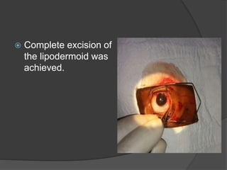
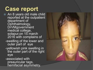
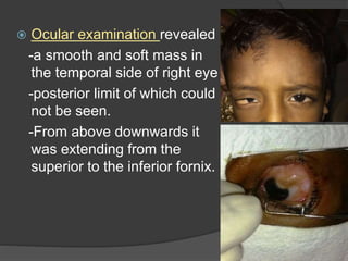
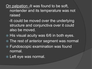
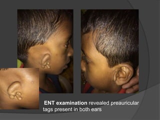
GOLDENHAR SYNDROME-
A CASE REPORT
By Dr. Sonali Ashok Bhosale
(Junior resident ,Dr V.M.Government medical
college, solapur )
INTRODUCTION
 Goldenhar syndrome is a birth defect
resulting from the maldevelopment of the first
two branchial arches with incomplete
development of the ear, nose, soft palate, lip
and mandible.
 It is unilateral in 70-80% of the cases
 Prevalence rate is 1-9/per 100000 and
 Incidence rate is 1 per 25000-45000 births
 Male is more commonly affected than the
female (ratio 2:1).
 Ocular anomalies occur in about 50% of
the cases.
 Epibulbar dermoid and lipodermoid are
the most common.
 Limbal dermoid or lipodermoid are
mainly located in the inferotemporal
region of the eye.
 Coloboma of the upper eyelid may be
present.
 Extraocular defects are reported in 65%
of the cases and include
 Pre-auricular tags, microtia, anotia,
conductive hearing loss.
 Vertebral anomalies are combination of
hemivertebra, fused ribs, kyphosis and
scoliosis.
 Additional features such as cardiac,
genito-urinary and pulmonary systems
can also be affected.
Case report
 An 8 years old male child
reported at the outpatient
department of
Ophthalmology,
DrVMgovernment
medical college,
solapur,on 15 march
2016 with complains of
-swelling of the lower and
outer part of eye
-yellowish pink swelling in
the outer part of the right
eye
-associated with
preauricular tags,
hemifacial asymmetry.
 Ocular examination revealed
-a smooth and soft mass in
the temporal side of right eye
-posterior limit of which could
not be seen.
-From above downwards it
was extending from the
superior to the inferior fornix.
On palpation -It was found to be soft,
nontender and its temperature was not
raised
-It could be moved over the underlying
structure and conjunctiva over it could
also be moved.
 His visual acuity was 6/6 in both eyes.
 The rest of anterior segment was normal
 Fundoscopic examination was found
normal.
 Left eye was normal.
 The clinical diagnosis was
Dermolipoma, which was confirmed by a
histological examination of an excised
portion.
ENT examination revealed preauricular
tags present in both ears
 No other abnormalities were detected.
 Systemic examination like cardiovascular,
pulmonary and genito-urinary systems was
done but no abnormalities were detected.
 The laboratory investigations were within
normal limits.
 Electrocardiography was normal,
 X-ray chest (postero anterior view) showed
nothing abnormality detected,
 X-ray of the vertebral column was normal.
 Complete excision of
the lipodermoid was
achieved.
Conclusion
 This case has been presented to
increase the awareness of this rare
entity.
 To highlight the clinical findings and
association with various systemic
conditions.
 Multidisciplinary treatment approach
and long term follow up is required to
monitor the growth and development of
such patients.

Goldenhar Syndrome-A Case Report

  • 1.
    GOLDENHAR SYNDROME- A CASEREPORT By Dr. Sonali Ashok Bhosale (Junior resident ,Dr V.M.Government medical college, solapur )
  • 2.
    INTRODUCTION  Goldenhar syndromeis a birth defect resulting from the maldevelopment of the first two branchial arches with incomplete development of the ear, nose, soft palate, lip and mandible.  It is unilateral in 70-80% of the cases  Prevalence rate is 1-9/per 100000 and  Incidence rate is 1 per 25000-45000 births  Male is more commonly affected than the female (ratio 2:1).
  • 3.
     Ocular anomaliesoccur in about 50% of the cases.  Epibulbar dermoid and lipodermoid are the most common.  Limbal dermoid or lipodermoid are mainly located in the inferotemporal region of the eye.  Coloboma of the upper eyelid may be present.
  • 4.
     Extraocular defectsare reported in 65% of the cases and include  Pre-auricular tags, microtia, anotia, conductive hearing loss.  Vertebral anomalies are combination of hemivertebra, fused ribs, kyphosis and scoliosis.  Additional features such as cardiac, genito-urinary and pulmonary systems can also be affected.
  • 5.
    Case report  An8 years old male child reported at the outpatient department of Ophthalmology, DrVMgovernment medical college, solapur,on 15 march 2016 with complains of -swelling of the lower and outer part of eye -yellowish pink swelling in the outer part of the right eye -associated with preauricular tags, hemifacial asymmetry.
  • 6.
     Ocular examinationrevealed -a smooth and soft mass in the temporal side of right eye -posterior limit of which could not be seen. -From above downwards it was extending from the superior to the inferior fornix.
  • 7.
    On palpation -Itwas found to be soft, nontender and its temperature was not raised -It could be moved over the underlying structure and conjunctiva over it could also be moved.  His visual acuity was 6/6 in both eyes.  The rest of anterior segment was normal  Fundoscopic examination was found normal.  Left eye was normal.
  • 8.
     The clinicaldiagnosis was Dermolipoma, which was confirmed by a histological examination of an excised portion.
  • 9.
    ENT examination revealedpreauricular tags present in both ears
  • 10.
     No otherabnormalities were detected.  Systemic examination like cardiovascular, pulmonary and genito-urinary systems was done but no abnormalities were detected.  The laboratory investigations were within normal limits.  Electrocardiography was normal,  X-ray chest (postero anterior view) showed nothing abnormality detected,  X-ray of the vertebral column was normal.
  • 11.
     Complete excisionof the lipodermoid was achieved.
  • 12.
    Conclusion  This casehas been presented to increase the awareness of this rare entity.  To highlight the clinical findings and association with various systemic conditions.  Multidisciplinary treatment approach and long term follow up is required to monitor the growth and development of such patients.