Downloaded 295 times


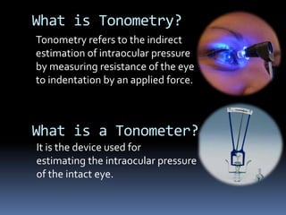


The document discusses various techniques used to measure intraocular pressure (IOP), including: 1) Digital tonometry which involves applying gentle pressure to the eyeball with a finger, though it is subjective. 2) Indentation tonometry like the Schiotz tonometer which measures corneal indentation from a known weight on a plunger, though readings require correction for ocular rigidity. 3) Applanation tonometry like the Goldmann tonometer which flattens a small corneal area to balance internal and external pressure, providing a more accurate IOP reading less influenced by ocular rigidity factors.


