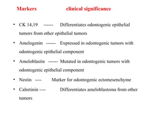
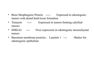
RECENT CONCEPTSRECENT CONCEPTS
Contents
1. Developmental disturbances
2. Cysts and tumours of odontogenic origin.
3. Benign and malignant tumours
4. Salivary gland lesions
5. fibro-osseous lesions
6. Infections in oral cavity
Recent Concepts in Developmental Disturbances
Previous classification: shields classification
DI Type I - Osteogenesis Imperfecta With Opalsent Dentin
DI Type II – Isolated Opalsent Teeth- Herditary Opalsent Teeth
DI Type III - Isolated Opalsent Teeth – Brandywine Isolate
REVISED CLASSIFICATIONREVISED CLASSIFICATION
DENTINOGENESIS TYPE I : OPALESENT DENTIN - DI
WITHOUT OSTEOGENESIS IMPERFECTA. SHIELDS TYPE II.
DENTINOGENESIS IMPERFECTA TYPE II : BRANDYWINE
TYPE, SHIELDS CLASSIFICATION TYPE III
Identical pattern of expression was seen in both type 2 and 3
Single mutation in DSPP shows both phenotypes
TYPE I TYPE II
FISSURAL CYSTS
Globulomaxillary cyst ?????
The originally described globulomaxillary cyst was acclaimed to be a
fissural cyst arising from epithelium entrapped during fusion of the
globular portion of the medial nasal process with the maxillary process.
ARGUMENTS:
1) Fusion of facial processes does occur and epithelium is
entrapped between medial nasal, lateral nasal, and maxilla processes.
The term “fusion” is misleading, i.e., On the basis on this view,
development of the anterior maxilla occurs by the merging of growth
centers rather than fusion of facial processes. So no ectodermal
entrapment can occur, and therefore the globulomaxillary cyst cannot
ensue.
Histologically, the cyst is lined by ciliated columnar or
stratified squamous epithelium, and the supporting fibrous
capsule exhibiting an inflammatory cell infiltrate
Cysts in the globulomaxillary area have reportedly been lined
by respiratory epithelium. This observation could be explained on
basis of the close proximity to the sinonasal cavity and also could
be epithelial metaplasia, as respiratory epithelium lining has been
reported in inflammatory cysts in the mandible.
3) Globulomaxillary cyst should be considered the intraosseous
counterpart of the nasolabial cyst. There is no any scientific
justification of this assumption. On the other hand, there is general
consensus among oral pathologists that nasolabial cyst does arise
from ectopic epithelium of the lower portion of the nasolacrimal
duct.
In conclusion, there is no scientific evidence supporting the
existence of globulomaxillary cyst, therefore, this entity should no
longer be accepted as a pathologic diagnosis
Galal Omami GLOBULOMAXILLARY CYST: GAME OVER!
Median rhomboid glossitis
Posterior midline atrophic candidiasis
• Smooth , erythematous mucosa , lacking in papilla or taste buds.
Currently the etiology of is no longer thought to be a developmental
anomoly but persistent inflammatory process secondary to chronic
infection by candida albicans with a location anterior to the
circumvallate papillae at the junction between the anterior 2/3 and
posterior 1/3 .
It is also a degenerative process
What caused this symptomatic change on tounge? Dermatol. Vol 15-issue 8 aug 2007.
Recent Concepts in Odontogenic Cysts and
Tumours
• Keratocystic Odontogenic Tumour : Current concepts
• Calcifying epithelial odontogenic cyst : cyst or tumour
• Current Classification in Odontogenic Tumours
• Ameloblastoma Pathogenesis
Keratocystic Odontogenic Tumour
• Keratocystic Odontogenic Tumor (KCOTs) is defined asKeratocystic Odontogenic Tumor (KCOTs) is defined as
benign, odontogenic, uni- or multicystic intraosseous tumors,benign, odontogenic, uni- or multicystic intraosseous tumors,
with characteristic parakeratinized squamous epithelium lining,with characteristic parakeratinized squamous epithelium lining,
having a potential for aggressive and infiltrative growth .having a potential for aggressive and infiltrative growth .
(WHO 2005)(WHO 2005)
• KCOTs also exhibit some cyst-like features, includingKCOTs also exhibit some cyst-like features, including
response to decompression .response to decompression .
• Tumoural nature of this lesion remains the subject of debateTumoural nature of this lesion remains the subject of debate
among investigatorsamong investigators
IOSR Journal of Dental and Medical Sciences (IOSR-JDMS)Volume 9, Issue 2
Etiology:
Remnants of dental lamina, and also proliferations of the
basal cell layer of oral epithelium, are considered as
possible sources of epithelial cell proliferation, which
proliferate into the deeper tissues and form microcysts,
suggesting that KCOTs should be considered as hamartomas.
Pathogenesis:
• Behaviour : The KCOT is locally destructive and highly
recurrent.
• Histopathology: The basal layer of the KCOT budding into
connective tissue in the form of daughter cysts and mitotic
figures are frequently found in the supra basal layers.
Keratocystic Odontogenic Tumour : Reclassification of the Odontogenic Keratocyst from Cyst to
Tumour J CDA jcda March 2008, Vol. 74, No. 2 .
Daughter cysts in the wall of a KCOT
Genetic factors in pathogenesis
PTCH (“patched”), a tumour suppressor gene involved in both
NBCCS and sporadic KCOTs on chromosome 9q22.3-q31,
normally forms a receptor complex with oncogene SMO
(“smoothened”) for the SHH (“sonic hedgehog”) ligand . PTCH
binding to SMO inhibits growth-signal transduction. SHH binding
to PTCH releases this inhibition. If normal functioning of PTCH is
lost, the proliferation-stimulating effects of SMO are permitted to
predominate.
• Loss of heterozygosity (LOH) and frequency of allelic
loss for several tumor-suppressor genes p53 and PTCH
gene in sporadic KCOTs, associated with the presence of
satellite microcysts in KCOT walls.
• Activation of H-ras oncogene in KCOTs was
demonstrated .
• Also, recent study indicated that alterations in BIRC5
gene, encoding antiapoptotic protein survivin, may
contribute to the pathogenesis of KCOTs.
• Genetic defects in pathogenesis of KCOTs support the
opinion of the tumoral nature of these lesions.
• Keratocystic Odontogenic Tumors – Clinical and Molecular Features A Textbook
of Advanced Oral and Maxillofacial Surgery
Is a heterogeneous group of lesion existing
either as cystic or solid variant.
• In 2005, WHO renamed the lesion as
calcifying cystic odontogenic tumor
Calcifying epithelial odontogenic cyst: cyst or tumour ?
Gorlin cyst, Calcifying ghost cell odontogenic tumour,
Cystic calcifying odontogenic tumor
Calcifying ghost cell odontogenic cyst: A review on terminologies and
classifications Journal of Oral and Maxillofacial Pathology Vol. 16 Issue 3 Sep - Dec
2012
PRÆTORIUS 2006, PERSONAL COMMUNICATIONPRÆTORIUS 2006, PERSONAL COMMUNICATION
Group 1- ‘Simple’ cysts
1. Calcifying odontogenic cyst (COC)
Group 2- Cysts associated with odontogenic hamartomas or
benign neoplasms: calcifying cystic odontogenic tumours
(CCOT). The following combinations have been published:
1. CCOT associated with an odontome
2. CCOT associated with adenomatoid odontogenic tumor
3. CCOT associated with ameloblastoma
4. CCOT associated with ameloblastic fibroma
5. CCOT associated with ameloblastic fibro-odontoma
6. CCOT associated with odonto-ameloblastoma
7. CCOT associated with odontogenic myxofibroma
Group 3 -Solid benign odontogenic neoplasms with similar cell
morphology to that in the COC, and with dentinoid formation
1. Dentinogenic ghost cell tumour
Group4 -Malignant odontogenic neoplasms with features similar to
those of the dentinogenic ghost cell tumour
1. Ghost cell odontogenic carcinoma
Classification proposed by Toida (1998)
Cyst:Cyst: Calcifying ghost cell odontogenic cyst (CGCOC)
Neoplasm:Neoplasm:
• Benign: Calcifying ghost cell odontogenic tumor
(CGCOT)
• Cystic variant: Cystic CGCOT
• Solid variant: Solid CGCOT
• Malignant: Malignant CGCOT
Combined lesionCombined lesion: Each of the categories described
above
CGCOC, CGCOT, malignant (CGCOT) associated
with the following lesions:
• a. Odontoma
• b. Ameloblastoma
• c. Other odontogenic lesions
oDONTOGENIC TUMOURSoDONTOGENIC TUMOURS
WHO histological classification of odontogenic tumours (WHO
2005)
MALIGNANT TUMOURS
Odontogenic carcinomas
Metastasizing (malignant) ameloblastoma
Ameloblastic carcinoma - primary type
Ameloblastic carcinoma - secondary type Intraosseous
(dedifferentiated)
Ameloblastic carcinoma - secondary type Peripheral (dedifferentiated)
Primary intraosseous squamous cell carcinoma - solid type
Primary intraosseous squamous cell carcinoma derived from keratocystic
odontogenic tumour
Primary intraosseous squamous cell carcinoma derived from odontogenic
cysts
Clear cell odontogenic carcinoma
Ghost cell odontogenic carcinoma
John M. Wright • Edward W. Odell. Odontogenic Tumors, WHO 2005:Head and Neck Pathol
(2014) 8:373–382
Odontogenic sarcomas
• Ameloblastoma fibrosarcoma
• Ameloblastic fibrodentino-and fibro-odontosarcoma
BENIGN TUMOURS
Odontogenic epithelium with mature, fibrous stroma without
Odontogenic ectomesenchyme
Ameloblastoma, solid/multicystic type
Ameloblastoma, extraosseous/peripheral type
Ameloblastoma, desmoplastic type
Ameloblastoma, unicystic type
Squamous odontogenic tumour
Calcifying epithelial odontogenic tumour
Adenomatoid odontogenic tumour
Keratocystic odontogenic tumour
Odontogenic epithelium with odontogenic
ectomesenchyme, with or without hard tissue formation
• Ameloblastic fibroma
• Ameloblastic fibrodentinoma
• Ameloblastic fibroodontoma
Odontoma
I .Odontoma, complex type
II. Odontoma, compound type
• Odontoameloblastoma
• Calcifying cystic odontogenic tumour
• Dentinogenic ghost cell tumour
Odontogenic ectomesenchyme with or without included
odontogenic epithelium
• Odontogenic fibroma
epithelium poor or ‘‘simple type
epithelium rich or ‘‘WHO type’’.
• Myxoma( myxofibroma)
• Cementoblastoma( benign cementoblastoma,true cementoma)
Bone related lesionsBone related lesions
• Osseous dysplasias
• Fibrous dysplasias
• cherubism
• Ossifying fibromas
• Central giant cell granulomas
• Aneurysmal bone cysts
• Simple bone cyst
Other tumoursOther tumours
• Melanotic neuroectodermal tumour
AMELOBLASTOMA PATHOGENESIS
molecular mechanisms
The overexpression and under expression of signaling molecules
may play an important role in tumorogenesis and oncogenesis
Locally invasive behaviour and recurrence
CLONALITY:CLONALITY:
The pathogenesis of tumours is the determination of its clonal
pattern. Recently, has been demonstrated that solid ⁄
multicystic ameloblastoma as monoclonal in origin.
TUMOR SUPPERSSOR GENESTUMOR SUPPERSSOR GENES
• Increased reactivity for p53 has been detected in ameloblastomas
• Overexpression of MDM2 and p14ARF has been detected in
GROWTH FACTORSGROWTH FACTORS
EGFR and TGF -β expression in ameloblastomas, are involved in
tumorgenesis of ameloblastomas
CELL CYCLE REGULATORSCELL CYCLE REGULATORS
Expression of cyclin D1, p16
, p21
, and p27
is well preserved when
compared with tooth germs, suggesting that the proliferation of
odontogenic epithelial cells is strictly controlled by these cell cycle
regulators
J Oral Pathol Med (2010) 39: 585–591
HARD TISSUE RELATED PROTIENSHARD TISSUE RELATED PROTIENS
• Mutations of ameloblastin gene have been detected in
ameloblastomas.
• Ameloblastomas have shown expression of several integrins
and CD44 predominantly at epithelial–mesenchymal interfaces,
suggesting that these cell adhesion molecules might mediate
parenchymal–stromal interactions in the odontogenic tumors
J Oral Pathol Med (2010) 39: 585–591
MATRIX DEGRADING PROTEINASESMATRIX DEGRADING PROTEINASES
Ameloblastomas express MMPs-1, 2, and 9
Degrade extracellular matrix and basement
membrane components
Their inhibitors, tissue inhibitors of
metalloproteinases (TIMPs)-1 and -2, have
also been recognized in ameloblastomas.
These features suggest that matrix-
degrading enzymes may contribute to
the local invasiveness of odontogenic
tumors.
OSTEOLYTIC CYTOKINESOSTEOLYTIC CYTOKINES
• Inflammatory cytokines with osteolytic
activity, such as IL-1, IL-6, and TNF-
α, are synthesized in ameloblastomas
• Benign and malignant ameloblastomas
express RANKL and OPG
predominantly in stromal cells rather
than tumor cells
• These observations suggest that
osteolytic cytokines have a role in
local bone resorption during the
progression of odontogenic tumors.
J Oral Pathol Med (2010) 39: 585–591 Odontogenic tumors & allied lesions- Reichart
Odontogenic Carcinomas:
• Odontogenic carcinomas are the malignant epithelial odontogenic
neoplasms. These tumours are believed to take origin from the
epithelial components of the odontogenic apparatus.
• The cell rests of Malassez, reduced enamel epithelium, the rests
of Serres in the gingiva, and the linings of odontogenic cysts
represent the precursor cells for odontogenic carcinoma.
• Pathogenesis and Nomenclature of Odontogenic Carcinomas: Revisited . journal of
oncology march 2014
Classification of odontogenic carcinomas ( L J Slater)Classification of odontogenic carcinomas ( L J Slater)
• Odontogenic carcinomas
1. metastasizing ameloblastoma
2. Ameloblastic carcinoma
a. carcinoma ex ameloblastoma
b. De novo
3. Primary intraosseous carcinoma
a. Solid
b. Cystic
c. central mucoepidermoid carcinoma
4. ghost cell odontogenic carcinoma
5. Clear cell odontogenic carcinoma
Malignant Ameloblastoma
• Described as “ benign neoplasm in which the features of an
ameloblastoma are shown by the primary growth in the jaws and
by any metastatic growth
• Clinical behaviour justifies the diagnosis of this lesion
Ameloblastic carcinomaAmeloblastic carcinoma combines the
histological features of Ameloblastoma with
cytological atypia.
• The tumour may arise de novo, in which
case it is called a primary type, or from a
preexisting benign odontogenic tumour it is
called secondary.
•Cortical expansion and spread into adjacent
soft tissues is seen
Molecular markers of cell adhesion in ameloblastomas. An updateMolecular markers of cell adhesion in ameloblastomas. An update
• Cellular adhesion potentially plays an important role in the
manifestation of characteristics and in the tumor biology of
ameloblastomas.
• Med Oral Patol Oral Cir Bucal. 2014 Jan 1;19 (1):e8-14.
E-cadherin
Modulator during development tooth, participate in morphodifferentation of the
odontogenic epithelium of ameloblastomas. Promotes the adhesion between
distant cells
catenins- alpha,beta,gammacatenins- alpha,beta,gamma
Participates in cell-cell adhesion and in Wnt signaling pathway during
odontogenesis, especially at the bell stage.
SDC-1SDC-1
Plays an important role in the maintenance of proliferation and movement in
dental follicle cells.
CD56CD56
Maybe have an important role morphoregulatory during tooth molar development
.
CD147CD147
Possible This protein participates in perivascular cells, may
promote the capability to differentiation in odontoblast
Integrinntegrin
Important component in organization and tension of the
cytoskeleton and that in turn strongly influence in intracellular
phenomena that possibly are implicated in growth, adhesion,
migration, proliferation, apoptosis and cellular morphology
ClaudinsClaudins
• May particpate in activation of various MMPs leading to
controlled enamel matrix formation.
Odontogenic Tumor Markers - An OverviewOdontogenic Tumor Markers - An Overview
Tumor markers are molecules that help the pathologists for
confirmatory diagnosis of histopathologically confounding
lesions
Journal of International Oral Health. Mar-Apr 2013; 5(2):59-69
Markers clinical significance
• CK 14,19 ------ Differentiates odontogenic epithelial
tumors from other epithelial tumors
• Amelogenin ------ Expressed in odontogenic tumors with
odontogenic epithelial component
• Ameloblastin ------ Mutated in odontogenic tumors with
odontogenic epithelial component
• Nestin ---- Marker for odontogenic ectomesenchyme
• Calretinin ---- Differentiates ameloblastoma from other
tumors
• Bone Morphogenic Protein ----- Expressed in odontogenic
tumors with dental hard tissue formation
• Tenascin ----- Expressed in tumors forming calcified
masses
• HMGA2 ---- Over expression in odontogenic mesenchymal
tumors
• Basement membrane proteins : Laminin 1 ---- Marker for
odontogenic epithelium
Current Concepts in Benign and MalignantCurrent Concepts in Benign and Malignant
TumoursTumours
ORAL POTENTIALLY MALIGNANT DISORDERS
(OPMDS)
Precancerous potentially malignant disordersPrecancerous potentially malignant disorders
• In 1978, working group of WHO classified ‘precancer’ into
‘lesions’ and ‘conditions’ with following definitions
• A precancerous lesionprecancerous lesion is ‘a morphologically altered tissue in
which oral cancer is more likely to occur than in its apparently
normal counterpart’
• A precancerous conditionprecancerous condition is ‘a generalized state associated with
a significantly increased risk of cancer’.
• WHO monograph of Head and Neck Tumors (2005) uses the term
‘epithelial precursor lesions’.‘epithelial precursor lesions’.
• It defined it as ‘altered epithelium with an increased likelihood for
progression to squamous cell carcinoma’.
• It is also mentioned that word ‘altered’ in the definition means
epithelial dysplasia.
• Development of second primary tumor at clinically normal
mucosa supports the contention.
LeukoplakiaLeukoplakia is purely a clinical terminology and histopathologically
it is reported as epithelial dysplasia.
• WHO in 2005 proposed five grades of epithelial dysplasia
1. Squamous Hyperplasia – benign lesion.
2. Mild Dysplasia – better prognosis.
3. Moderate Dysplasia.
4. Severe Dysplasia.
5. Carcinoma In-situ – poor prognosis.
• It has been recently proposed to modify the above 5- tier system
into a binary system of ‘high risk’ and ‘low risk’binary system of ‘high risk’ and ‘low risk’ lesions to
improve clinical management of these lesions
Oral squamous cell carcinoma
• Staging: It is the extent of spread of tumor within patients
• Grading : It is defined as macroscopic and microscopic degree of
differentiation of tumor.
• TNM staging system has been used clinically estimating response
to therapy and survival.
• TNM system does not provide any information on the biologic
characteristics- and thus an aggressive clinical behaviour of the
tumor.
Broder systemBroder system
• Does not provide any correlation between
degree of differentiation and prognosis
Jacobsson et al system includes :
Morphologic parameters
Tumor-host relationship as estimated by
parameters
Fisher (1975):Fisher (1975):
Slight modification of JAKOBSSON et al.
Lund et al (1975)
Modified grading system of Jakobsson et al. by presenting a more
exact definition of each parameter and grade and by introducing a
histologic score, defined a total sum of points divided by the number of
parameters evaluated.
Bryne’s (1989, 1992) (ITF) Invasive Tumor FrontBryne’s (1989, 1992) (ITF) Invasive Tumor Front
Grading System:Grading System:
• Parameters were measured in the deepest invasive margins.
• Did not measure the entire thickness of the tumor
Histologic grading of malignancy of tumor cell population
Morphologic
Parameters
1 2 3 4
Degree of
keratinization
Highly keratinized
(>50% of the cells)
Moderately
keratinized
(5-20% of the
cells)
Minimal
keratinization
(5-20% of the
cells)
No keratinization (0-
5%)
Nuclear
polymorphism
Little nuclear
polymorphism
(>75% mature
cells)
Moderately
abundant nuclear
polymorphism (50-
75% mature cells)
Abundant nuclear
polymorphism
(25-50% mature
cells)
Extreme nuclear
polymorphism (0-25%
mature cells)
Number of
mitoses/HPF
0-1 2-3 4-5 >5
Morphologic
parameters
1 2 3 4
Pattern of
invasion
Pushing, well
delineated
infiltrating
borders
Infiltrating, solid
cords, bands and
or strands
Small groups or
cords of
infiltrating cells
(n > 15)
Marked and
widespread
Cellular
dissociation in
small groups of
cells(n<15) and
or in single cells
Grade I: 4-8
Grade II: 9-12
Grade III: 13-16
Oral cavity squamous cell carcinoma classification using the (TNM)
classification system.
Primary Tumor Regional Lymph Nodes Distant Metastasis
• TX cannot be assessed NX cannot be assessed M0 absent
• T0 no evidence of tumors N0 none M1
present
• Tis carcinoma in situ N1 single, ipsilateral, < 3 cm
• T1 < 2cm N2a single, ipsilateral, 3-6 cm
• T2 2-4 cm N2b multiple, ipsilateral, < 6 cm
• T3 > 4 cm N2c bilateral or contralateral, < 6 cm
• T4a invades adjacent structure N3 > 6 cm
• T4b invades distant structures
Staging system of oral cavity squamous cell carcinoma
Stage T N M
0 Tis N0 M0
I T1 N0 M0
II T2 N0 M0
III T1-3 N1 M0
T3 N0 M0
IV T1-3 N2 M0
T4 N1-2 M0
T1-3 N0-3 M0
T1-3 N0-3 M1
Oral Cavity Squamous Cell Carcinoma – An Overview

Recent concepts in omfp part 1

  • 1.
  • 2.
    Contents 1. Developmental disturbances 2.Cysts and tumours of odontogenic origin. 3. Benign and malignant tumours 4. Salivary gland lesions 5. fibro-osseous lesions 6. Infections in oral cavity
  • 3.
    Recent Concepts inDevelopmental Disturbances Previous classification: shields classification DI Type I - Osteogenesis Imperfecta With Opalsent Dentin DI Type II – Isolated Opalsent Teeth- Herditary Opalsent Teeth DI Type III - Isolated Opalsent Teeth – Brandywine Isolate REVISED CLASSIFICATIONREVISED CLASSIFICATION DENTINOGENESIS TYPE I : OPALESENT DENTIN - DI WITHOUT OSTEOGENESIS IMPERFECTA. SHIELDS TYPE II. DENTINOGENESIS IMPERFECTA TYPE II : BRANDYWINE TYPE, SHIELDS CLASSIFICATION TYPE III Identical pattern of expression was seen in both type 2 and 3 Single mutation in DSPP shows both phenotypes
  • 4.
  • 5.
    FISSURAL CYSTS Globulomaxillary cyst????? The originally described globulomaxillary cyst was acclaimed to be a fissural cyst arising from epithelium entrapped during fusion of the globular portion of the medial nasal process with the maxillary process. ARGUMENTS: 1) Fusion of facial processes does occur and epithelium is entrapped between medial nasal, lateral nasal, and maxilla processes. The term “fusion” is misleading, i.e., On the basis on this view, development of the anterior maxilla occurs by the merging of growth centers rather than fusion of facial processes. So no ectodermal entrapment can occur, and therefore the globulomaxillary cyst cannot ensue.
  • 6.
    Histologically, the cystis lined by ciliated columnar or stratified squamous epithelium, and the supporting fibrous capsule exhibiting an inflammatory cell infiltrate Cysts in the globulomaxillary area have reportedly been lined by respiratory epithelium. This observation could be explained on basis of the close proximity to the sinonasal cavity and also could be epithelial metaplasia, as respiratory epithelium lining has been reported in inflammatory cysts in the mandible.
  • 7.
    3) Globulomaxillary cystshould be considered the intraosseous counterpart of the nasolabial cyst. There is no any scientific justification of this assumption. On the other hand, there is general consensus among oral pathologists that nasolabial cyst does arise from ectopic epithelium of the lower portion of the nasolacrimal duct. In conclusion, there is no scientific evidence supporting the existence of globulomaxillary cyst, therefore, this entity should no longer be accepted as a pathologic diagnosis Galal Omami GLOBULOMAXILLARY CYST: GAME OVER!
  • 8.
    Median rhomboid glossitis Posteriormidline atrophic candidiasis • Smooth , erythematous mucosa , lacking in papilla or taste buds. Currently the etiology of is no longer thought to be a developmental anomoly but persistent inflammatory process secondary to chronic infection by candida albicans with a location anterior to the circumvallate papillae at the junction between the anterior 2/3 and posterior 1/3 . It is also a degenerative process What caused this symptomatic change on tounge? Dermatol. Vol 15-issue 8 aug 2007.
  • 9.
    Recent Concepts inOdontogenic Cysts and Tumours • Keratocystic Odontogenic Tumour : Current concepts • Calcifying epithelial odontogenic cyst : cyst or tumour • Current Classification in Odontogenic Tumours • Ameloblastoma Pathogenesis
  • 10.
    Keratocystic Odontogenic Tumour •Keratocystic Odontogenic Tumor (KCOTs) is defined asKeratocystic Odontogenic Tumor (KCOTs) is defined as benign, odontogenic, uni- or multicystic intraosseous tumors,benign, odontogenic, uni- or multicystic intraosseous tumors, with characteristic parakeratinized squamous epithelium lining,with characteristic parakeratinized squamous epithelium lining, having a potential for aggressive and infiltrative growth .having a potential for aggressive and infiltrative growth . (WHO 2005)(WHO 2005) • KCOTs also exhibit some cyst-like features, includingKCOTs also exhibit some cyst-like features, including response to decompression .response to decompression . • Tumoural nature of this lesion remains the subject of debateTumoural nature of this lesion remains the subject of debate among investigatorsamong investigators IOSR Journal of Dental and Medical Sciences (IOSR-JDMS)Volume 9, Issue 2
  • 11.
    Etiology: Remnants of dentallamina, and also proliferations of the basal cell layer of oral epithelium, are considered as possible sources of epithelial cell proliferation, which proliferate into the deeper tissues and form microcysts, suggesting that KCOTs should be considered as hamartomas. Pathogenesis: • Behaviour : The KCOT is locally destructive and highly recurrent. • Histopathology: The basal layer of the KCOT budding into connective tissue in the form of daughter cysts and mitotic figures are frequently found in the supra basal layers. Keratocystic Odontogenic Tumour : Reclassification of the Odontogenic Keratocyst from Cyst to Tumour J CDA jcda March 2008, Vol. 74, No. 2 .
  • 12.
    Daughter cysts inthe wall of a KCOT
  • 13.
    Genetic factors inpathogenesis PTCH (“patched”), a tumour suppressor gene involved in both NBCCS and sporadic KCOTs on chromosome 9q22.3-q31, normally forms a receptor complex with oncogene SMO (“smoothened”) for the SHH (“sonic hedgehog”) ligand . PTCH binding to SMO inhibits growth-signal transduction. SHH binding to PTCH releases this inhibition. If normal functioning of PTCH is lost, the proliferation-stimulating effects of SMO are permitted to predominate.
  • 14.
    • Loss ofheterozygosity (LOH) and frequency of allelic loss for several tumor-suppressor genes p53 and PTCH gene in sporadic KCOTs, associated with the presence of satellite microcysts in KCOT walls. • Activation of H-ras oncogene in KCOTs was demonstrated . • Also, recent study indicated that alterations in BIRC5 gene, encoding antiapoptotic protein survivin, may contribute to the pathogenesis of KCOTs. • Genetic defects in pathogenesis of KCOTs support the opinion of the tumoral nature of these lesions. • Keratocystic Odontogenic Tumors – Clinical and Molecular Features A Textbook of Advanced Oral and Maxillofacial Surgery
  • 15.
    Is a heterogeneousgroup of lesion existing either as cystic or solid variant. • In 2005, WHO renamed the lesion as calcifying cystic odontogenic tumor Calcifying epithelial odontogenic cyst: cyst or tumour ? Gorlin cyst, Calcifying ghost cell odontogenic tumour, Cystic calcifying odontogenic tumor Calcifying ghost cell odontogenic cyst: A review on terminologies and classifications Journal of Oral and Maxillofacial Pathology Vol. 16 Issue 3 Sep - Dec 2012
  • 16.
    PRÆTORIUS 2006, PERSONALCOMMUNICATIONPRÆTORIUS 2006, PERSONAL COMMUNICATION Group 1- ‘Simple’ cysts 1. Calcifying odontogenic cyst (COC) Group 2- Cysts associated with odontogenic hamartomas or benign neoplasms: calcifying cystic odontogenic tumours (CCOT). The following combinations have been published: 1. CCOT associated with an odontome 2. CCOT associated with adenomatoid odontogenic tumor 3. CCOT associated with ameloblastoma 4. CCOT associated with ameloblastic fibroma 5. CCOT associated with ameloblastic fibro-odontoma 6. CCOT associated with odonto-ameloblastoma 7. CCOT associated with odontogenic myxofibroma
  • 17.
    Group 3 -Solidbenign odontogenic neoplasms with similar cell morphology to that in the COC, and with dentinoid formation 1. Dentinogenic ghost cell tumour Group4 -Malignant odontogenic neoplasms with features similar to those of the dentinogenic ghost cell tumour 1. Ghost cell odontogenic carcinoma
  • 18.
    Classification proposed byToida (1998) Cyst:Cyst: Calcifying ghost cell odontogenic cyst (CGCOC) Neoplasm:Neoplasm: • Benign: Calcifying ghost cell odontogenic tumor (CGCOT) • Cystic variant: Cystic CGCOT • Solid variant: Solid CGCOT • Malignant: Malignant CGCOT Combined lesionCombined lesion: Each of the categories described above CGCOC, CGCOT, malignant (CGCOT) associated with the following lesions: • a. Odontoma • b. Ameloblastoma • c. Other odontogenic lesions
  • 19.
    oDONTOGENIC TUMOURSoDONTOGENIC TUMOURS WHOhistological classification of odontogenic tumours (WHO 2005) MALIGNANT TUMOURS Odontogenic carcinomas Metastasizing (malignant) ameloblastoma Ameloblastic carcinoma - primary type Ameloblastic carcinoma - secondary type Intraosseous (dedifferentiated) Ameloblastic carcinoma - secondary type Peripheral (dedifferentiated) Primary intraosseous squamous cell carcinoma - solid type Primary intraosseous squamous cell carcinoma derived from keratocystic odontogenic tumour Primary intraosseous squamous cell carcinoma derived from odontogenic cysts Clear cell odontogenic carcinoma Ghost cell odontogenic carcinoma John M. Wright • Edward W. Odell. Odontogenic Tumors, WHO 2005:Head and Neck Pathol (2014) 8:373–382
  • 20.
    Odontogenic sarcomas • Ameloblastomafibrosarcoma • Ameloblastic fibrodentino-and fibro-odontosarcoma BENIGN TUMOURS Odontogenic epithelium with mature, fibrous stroma without Odontogenic ectomesenchyme Ameloblastoma, solid/multicystic type Ameloblastoma, extraosseous/peripheral type Ameloblastoma, desmoplastic type Ameloblastoma, unicystic type Squamous odontogenic tumour Calcifying epithelial odontogenic tumour Adenomatoid odontogenic tumour Keratocystic odontogenic tumour
  • 21.
    Odontogenic epithelium withodontogenic ectomesenchyme, with or without hard tissue formation • Ameloblastic fibroma • Ameloblastic fibrodentinoma • Ameloblastic fibroodontoma Odontoma I .Odontoma, complex type II. Odontoma, compound type • Odontoameloblastoma • Calcifying cystic odontogenic tumour • Dentinogenic ghost cell tumour
  • 22.
    Odontogenic ectomesenchyme withor without included odontogenic epithelium • Odontogenic fibroma epithelium poor or ‘‘simple type epithelium rich or ‘‘WHO type’’. • Myxoma( myxofibroma) • Cementoblastoma( benign cementoblastoma,true cementoma)
  • 23.
    Bone related lesionsBonerelated lesions • Osseous dysplasias • Fibrous dysplasias • cherubism • Ossifying fibromas • Central giant cell granulomas • Aneurysmal bone cysts • Simple bone cyst Other tumoursOther tumours • Melanotic neuroectodermal tumour
  • 24.
    AMELOBLASTOMA PATHOGENESIS molecular mechanisms Theoverexpression and under expression of signaling molecules may play an important role in tumorogenesis and oncogenesis Locally invasive behaviour and recurrence CLONALITY:CLONALITY: The pathogenesis of tumours is the determination of its clonal pattern. Recently, has been demonstrated that solid ⁄ multicystic ameloblastoma as monoclonal in origin. TUMOR SUPPERSSOR GENESTUMOR SUPPERSSOR GENES • Increased reactivity for p53 has been detected in ameloblastomas • Overexpression of MDM2 and p14ARF has been detected in
  • 25.
    GROWTH FACTORSGROWTH FACTORS EGFRand TGF -β expression in ameloblastomas, are involved in tumorgenesis of ameloblastomas CELL CYCLE REGULATORSCELL CYCLE REGULATORS Expression of cyclin D1, p16 , p21 , and p27 is well preserved when compared with tooth germs, suggesting that the proliferation of odontogenic epithelial cells is strictly controlled by these cell cycle regulators J Oral Pathol Med (2010) 39: 585–591
  • 26.
    HARD TISSUE RELATEDPROTIENSHARD TISSUE RELATED PROTIENS • Mutations of ameloblastin gene have been detected in ameloblastomas. • Ameloblastomas have shown expression of several integrins and CD44 predominantly at epithelial–mesenchymal interfaces, suggesting that these cell adhesion molecules might mediate parenchymal–stromal interactions in the odontogenic tumors J Oral Pathol Med (2010) 39: 585–591
  • 27.
    MATRIX DEGRADING PROTEINASESMATRIXDEGRADING PROTEINASES Ameloblastomas express MMPs-1, 2, and 9 Degrade extracellular matrix and basement membrane components Their inhibitors, tissue inhibitors of metalloproteinases (TIMPs)-1 and -2, have also been recognized in ameloblastomas. These features suggest that matrix- degrading enzymes may contribute to the local invasiveness of odontogenic tumors.
  • 28.
    OSTEOLYTIC CYTOKINESOSTEOLYTIC CYTOKINES •Inflammatory cytokines with osteolytic activity, such as IL-1, IL-6, and TNF- α, are synthesized in ameloblastomas • Benign and malignant ameloblastomas express RANKL and OPG predominantly in stromal cells rather than tumor cells • These observations suggest that osteolytic cytokines have a role in local bone resorption during the progression of odontogenic tumors. J Oral Pathol Med (2010) 39: 585–591 Odontogenic tumors & allied lesions- Reichart
  • 29.
    Odontogenic Carcinomas: • Odontogeniccarcinomas are the malignant epithelial odontogenic neoplasms. These tumours are believed to take origin from the epithelial components of the odontogenic apparatus. • The cell rests of Malassez, reduced enamel epithelium, the rests of Serres in the gingiva, and the linings of odontogenic cysts represent the precursor cells for odontogenic carcinoma. • Pathogenesis and Nomenclature of Odontogenic Carcinomas: Revisited . journal of oncology march 2014
  • 30.
    Classification of odontogeniccarcinomas ( L J Slater)Classification of odontogenic carcinomas ( L J Slater) • Odontogenic carcinomas 1. metastasizing ameloblastoma 2. Ameloblastic carcinoma a. carcinoma ex ameloblastoma b. De novo 3. Primary intraosseous carcinoma a. Solid b. Cystic c. central mucoepidermoid carcinoma 4. ghost cell odontogenic carcinoma 5. Clear cell odontogenic carcinoma
  • 31.
    Malignant Ameloblastoma • Describedas “ benign neoplasm in which the features of an ameloblastoma are shown by the primary growth in the jaws and by any metastatic growth • Clinical behaviour justifies the diagnosis of this lesion
  • 32.
    Ameloblastic carcinomaAmeloblastic carcinomacombines the histological features of Ameloblastoma with cytological atypia. • The tumour may arise de novo, in which case it is called a primary type, or from a preexisting benign odontogenic tumour it is called secondary. •Cortical expansion and spread into adjacent soft tissues is seen
  • 33.
    Molecular markers ofcell adhesion in ameloblastomas. An updateMolecular markers of cell adhesion in ameloblastomas. An update • Cellular adhesion potentially plays an important role in the manifestation of characteristics and in the tumor biology of ameloblastomas. • Med Oral Patol Oral Cir Bucal. 2014 Jan 1;19 (1):e8-14.
  • 35.
    E-cadherin Modulator during developmenttooth, participate in morphodifferentation of the odontogenic epithelium of ameloblastomas. Promotes the adhesion between distant cells catenins- alpha,beta,gammacatenins- alpha,beta,gamma Participates in cell-cell adhesion and in Wnt signaling pathway during odontogenesis, especially at the bell stage. SDC-1SDC-1 Plays an important role in the maintenance of proliferation and movement in dental follicle cells. CD56CD56 Maybe have an important role morphoregulatory during tooth molar development .
  • 36.
    CD147CD147 Possible This proteinparticipates in perivascular cells, may promote the capability to differentiation in odontoblast Integrinntegrin Important component in organization and tension of the cytoskeleton and that in turn strongly influence in intracellular phenomena that possibly are implicated in growth, adhesion, migration, proliferation, apoptosis and cellular morphology ClaudinsClaudins • May particpate in activation of various MMPs leading to controlled enamel matrix formation.
  • 37.
    Odontogenic Tumor Markers- An OverviewOdontogenic Tumor Markers - An Overview Tumor markers are molecules that help the pathologists for confirmatory diagnosis of histopathologically confounding lesions Journal of International Oral Health. Mar-Apr 2013; 5(2):59-69
  • 38.
    Markers clinical significance •CK 14,19 ------ Differentiates odontogenic epithelial tumors from other epithelial tumors • Amelogenin ------ Expressed in odontogenic tumors with odontogenic epithelial component • Ameloblastin ------ Mutated in odontogenic tumors with odontogenic epithelial component • Nestin ---- Marker for odontogenic ectomesenchyme • Calretinin ---- Differentiates ameloblastoma from other tumors
  • 39.
    • Bone MorphogenicProtein ----- Expressed in odontogenic tumors with dental hard tissue formation • Tenascin ----- Expressed in tumors forming calcified masses • HMGA2 ---- Over expression in odontogenic mesenchymal tumors • Basement membrane proteins : Laminin 1 ---- Marker for odontogenic epithelium
  • 40.
    Current Concepts inBenign and MalignantCurrent Concepts in Benign and Malignant TumoursTumours
  • 41.
    ORAL POTENTIALLY MALIGNANTDISORDERS (OPMDS) Precancerous potentially malignant disordersPrecancerous potentially malignant disorders • In 1978, working group of WHO classified ‘precancer’ into ‘lesions’ and ‘conditions’ with following definitions • A precancerous lesionprecancerous lesion is ‘a morphologically altered tissue in which oral cancer is more likely to occur than in its apparently normal counterpart’ • A precancerous conditionprecancerous condition is ‘a generalized state associated with a significantly increased risk of cancer’.
  • 42.
    • WHO monographof Head and Neck Tumors (2005) uses the term ‘epithelial precursor lesions’.‘epithelial precursor lesions’. • It defined it as ‘altered epithelium with an increased likelihood for progression to squamous cell carcinoma’. • It is also mentioned that word ‘altered’ in the definition means epithelial dysplasia. • Development of second primary tumor at clinically normal mucosa supports the contention.
  • 43.
    LeukoplakiaLeukoplakia is purelya clinical terminology and histopathologically it is reported as epithelial dysplasia. • WHO in 2005 proposed five grades of epithelial dysplasia 1. Squamous Hyperplasia – benign lesion. 2. Mild Dysplasia – better prognosis. 3. Moderate Dysplasia. 4. Severe Dysplasia. 5. Carcinoma In-situ – poor prognosis. • It has been recently proposed to modify the above 5- tier system into a binary system of ‘high risk’ and ‘low risk’binary system of ‘high risk’ and ‘low risk’ lesions to improve clinical management of these lesions
  • 44.
    Oral squamous cellcarcinoma • Staging: It is the extent of spread of tumor within patients • Grading : It is defined as macroscopic and microscopic degree of differentiation of tumor. • TNM staging system has been used clinically estimating response to therapy and survival. • TNM system does not provide any information on the biologic characteristics- and thus an aggressive clinical behaviour of the tumor.
  • 45.
    Broder systemBroder system •Does not provide any correlation between degree of differentiation and prognosis Jacobsson et al system includes : Morphologic parameters Tumor-host relationship as estimated by parameters Fisher (1975):Fisher (1975): Slight modification of JAKOBSSON et al. Lund et al (1975) Modified grading system of Jakobsson et al. by presenting a more exact definition of each parameter and grade and by introducing a histologic score, defined a total sum of points divided by the number of parameters evaluated.
  • 46.
    Bryne’s (1989, 1992)(ITF) Invasive Tumor FrontBryne’s (1989, 1992) (ITF) Invasive Tumor Front Grading System:Grading System: • Parameters were measured in the deepest invasive margins. • Did not measure the entire thickness of the tumor
  • 47.
    Histologic grading ofmalignancy of tumor cell population Morphologic Parameters 1 2 3 4 Degree of keratinization Highly keratinized (>50% of the cells) Moderately keratinized (5-20% of the cells) Minimal keratinization (5-20% of the cells) No keratinization (0- 5%) Nuclear polymorphism Little nuclear polymorphism (>75% mature cells) Moderately abundant nuclear polymorphism (50- 75% mature cells) Abundant nuclear polymorphism (25-50% mature cells) Extreme nuclear polymorphism (0-25% mature cells) Number of mitoses/HPF 0-1 2-3 4-5 >5
  • 48.
    Morphologic parameters 1 2 34 Pattern of invasion Pushing, well delineated infiltrating borders Infiltrating, solid cords, bands and or strands Small groups or cords of infiltrating cells (n > 15) Marked and widespread Cellular dissociation in small groups of cells(n<15) and or in single cells Grade I: 4-8 Grade II: 9-12 Grade III: 13-16
  • 49.
    Oral cavity squamouscell carcinoma classification using the (TNM) classification system. Primary Tumor Regional Lymph Nodes Distant Metastasis • TX cannot be assessed NX cannot be assessed M0 absent • T0 no evidence of tumors N0 none M1 present • Tis carcinoma in situ N1 single, ipsilateral, < 3 cm • T1 < 2cm N2a single, ipsilateral, 3-6 cm • T2 2-4 cm N2b multiple, ipsilateral, < 6 cm • T3 > 4 cm N2c bilateral or contralateral, < 6 cm • T4a invades adjacent structure N3 > 6 cm • T4b invades distant structures
  • 50.
    Staging system oforal cavity squamous cell carcinoma Stage T N M 0 Tis N0 M0 I T1 N0 M0 II T2 N0 M0 III T1-3 N1 M0 T3 N0 M0 IV T1-3 N2 M0 T4 N1-2 M0 T1-3 N0-3 M0 T1-3 N0-3 M1 Oral Cavity Squamous Cell Carcinoma – An Overview