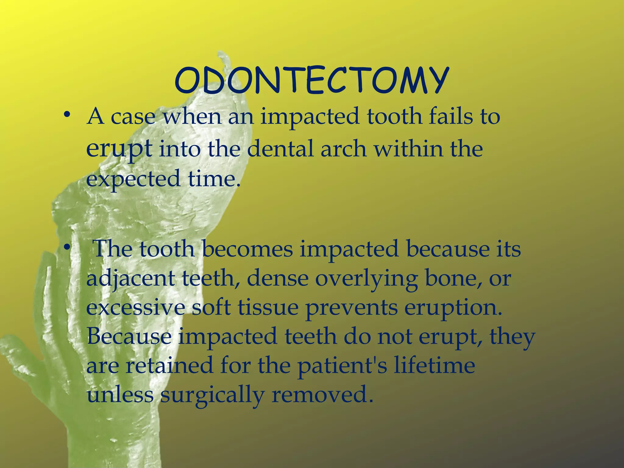
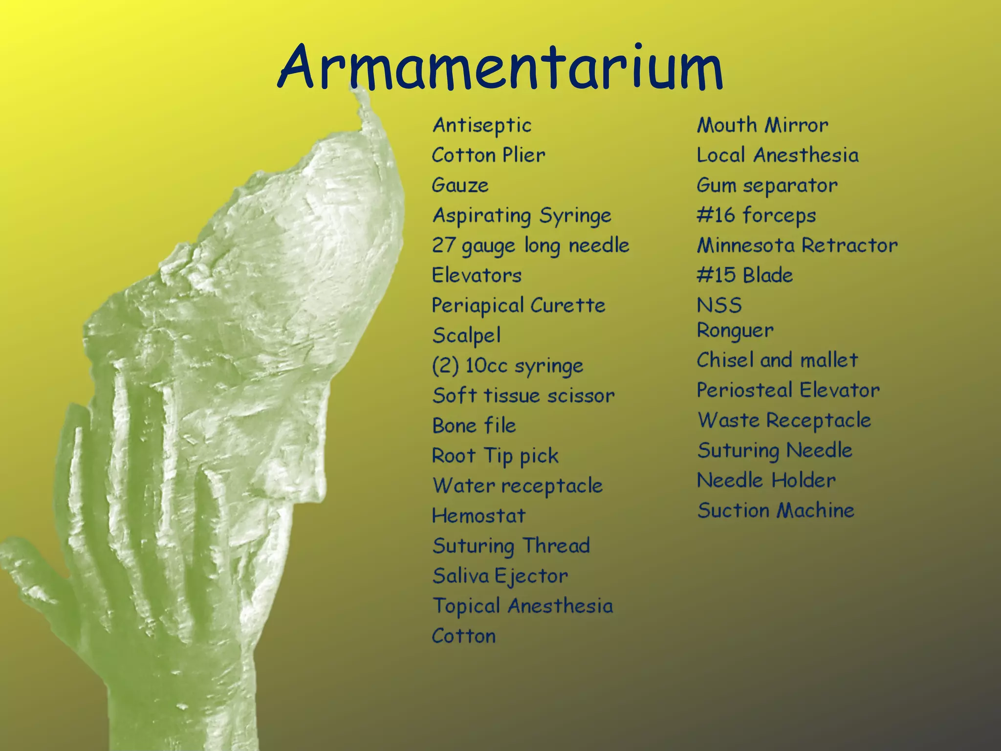
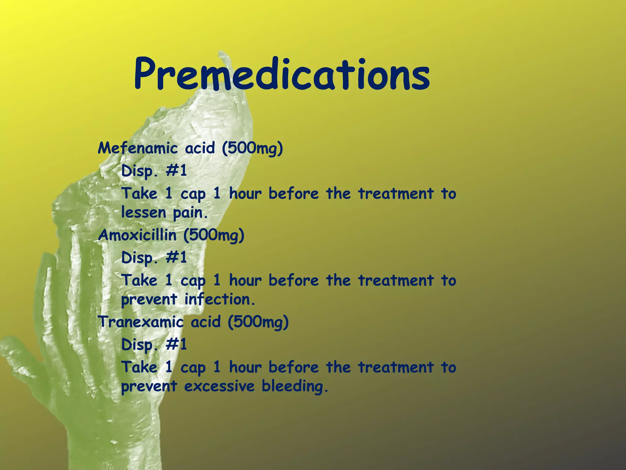
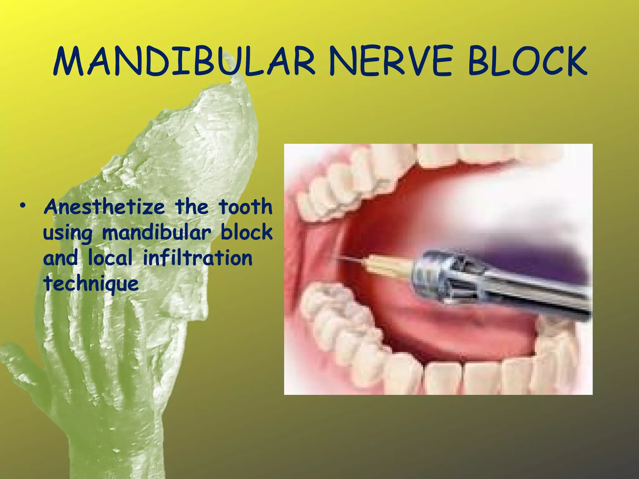
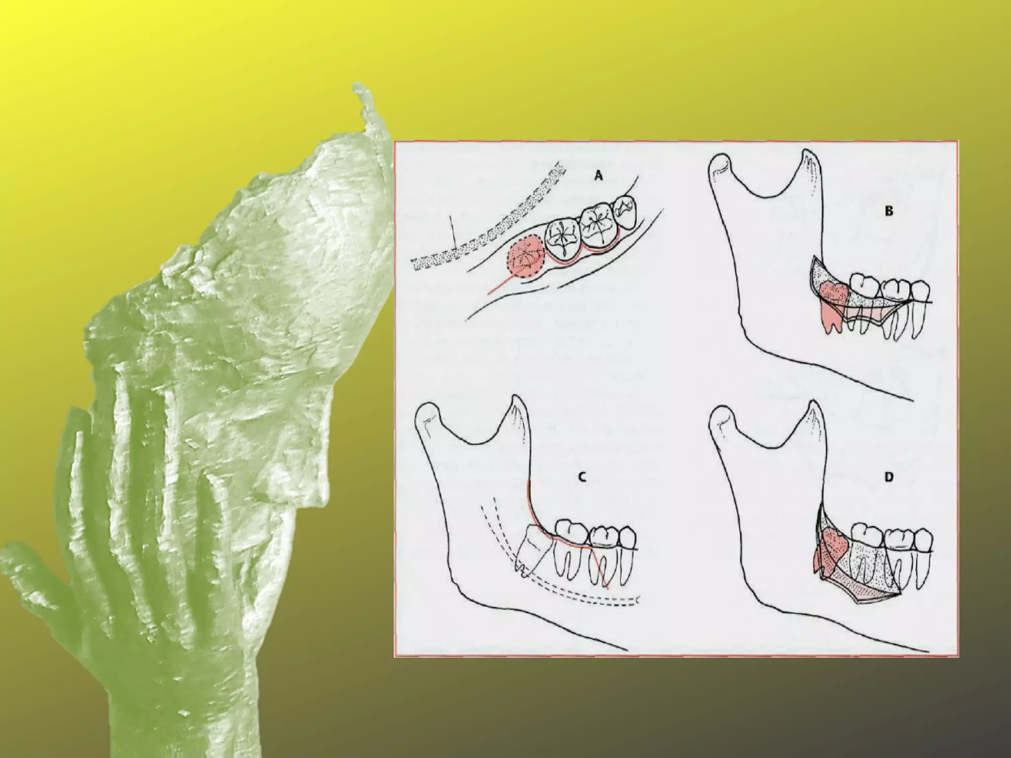
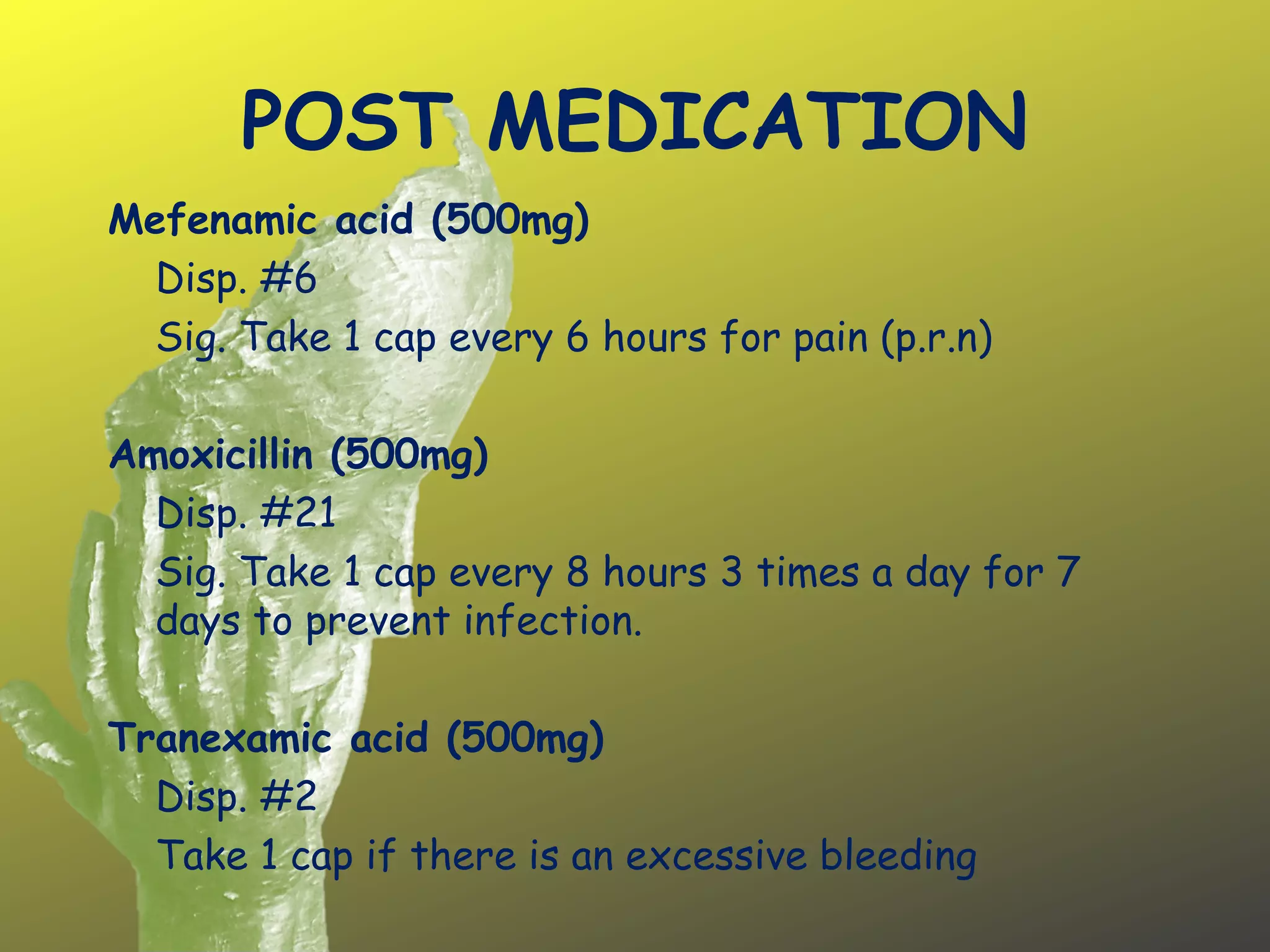
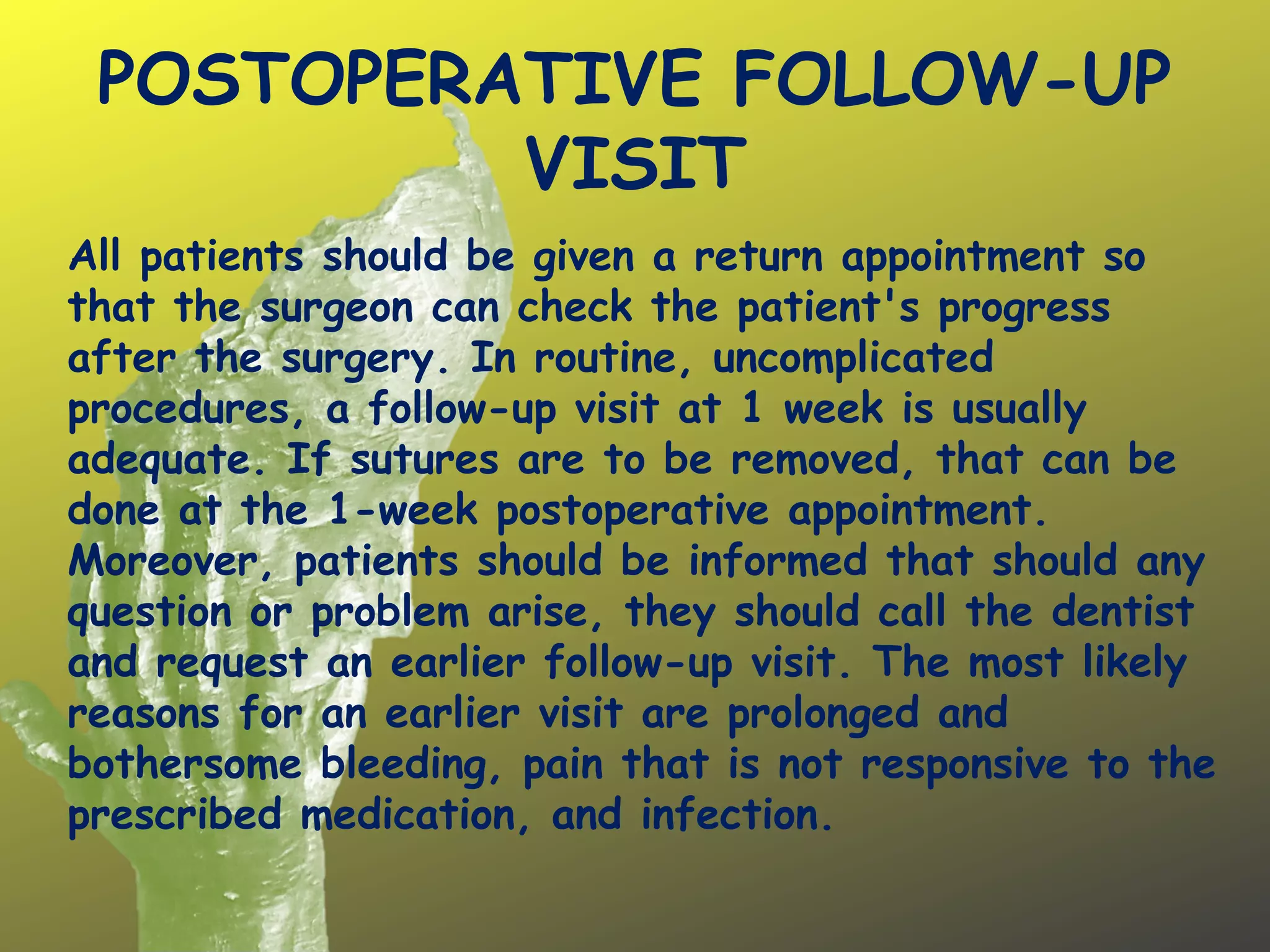
ODONTECTOMY
Chica, Danna Paula Louise
ODONTECTOMY
• A case when an impacted tooth fails to
erupt into the dental arch within the
expected time.
• The tooth becomes impacted because its
adjacent teeth, dense overlying bone, or
excessive soft tissue prevents eruption.
Because impacted teeth do not erupt, they
are retained for the patient's lifetime
unless surgically removed.
Armamentarium
Premedications
 
Mefenamic acid (500mg)
Disp. #1
Take 1 cap 1 hour before the treatment to
lessen pain.
Amoxicillin (500mg)
Disp. #1
Take 1 cap 1 hour before the treatment to
prevent infection.
Tranexamic acid (500mg)
Disp. #1
Take 1 cap 1 hour before the treatment to
prevent excessive bleeding.
 
Procedure
1.Aseptic Technique-minimizes wound contamination
by pathogens through the following:
a. Sterilization of Instruments
b. Operatory Disinfections
c. Surgical Staff Preparation
2.Pain and Anxiety Control
a. Dry the mucosa and apply topical anes, on surgical area
b. Admin. Local anes, using Mandibular Block Tech.
3.Flap Design
a. Full thickness flap will be used to reflect the soft tissue for
removal of impacted molar.
SURGICAL PROCEDURE
1. To have an adequate area of exposure, incise
the tissue from retromolar area down to the
bone making it a full thickness flap.
2. The incision will run from ramus area for
posterior extension.
3. The posterior extension should diverge laterally
to avoid lingual nerve injury.
4. Reflect the incision laterally to expose the
underlying bone covering the impactd 3rd
molar.
MANDIBULAR NERVE BLOCK
• Anesthetize the tooth
using mandibular block
and local infiltration
technique
SURGICAL PROCEDURE
1. Bone removal
 Assess the need and extent of the bone to be removed.
 Remove bone on buccal cortical plate using surgical bur and handpiece to
expose the greatest convexity of the crown.
 Irrigate to remove debris and to avoid overheating due to constant
friction.
1. Using surgical bur, exposed crown is then cut up to the ¾ portion of
the crown. This creates a slot wherein the angular elevator is
inserted and then rotated to completely split the tooth. Coronal
part is delivered first out of the socket using angular elevator from
a mesiobucccal direction. Using a cryer elevator, the apical portion
is then luxated out of the socket.
2. Curette the socket and remove the follicular sac.
3. Smoothen the sharp and bony spicules using a bone file.
4. Irrigate the area using NSS then suction.
SURGICAL PROCEDURE
6. Place appropriate amount of gel foam on the socket
for promotion of hemostasis on the area.
7. Prepare for suturing. Stabilize loose tissue forceps.
Coaptate the loose and movable tissue.
8. Suture with sterile suturing material. Suture design is
multiple interrupted sutures.
9. Provide instructions for post operative phase to the
patient.
10.Recall after 1 week.
The preferred incision for the removal of an impacted mandibular
third molar is an envelope incision that extends from the mesial
papilla of the mandibular first molar, around the necks of the
teeth to the distobuccal line angle of the second molar, and then
posteriorly to and laterally up the anterior border of the mandible
 
A, Envelope incision is most commonly used to reflect soft tissue for
removal of impacted third molar. Posterior extension of incision
should laterally diverge to avoid injury to lingual nerve.
B, Envelope incision is laterally reflected to expose bone overlying
impacted tooth.
C, When three-cornered flap is made, a releasing incision is made at
mesial aspect of second molar.
D, When soft tissue flap is reflected by means of a releasing
incision, greater visibility is possible, especially at apical aspect of
surgical field.
POST OPERATIVE
INSTRUCTION
Relax after surgery. Physical activity may increase bleeding.
Have a soft diet and gradually add solid foods to your diet
as healing progresses.
Do not drink alcohol or hot fluids such as tea or coffee and
avoids spicy foods until the gum is fully healed.
Many surgeons recommend the use of ice packs on the face
to help prevent postoperative swelling.
Avoid smoking
After the first day, gently rinse your mouth with warm salt
water several times a day to reduce swelling and relieve
pain.
Avoid rubbing the area with your tongue or touching it with
your fingers.
POST MEDICATION
Mefenamic acid (500mg)
Disp. #6
Sig. Take 1 cap every 6 hours for pain (p.r.n)
Amoxicillin (500mg)
Disp. #21
Sig. Take 1 cap every 8 hours 3 times a day for 7
days to prevent infection.
Tranexamic acid (500mg)
Disp. #2
Take 1 cap if there is an excessive bleeding
POSTOPERATIVE FOLLOW-UP
VISIT
All patients should be given a return appointment so
that the surgeon can check the patient's progress
after the surgery. In routine, uncomplicated
procedures, a follow-up visit at 1 week is usually
adequate. If sutures are to be removed, that can be
done at the 1-week postoperative appointment.
Moreover, patients should be informed that should any
question or problem arise, they should call the dentist
and request an earlier follow-up visit. The most likely
reasons for an earlier visit are prolonged and
bothersome bleeding, pain that is not responsive to the
prescribed medication, and infection.
PATIENT’S INFORMATION
PATIENT NAME:
ADDRESS:
AGE:
SEX: Female
PATIENT’S INFORMATION
MEDICAL HISTORY: The patient is
healthy and no history of any diseases.
PATIENT’S INFORMATION
DENTAL HISTORY: The patient’s last
dental visit was last 2012.
Class I occlusion
FRONT VIEW
SIDE VIEW
INTRAORAL

Odontectomy

  • 1.
  • 2.
    ODONTECTOMY • A casewhen an impacted tooth fails to erupt into the dental arch within the expected time. • The tooth becomes impacted because its adjacent teeth, dense overlying bone, or excessive soft tissue prevents eruption. Because impacted teeth do not erupt, they are retained for the patient's lifetime unless surgically removed.
  • 3.
  • 4.
    Premedications   Mefenamic acid (500mg) Disp.#1 Take 1 cap 1 hour before the treatment to lessen pain. Amoxicillin (500mg) Disp. #1 Take 1 cap 1 hour before the treatment to prevent infection. Tranexamic acid (500mg) Disp. #1 Take 1 cap 1 hour before the treatment to prevent excessive bleeding.  
  • 5.
    Procedure 1.Aseptic Technique-minimizes woundcontamination by pathogens through the following: a. Sterilization of Instruments b. Operatory Disinfections c. Surgical Staff Preparation 2.Pain and Anxiety Control a. Dry the mucosa and apply topical anes, on surgical area b. Admin. Local anes, using Mandibular Block Tech. 3.Flap Design a. Full thickness flap will be used to reflect the soft tissue for removal of impacted molar.
  • 6.
    SURGICAL PROCEDURE 1. Tohave an adequate area of exposure, incise the tissue from retromolar area down to the bone making it a full thickness flap. 2. The incision will run from ramus area for posterior extension. 3. The posterior extension should diverge laterally to avoid lingual nerve injury. 4. Reflect the incision laterally to expose the underlying bone covering the impactd 3rd molar.
  • 7.
    MANDIBULAR NERVE BLOCK •Anesthetize the tooth using mandibular block and local infiltration technique
  • 8.
    SURGICAL PROCEDURE 1. Boneremoval  Assess the need and extent of the bone to be removed.  Remove bone on buccal cortical plate using surgical bur and handpiece to expose the greatest convexity of the crown.  Irrigate to remove debris and to avoid overheating due to constant friction. 1. Using surgical bur, exposed crown is then cut up to the ¾ portion of the crown. This creates a slot wherein the angular elevator is inserted and then rotated to completely split the tooth. Coronal part is delivered first out of the socket using angular elevator from a mesiobucccal direction. Using a cryer elevator, the apical portion is then luxated out of the socket. 2. Curette the socket and remove the follicular sac. 3. Smoothen the sharp and bony spicules using a bone file. 4. Irrigate the area using NSS then suction.
  • 9.
    SURGICAL PROCEDURE 6. Placeappropriate amount of gel foam on the socket for promotion of hemostasis on the area. 7. Prepare for suturing. Stabilize loose tissue forceps. Coaptate the loose and movable tissue. 8. Suture with sterile suturing material. Suture design is multiple interrupted sutures. 9. Provide instructions for post operative phase to the patient. 10.Recall after 1 week.
  • 10.
    The preferred incisionfor the removal of an impacted mandibular third molar is an envelope incision that extends from the mesial papilla of the mandibular first molar, around the necks of the teeth to the distobuccal line angle of the second molar, and then posteriorly to and laterally up the anterior border of the mandible   A, Envelope incision is most commonly used to reflect soft tissue for removal of impacted third molar. Posterior extension of incision should laterally diverge to avoid injury to lingual nerve. B, Envelope incision is laterally reflected to expose bone overlying impacted tooth. C, When three-cornered flap is made, a releasing incision is made at mesial aspect of second molar. D, When soft tissue flap is reflected by means of a releasing incision, greater visibility is possible, especially at apical aspect of surgical field.
  • 12.
    POST OPERATIVE INSTRUCTION Relax aftersurgery. Physical activity may increase bleeding. Have a soft diet and gradually add solid foods to your diet as healing progresses. Do not drink alcohol or hot fluids such as tea or coffee and avoids spicy foods until the gum is fully healed. Many surgeons recommend the use of ice packs on the face to help prevent postoperative swelling. Avoid smoking After the first day, gently rinse your mouth with warm salt water several times a day to reduce swelling and relieve pain. Avoid rubbing the area with your tongue or touching it with your fingers.
  • 13.
    POST MEDICATION Mefenamic acid(500mg) Disp. #6 Sig. Take 1 cap every 6 hours for pain (p.r.n) Amoxicillin (500mg) Disp. #21 Sig. Take 1 cap every 8 hours 3 times a day for 7 days to prevent infection. Tranexamic acid (500mg) Disp. #2 Take 1 cap if there is an excessive bleeding
  • 14.
    POSTOPERATIVE FOLLOW-UP VISIT All patientsshould be given a return appointment so that the surgeon can check the patient's progress after the surgery. In routine, uncomplicated procedures, a follow-up visit at 1 week is usually adequate. If sutures are to be removed, that can be done at the 1-week postoperative appointment. Moreover, patients should be informed that should any question or problem arise, they should call the dentist and request an earlier follow-up visit. The most likely reasons for an earlier visit are prolonged and bothersome bleeding, pain that is not responsive to the prescribed medication, and infection.
  • 15.
  • 16.
    PATIENT’S INFORMATION MEDICAL HISTORY:The patient is healthy and no history of any diseases.
  • 17.
    PATIENT’S INFORMATION DENTAL HISTORY:The patient’s last dental visit was last 2012. Class I occlusion
  • 18.
  • 19.
  • 20.