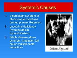
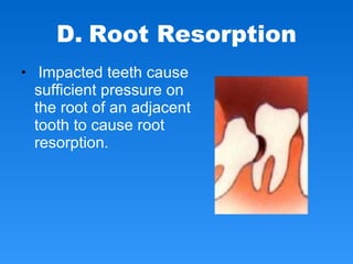
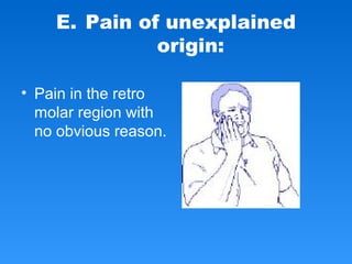
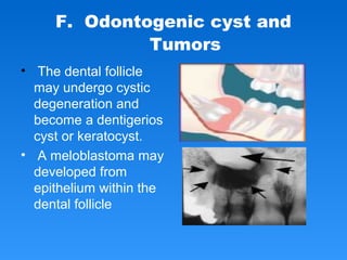
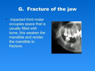
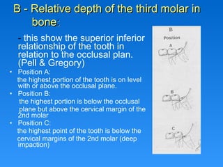
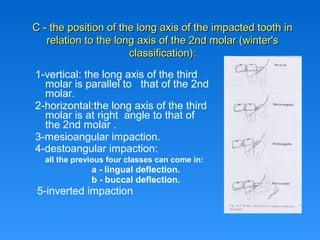
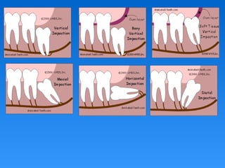
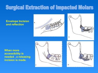
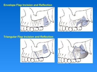
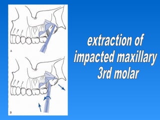
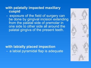
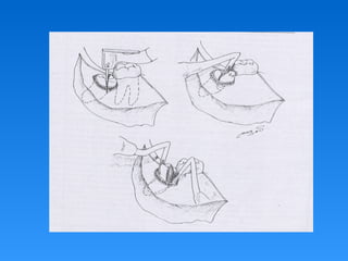
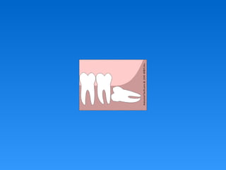
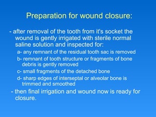
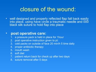
DENTAL COLLEGE
AZAMGARH
DEPT OF ORAL AND
MAXILLOFACIAL SURGERY
Impaction
PRESENTED BY
Tawseef ahmed
GUIDED BY
Dr PARTH SARTHI DIXIT
Definition :-
is a tooth that fails to erupt into its normal
functioning position in the dental arch
within the expected time.
The term Unerupted includes both
impacted teeth and teeth that are in the
process of erupting.
Causes of impaction
Systemic Causes
A. a hereditary syndrom of
cliedocranial dysistosis
termed primary Retention.
B. endocrinal deficiency
(hypothyrodism,
hypopituitarism).
C. febrile disease, down
syndrom, irradiation (all
cause multiple teeth
impaction).
Local Factors
A. prolonged deciduous tooth retention
B. malposed tooth germ
C. arch length deficiency
D. odontoginic tumors abnormal eruption
path
E. cleft lip and palate
frequency of impaction
- The order of frequency of impacted
teeth is as follow:-
frequency of impaction
1. mandibular 3rd molar
2. maxillary 3rd molar
3. maxillary cuspid
4. mandibular cuspid
5. Mandibular premolar
6. maxillary premolars
7. maxillary central and lateral incisors
Complication of
impacted teeth
(indication for removal):
A. PERICORONITIS
• when a tooth is partially
impacted with a large
amount of soft tissue over
the axial and occlusal
surfaces, the patient
frequently has
one or more episodes of
pericronitis.
Definition of pericoronitis
• is an infection of the soft
tissue around the crown of
partially impacted tooth and
is caused by the normal
oral flora.
• When third molar is
impacted or partially
impacted ,the bacteria
that cause dental
caries can be
exposed to the distal
aspect of the 2nd
molar, as well as to
third molar
B. Dental Caries
• Erupted teeth adjacent to
impacted teeth are
predisposed to periodontal
disease.
• As it decrease amount of
bone on the distal aspect of
adjacent 2nd molar, with
deep periodontal pocket on
the distal aspect of the 2nd
molar.
C. Periodontal Disease
• Impacted teeth cause
sufficient pressure on
the root of an adjacent
tooth to cause root
resorption.
D. Root Resorption
E. Pain of unexplained
origin:
• Pain in the retro
molar region with
no obvious reason.
F. Odontogenic cyst and
Tumors
• The dental follicle
may undergo cystic
degeneration and
become a dentigerios
cyst or keratocyst.
• A meloblastoma may
developed from
epithelium within the
dental follicle
G. Fracture of the jaw
• impacted third molar
occupies space that is
usually filled with
bone, this weaken the
mandible and render
the mandible to
fracture.
Contraindication for
removal of impacted
teeth:
1. extreme of age:
- as the bone become highly calcified, less
flexible, less likely to bend under force of
tooth extraction
the result ,bone more surgically removed to
displace tooth from its socket and less post
operative sequla
1. compromised medical status:
2. probable excessive damage to adjacent
structure:
Classification system
of impacted teeth
- this is done to help dentist in evaluation of
the extent of the surgical procedure and in
the planning of this procedure.
1-Classification of impacted
mandibular third molar:
A - Relation of the tooth to the ascending
ramus of the mandible and to the distal
surface of the 2nd molar: (Pell
&Gregory)
– this show the anterioposterior relationship of the
tooth to the arch and the amount of resistance
offered by the bone of the ascending ramus that
may influence the tooth removal
Class1
• the space between
the anterior part of the
ascending ramus and
the distal surface of
the 2nd molar is
sufficient to
accommodate the
mesiodistal diameter
of the crown of the
third molar.
Class2
• the space between
the anterior part of the
ascending ramus and
distal surface of the
2nd molar is less than
the mesiodistal
diameter of the crown
of the third molar (part
of the tooth located
within the ramus)
Class3
• all the third molar is
located within the
ascending ramus of
the mandible.
- this show the superior inferior
relationship of the tooth in
relation to the occlusal plan.
(Pell & Gregory)
• Position A:
the highest portion of the tooth is on level
with or above the occlusal plane.
• Position B:
the highest portion is below the occlusal
plane but above the cervical margin of the
2nd molar
• Position C:
the highest point of the tooth is below the
cervical margins of the 2nd molar (deep
impaction)
B - Relative depth of the third molar inB - Relative depth of the third molar in
bonebone::
1-vertical: the long axis of the third
molar is parallel to that of the 2nd
molar.
2-horizontal:the long axis of the third
molar is at right angle to that of
the 2nd molar .
3-mesioangular impaction.
4-destoangular impaction:
all the previous four classes can come in:
a - lingual deflection.
b - buccal deflection.
5-inverted impaction
C - the position of the long axis of the impacted tooth inC - the position of the long axis of the impacted tooth in
relation to the long axis of the 2nd molar (winter'srelation to the long axis of the 2nd molar (winter's
classification):classification):
2 -Classification of impacted
maxillary third molar:
1. The relationship of the tooth to occlusal plane of the
2nd molar (as before)
2. The relationship of tooth to maxillary sinus :
a-sinus approximation :
(s.a) where no bone or very thin bone exist
between the impacted teeth and floor of sinus.
b-no sinus approximation :
(n.s.a) where 2 mm or more of bone exist
between the floor of sinus and impacted teeth.
3-Classification of impacted
maxillary cuspids:
• Class1:
palatally impacted cuspids ,these could be in vertical,
horizontal, semivertical position.
• Class2:
labialy impacted cuspide which could be in vertical,
horizontal, semivertical.
• Class3:
impacted cuspid located both in the palatal and labial
surfaces.
• Class4:
impacted cuspid that are present in an edentulous
maxilla and may assume any of the previous three
classes.
Surgical removal of
impacted teeth:
1- Proper radiographic and clinical evaluation of
the condition:
A- periapical radiograph
B- occlusal radiograph
C- panoramic radiograph
2- Classification of impaction to help in planning
the surgical procedure:
3- Selection of the time for surgical procedure:
 surgical removal of impacted third molar is not as a
surgical emergency, it is an elective procedure which
shouldn't be postponed for along period of time until
several complication arises.
4- The condition should be explained to patient in
a simple easy way directing his attention to
possible complication that may arise from
leaving tooth in position
5- Surgical removal can be made under local
anesthesia as well as general anesthesia the
choice of the anesthetic technique depends on:
a- general condition of the patient and his ability
psychologically and physically take the procedure. in
very apprehensive patient, general anesthesia is
preferred.
b- position of impaction and extent of surgical procedure
c- patient co-operation
d- number of impaction that will be removed in the
setting
the surgical procedure is divided
into following stages:
A- elevation of an adequate
mucoperosteal flap to expose the
field of surgery:
Pyramidal flap used in all third molar
impaction, the anterior incision of
the flap could extend from the distal
aspect to 2nd molar running at 45
degree angel and extend to the
mucobucal fold.
In deep impaction ,a bigger flap is
advisable. the anterior incision could
start from the mesial aspect of 2nd
molar
1- gaining access to impacted tooth:
Envelope Incision
and reflection
When more
accessibility is
needed , a releasing
incision is made.
Envelope Flap Incision and Reflection
Triangular Flap Incision and Reflection
with palatally impacted maxillary
cuspid
- exposure of the field of surgery can
be done by gingival incision extending
from the palatal side of premolar in
one side to other side all around the
palatal gingiva of the present teeth.
with labially placed impaction
- a labial pyramidal flap is adequate
2- bone removal
This is done for :-
A- exposure of impaction
B- reduction of resistance
C- making a point for application of the elevator
Bone Removal With a
Fissure Surgical Bur
3- tooth delivery
1- total delivery by application of force using elevators:
a- mesial application of force :straight elevators and pot's
elevators.
b- buccal application of force :winter elevator
2-delivery of the tooth after tooth division :
- division is indicated to reduce resistance ,create a space or remove
interlocked cusps of the tooth
a- decapitation:- division of the crown of the tooth at cervical
margin level .
- indicated in horizontal mandibular and maxillary third molar
impaction and pallataly impacted maxillary cuspid
b- longitudinal tooth division:
- indicated when the impacted tooth has a widely divergent straight
roots, or when one root is straight and the other is curved
c- division of the interlocking cusp:
- this is done with mesioangular impaction ,removal of the inter
locking segment of the tooth usually located under the distal
surface of 2nd molar
Bone is removed with the surgical bur
to expose the whole crown
Decapitation is then performed
A purchase point is prepared in the
root, which is then removed with an
elevator
The second root is removed in the
same way
Preparation for wound closure:
- after removal of the tooth from it's socket the
wound is gently irrigated with sterile normal
saline solution and inspected for:
a- any remnant of the residual tooth sac is removed
b- remnant of tooth structure or fragments of bone
debris is gently removed
c- small fragments of the detached bone
d- sharp edges of interseptal or alveolar bone is
trimmed and smoothed
- then final irrigation and wound now is ready for
closure.
closure of the wound:
• well designed and properly reflected flap fall back easily
into place. using have circle a traumatic needle and 000
black silk suture to hold flap into place
• post operative care:
1. a pressure pack is held in place for 1hour
2. post operative instruction given to pt:
3. cold packs on outside of face 20 min/h 5 time daily
4. proper antibiotic therapy
5. mouth wash
6. soft diet
7. patient return back for check up after two days
8. suture removal after 5 days
Complication associated with
surgical removal pf impacted
tooth:
1- laceration of the soft tissue flap:
a-improper incision
b-improper elevation of the flap and improper retraction
this leads to delayed healing and sever discomfort
2- affection of the alveolar bone:
3- fracture of the jaw:
- in angle of mandible ,improper use of elevator with
uncontrolled force
4- fracture of tuberosity:
this occurs with erupted rather than unerupted tooth
due to improper use of force
5-comlication related to injury of adjacent structure:
a-injury to inferior alveolar canal:
- occurs in deeply seated vertical impaction, the nerve pass
between roots of impacted tooth .permanent numbness and
heamorraghe
b-damage to nasal floor:
- during surgical removal of impacted maxillary cuspid, profuse bleeding
from nasal mucosa
c- involvement of maxillary sinus:
- during removal of impacted maxillary third molar. oro anntral
fistula results
d- pushing of impacted tooth into maxillary sinus:
e- pushing of impacted maxillary molar into pterigopalatine fossa:
- uncontrolled mesial application of force in deep impaction
f- pushing impacted mandibular third molar into sub-mandibular
space:
- uncontrolled buccal application pf force and fracture of the
lingual plate
g-aspiration or swallowing of impacted tooth:
- with general anesthesia ,
post operative complication:
1. pain.
2. infection
3. heamoraghe
4. anesthesia or parenthesis of the lingual or inferior
alveolar nerve
5. trismus,limitation of jaw movement
6. osteomylitis
7. pain at tmj
8. pain on swallowing due to edema of pharynx and
hematoma formation.
Thank you

Impacted teeth

  • 1.
    DENTAL COLLEGE AZAMGARH DEPT OFORAL AND MAXILLOFACIAL SURGERY
  • 2.
  • 3.
    Definition :- is atooth that fails to erupt into its normal functioning position in the dental arch within the expected time. The term Unerupted includes both impacted teeth and teeth that are in the process of erupting.
  • 4.
  • 5.
    Systemic Causes A. ahereditary syndrom of cliedocranial dysistosis termed primary Retention. B. endocrinal deficiency (hypothyrodism, hypopituitarism). C. febrile disease, down syndrom, irradiation (all cause multiple teeth impaction).
  • 6.
    Local Factors A. prolongeddeciduous tooth retention B. malposed tooth germ C. arch length deficiency D. odontoginic tumors abnormal eruption path E. cleft lip and palate
  • 7.
    frequency of impaction -The order of frequency of impacted teeth is as follow:-
  • 8.
    frequency of impaction 1.mandibular 3rd molar 2. maxillary 3rd molar 3. maxillary cuspid 4. mandibular cuspid 5. Mandibular premolar 6. maxillary premolars 7. maxillary central and lateral incisors
  • 9.
  • 10.
    A. PERICORONITIS • whena tooth is partially impacted with a large amount of soft tissue over the axial and occlusal surfaces, the patient frequently has one or more episodes of pericronitis.
  • 11.
    Definition of pericoronitis •is an infection of the soft tissue around the crown of partially impacted tooth and is caused by the normal oral flora.
  • 12.
    • When thirdmolar is impacted or partially impacted ,the bacteria that cause dental caries can be exposed to the distal aspect of the 2nd molar, as well as to third molar B. Dental Caries
  • 13.
    • Erupted teethadjacent to impacted teeth are predisposed to periodontal disease. • As it decrease amount of bone on the distal aspect of adjacent 2nd molar, with deep periodontal pocket on the distal aspect of the 2nd molar. C. Periodontal Disease
  • 14.
    • Impacted teethcause sufficient pressure on the root of an adjacent tooth to cause root resorption. D. Root Resorption
  • 15.
    E. Pain ofunexplained origin: • Pain in the retro molar region with no obvious reason.
  • 16.
    F. Odontogenic cystand Tumors • The dental follicle may undergo cystic degeneration and become a dentigerios cyst or keratocyst. • A meloblastoma may developed from epithelium within the dental follicle
  • 17.
    G. Fracture ofthe jaw • impacted third molar occupies space that is usually filled with bone, this weaken the mandible and render the mandible to fracture.
  • 18.
  • 19.
    1. extreme ofage: - as the bone become highly calcified, less flexible, less likely to bend under force of tooth extraction the result ,bone more surgically removed to displace tooth from its socket and less post operative sequla 1. compromised medical status: 2. probable excessive damage to adjacent structure:
  • 20.
    Classification system of impactedteeth - this is done to help dentist in evaluation of the extent of the surgical procedure and in the planning of this procedure.
  • 21.
  • 22.
    A - Relationof the tooth to the ascending ramus of the mandible and to the distal surface of the 2nd molar: (Pell &Gregory) – this show the anterioposterior relationship of the tooth to the arch and the amount of resistance offered by the bone of the ascending ramus that may influence the tooth removal
  • 23.
    Class1 • the spacebetween the anterior part of the ascending ramus and the distal surface of the 2nd molar is sufficient to accommodate the mesiodistal diameter of the crown of the third molar.
  • 24.
    Class2 • the spacebetween the anterior part of the ascending ramus and distal surface of the 2nd molar is less than the mesiodistal diameter of the crown of the third molar (part of the tooth located within the ramus)
  • 25.
    Class3 • all thethird molar is located within the ascending ramus of the mandible.
  • 26.
    - this showthe superior inferior relationship of the tooth in relation to the occlusal plan. (Pell & Gregory) • Position A: the highest portion of the tooth is on level with or above the occlusal plane. • Position B: the highest portion is below the occlusal plane but above the cervical margin of the 2nd molar • Position C: the highest point of the tooth is below the cervical margins of the 2nd molar (deep impaction) B - Relative depth of the third molar inB - Relative depth of the third molar in bonebone::
  • 27.
    1-vertical: the longaxis of the third molar is parallel to that of the 2nd molar. 2-horizontal:the long axis of the third molar is at right angle to that of the 2nd molar . 3-mesioangular impaction. 4-destoangular impaction: all the previous four classes can come in: a - lingual deflection. b - buccal deflection. 5-inverted impaction C - the position of the long axis of the impacted tooth inC - the position of the long axis of the impacted tooth in relation to the long axis of the 2nd molar (winter'srelation to the long axis of the 2nd molar (winter's classification):classification):
  • 29.
    2 -Classification ofimpacted maxillary third molar:
  • 30.
    1. The relationshipof the tooth to occlusal plane of the 2nd molar (as before) 2. The relationship of tooth to maxillary sinus : a-sinus approximation : (s.a) where no bone or very thin bone exist between the impacted teeth and floor of sinus. b-no sinus approximation : (n.s.a) where 2 mm or more of bone exist between the floor of sinus and impacted teeth.
  • 31.
  • 32.
    • Class1: palatally impactedcuspids ,these could be in vertical, horizontal, semivertical position. • Class2: labialy impacted cuspide which could be in vertical, horizontal, semivertical. • Class3: impacted cuspid located both in the palatal and labial surfaces. • Class4: impacted cuspid that are present in an edentulous maxilla and may assume any of the previous three classes.
  • 33.
  • 34.
    1- Proper radiographicand clinical evaluation of the condition: A- periapical radiograph B- occlusal radiograph C- panoramic radiograph 2- Classification of impaction to help in planning the surgical procedure: 3- Selection of the time for surgical procedure:  surgical removal of impacted third molar is not as a surgical emergency, it is an elective procedure which shouldn't be postponed for along period of time until several complication arises.
  • 35.
    4- The conditionshould be explained to patient in a simple easy way directing his attention to possible complication that may arise from leaving tooth in position 5- Surgical removal can be made under local anesthesia as well as general anesthesia the choice of the anesthetic technique depends on: a- general condition of the patient and his ability psychologically and physically take the procedure. in very apprehensive patient, general anesthesia is preferred. b- position of impaction and extent of surgical procedure c- patient co-operation d- number of impaction that will be removed in the setting
  • 36.
    the surgical procedureis divided into following stages:
  • 37.
    A- elevation ofan adequate mucoperosteal flap to expose the field of surgery: Pyramidal flap used in all third molar impaction, the anterior incision of the flap could extend from the distal aspect to 2nd molar running at 45 degree angel and extend to the mucobucal fold. In deep impaction ,a bigger flap is advisable. the anterior incision could start from the mesial aspect of 2nd molar 1- gaining access to impacted tooth:
  • 38.
    Envelope Incision and reflection Whenmore accessibility is needed , a releasing incision is made.
  • 39.
    Envelope Flap Incisionand Reflection Triangular Flap Incision and Reflection
  • 41.
    with palatally impactedmaxillary cuspid - exposure of the field of surgery can be done by gingival incision extending from the palatal side of premolar in one side to other side all around the palatal gingiva of the present teeth. with labially placed impaction - a labial pyramidal flap is adequate
  • 42.
    2- bone removal Thisis done for :- A- exposure of impaction B- reduction of resistance C- making a point for application of the elevator
  • 43.
    Bone Removal Witha Fissure Surgical Bur
  • 44.
    3- tooth delivery 1-total delivery by application of force using elevators: a- mesial application of force :straight elevators and pot's elevators. b- buccal application of force :winter elevator 2-delivery of the tooth after tooth division : - division is indicated to reduce resistance ,create a space or remove interlocked cusps of the tooth a- decapitation:- division of the crown of the tooth at cervical margin level . - indicated in horizontal mandibular and maxillary third molar impaction and pallataly impacted maxillary cuspid b- longitudinal tooth division: - indicated when the impacted tooth has a widely divergent straight roots, or when one root is straight and the other is curved c- division of the interlocking cusp: - this is done with mesioangular impaction ,removal of the inter locking segment of the tooth usually located under the distal surface of 2nd molar
  • 45.
    Bone is removedwith the surgical bur to expose the whole crown Decapitation is then performed A purchase point is prepared in the root, which is then removed with an elevator The second root is removed in the same way
  • 48.
    Preparation for woundclosure: - after removal of the tooth from it's socket the wound is gently irrigated with sterile normal saline solution and inspected for: a- any remnant of the residual tooth sac is removed b- remnant of tooth structure or fragments of bone debris is gently removed c- small fragments of the detached bone d- sharp edges of interseptal or alveolar bone is trimmed and smoothed - then final irrigation and wound now is ready for closure.
  • 49.
    closure of thewound: • well designed and properly reflected flap fall back easily into place. using have circle a traumatic needle and 000 black silk suture to hold flap into place • post operative care: 1. a pressure pack is held in place for 1hour 2. post operative instruction given to pt: 3. cold packs on outside of face 20 min/h 5 time daily 4. proper antibiotic therapy 5. mouth wash 6. soft diet 7. patient return back for check up after two days 8. suture removal after 5 days
  • 50.
    Complication associated with surgicalremoval pf impacted tooth:
  • 51.
    1- laceration ofthe soft tissue flap: a-improper incision b-improper elevation of the flap and improper retraction this leads to delayed healing and sever discomfort 2- affection of the alveolar bone: 3- fracture of the jaw: - in angle of mandible ,improper use of elevator with uncontrolled force 4- fracture of tuberosity: this occurs with erupted rather than unerupted tooth due to improper use of force
  • 52.
    5-comlication related toinjury of adjacent structure: a-injury to inferior alveolar canal: - occurs in deeply seated vertical impaction, the nerve pass between roots of impacted tooth .permanent numbness and heamorraghe b-damage to nasal floor: - during surgical removal of impacted maxillary cuspid, profuse bleeding from nasal mucosa c- involvement of maxillary sinus: - during removal of impacted maxillary third molar. oro anntral fistula results d- pushing of impacted tooth into maxillary sinus: e- pushing of impacted maxillary molar into pterigopalatine fossa: - uncontrolled mesial application of force in deep impaction f- pushing impacted mandibular third molar into sub-mandibular space: - uncontrolled buccal application pf force and fracture of the lingual plate g-aspiration or swallowing of impacted tooth: - with general anesthesia ,
  • 53.
    post operative complication: 1.pain. 2. infection 3. heamoraghe 4. anesthesia or parenthesis of the lingual or inferior alveolar nerve 5. trismus,limitation of jaw movement 6. osteomylitis 7. pain at tmj 8. pain on swallowing due to edema of pharynx and hematoma formation.
  • 54.