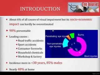
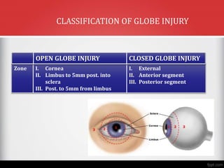
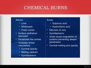
Ocular Injuries – Classification
& Management Protocol
Dr. Sujata Priyambada
Assistant Professor
Hi-tech Medical College & Hospital
INTRODUCTION
 About 6% of all causes of visual impairment but its socio-economic
impact can hardly be overestimated
 90% preventable
 Leading causes :
Road traffic accidents
Sport accidents
Consumer fireworks
Household chemicals
Workshop & factory
 Incidence more in <30 years, 85% males
 Nearly 48% at home
OBJECTIVES
 Classification of injury
 Evaluation of injured eye
 Emergency management
TYPES OF OCULAR TRAUMA
 Mechanical
 Chemical
 Thermal
 Combined
OCULAR TRAUMA TERMINOLOGY
 Need for standardization
 Birmingham Eye Trauma Terminology (BETT) has been
major advance
 Covers terminology of only mechanical injuries
 Tissue of reference is always entire globe
 Periocular injuries are not taken into consideration
BIRMINGHAM EYE TRAUMA TERMINOLOGY
 Eye wall : sclera & cornea
 Closed globe injury : no full thickness eye wall defect
 Open globe injury : full thickness defect
 Contusion : closed globe injury due
to blunt force
 Rupture as ‘inside –out’ injury
caused by blunt force
 Lacerating injury : full thickness defect caused by sharp force
 Penetrating : only entrance wound
 Perforating : both entrance & exit wounds present
 IOFB : entrance wound with FB lodged inside eye
PENETRATING
PERFORATING RUPTURE
BIRMINGHAM EYE TRAUMA TERMINOLOGY
OPEN GLOBE INJURY CLOSED GLOBE INJURY
Type A. Rupture
B. Penetrating
C. Perforating
D. IOFB
E. Combined
A. Contusion
B. Lamellar laceration
C. Superficial foreign body
D. Mixed
Grade A. >= 20/40
B. 20/50 – 20/100
C. 19/100 – 5/200
D. 4/200 – PL
E. NLP
- do -
Pupil A. Positive
B. Negative
- do -
BETT CLASSIFICATION OF GLOBE INJURY
Zone I. Cornea
II. Limbus to 5mm post. into
sclera
III. Post. to 5mm from limbus
I. External
II. Anterior segment
III. Posterior segment
OPEN GLOBE INJURY CLOSED GLOBE INJURY
CLASSIFICATION OF GLOBE INJURY
Scoring system to determine prognosis in open globe injuries
OCULAR TRAUMA SCORE
1. Determine raw points
2. Calculate sum total of raw points
3. Convert raw points into percentage chance of vision
OCULAR TRAUMA SCORE
GENERAL EVALUATION
 Accurate & thorough examination
 Appropriate sedation, Adequate analgesia & Protection
of open globe from further injury
 SYSTEMIC EVALUATION :
• Vital signs & mental status assessed immediately
• Once systemically stable proceed with history & ocular
examination
HISTORY
 HISTORY OF EVENTS
 TYPE OF INJURY
 TIME OF THE INJURY
 VISUAL ACUITY PRIOR TO THE INJURY
 SPECIFIC SYMPTOMS
 PAST OCULAR OR SURGICAL HISTORY
 TETANUS IMMUNIZATION
A. VISUAL ACUITY :
• Best predictor of final visual outcome
• Pin hole acuity is indicated
• Check one eye at a time
B. CONFRONTATIONAL VISUAL FIELD :
• Static finger counting as rapid way of grossly assessing
patient’s peripheral visual field
• Formal visual field performed after patient stable to
evaluate traumatic optic neuropathy
INITIAL EXAMINATION
C. PUPILLARY EXAMINATION :
• Normal pupillary function important as prognostic factor
• Swinging flashlight test using high intensity source such as
indirect ophthalmoscope
• RAPD indicates RD, Optic nerve damage
INITIAL EXAMINATION
D. IOP MEASUREMENT :
• Deferred in eyes with open injuries
• In closed injury important diagnostic tool
E. MOTILITY :
More in orbital injuries where muscle entrapment or injury
INITIAL EXAMINATION
F. Anterior segment evaluation :
 Always be conscious of multiple injuries to tissue
 Be extremely gentle
 Avoid pressure on traumatized eye
 Evaluation in stepwise manner starting from orbits &
lids to inner structures anatomically anterior to
posterior
INITIAL EXAMINATION
 Patient discouraged from sneezing in case of orbital fracture
 Small Lid lacerations may point towards occult penetrating injuries
 ‘Scleral ruptures can occur without overlying Conjunctival injury’
What to look for in initial examination ?
Fluorescein as important aid in
corneal lesion
Seidel’s test to locate
aqueous leak
Iris trans -
illumination defect
can locate point of
entry of FB
Pure corneal injury best
prognosis followed by
non –extensive
corneoscleral injury
Injuries of anterior sclera
more favourable than
posterior
Perforating injuries worst
prognosis
Location & extent of
penetrating injury
related to prognosis
POSTERIOR SEGMENT EVALUATION : Deferred in globe rupture
unless wound has been treated
DO NOT DILATE : HEAD TRAUMA
WHERE PUPIL EXAMINATION
NECESSARY FOR
NEURO-EVALUATION
From lens dislocation to cataract
1. Photo documentation in medico legal cases
2. Plain X-ray : screening tool for orbital fractures, IOFB
3. CT Scan : imaging bony structures, IOFB with
precision
4. MRI : better to image soft tissue but only in case of
non-magnetic foreign body
5. B-Scan
Role of imaging
Know when to Repair.
Know when to Refer.
Emergency management of ophthalmic
injuries
Emergency as a condition that requires timely
intervention to prevent further damage & delaying
treatment may lead to severe irreversible injury
A. Chemical burns
B. Expulsive choroidal haemorrhage
C. Open globe
D. Acute rise in IOP
E. Traumatic endophthalmitis
F. Orbital haemorrhage
Emergency management
CHEMICAL BURNS
CHEMICAL BURNS
GRADING OF CHEMICAL INJURY
GRADE PROGNOSIS FEATURES
I EXCELLENT CORNEA CLEAR
NO LIMBAL ISCHEMIA
II GOOD CORNEA HAZY BUT IRIS
DETAILS SEEN
LIMBAL ISCHEMIA < 180
III GUARDED HAZY CORNEA WITH NO
IRIS DETAILS
LIMBAL ISCHEMIA 180-270
IV VERY POOR OPAQUE CORNEA
LIMBAL ISCHEMIA > 270
EXPULSIVE
CHOROIDAL
HEMORRHAGE
Immediate closure of wound avoiding all
wound toilette
2 surgery after 5-12 days
OPEN GLOBE Prolapsed iris reposited
Sequence of closure is : limbus, cornea,
sclera by interrupted 10/0 suture
POST-TRAUMATIC
ENDOPHTHALMITIS
Vitrectomy to eradicate source of infection
followed by medical therapy
RISE IN IOP Cause assessed & treated along with
aqueous suppressants
ORBITAL
HEMORRHAGE
Immediate decompression
Emergency management
 Always record visual acuity
 Never think of the eye in isolation, always compare both
eyes & exclude life threatening or organ threatening
condition
 Initial evaluation & decision making paramount
 Encourage use of protective gears
Finally …….
Ocular Injuries – Classification & Management Protocol.pptx

Ocular Injuries – Classification & Management Protocol.pptx

  • 1.
    Ocular Injuries –Classification & Management Protocol Dr. Sujata Priyambada Assistant Professor Hi-tech Medical College & Hospital
  • 2.
    INTRODUCTION  About 6%of all causes of visual impairment but its socio-economic impact can hardly be overestimated  90% preventable  Leading causes : Road traffic accidents Sport accidents Consumer fireworks Household chemicals Workshop & factory  Incidence more in <30 years, 85% males  Nearly 48% at home
  • 3.
    OBJECTIVES  Classification ofinjury  Evaluation of injured eye  Emergency management
  • 4.
    TYPES OF OCULARTRAUMA  Mechanical  Chemical  Thermal  Combined
  • 5.
    OCULAR TRAUMA TERMINOLOGY Need for standardization  Birmingham Eye Trauma Terminology (BETT) has been major advance  Covers terminology of only mechanical injuries  Tissue of reference is always entire globe  Periocular injuries are not taken into consideration
  • 7.
    BIRMINGHAM EYE TRAUMATERMINOLOGY  Eye wall : sclera & cornea  Closed globe injury : no full thickness eye wall defect  Open globe injury : full thickness defect  Contusion : closed globe injury due to blunt force  Rupture as ‘inside –out’ injury caused by blunt force
  • 8.
     Lacerating injury: full thickness defect caused by sharp force  Penetrating : only entrance wound  Perforating : both entrance & exit wounds present  IOFB : entrance wound with FB lodged inside eye PENETRATING PERFORATING RUPTURE BIRMINGHAM EYE TRAUMA TERMINOLOGY
  • 9.
    OPEN GLOBE INJURYCLOSED GLOBE INJURY Type A. Rupture B. Penetrating C. Perforating D. IOFB E. Combined A. Contusion B. Lamellar laceration C. Superficial foreign body D. Mixed Grade A. >= 20/40 B. 20/50 – 20/100 C. 19/100 – 5/200 D. 4/200 – PL E. NLP - do - Pupil A. Positive B. Negative - do - BETT CLASSIFICATION OF GLOBE INJURY
  • 10.
    Zone I. Cornea II.Limbus to 5mm post. into sclera III. Post. to 5mm from limbus I. External II. Anterior segment III. Posterior segment OPEN GLOBE INJURY CLOSED GLOBE INJURY CLASSIFICATION OF GLOBE INJURY
  • 11.
    Scoring system todetermine prognosis in open globe injuries OCULAR TRAUMA SCORE
  • 12.
    1. Determine rawpoints 2. Calculate sum total of raw points 3. Convert raw points into percentage chance of vision OCULAR TRAUMA SCORE
  • 13.
    GENERAL EVALUATION  Accurate& thorough examination  Appropriate sedation, Adequate analgesia & Protection of open globe from further injury  SYSTEMIC EVALUATION : • Vital signs & mental status assessed immediately • Once systemically stable proceed with history & ocular examination
  • 14.
    HISTORY  HISTORY OFEVENTS  TYPE OF INJURY  TIME OF THE INJURY  VISUAL ACUITY PRIOR TO THE INJURY  SPECIFIC SYMPTOMS  PAST OCULAR OR SURGICAL HISTORY  TETANUS IMMUNIZATION
  • 15.
    A. VISUAL ACUITY: • Best predictor of final visual outcome • Pin hole acuity is indicated • Check one eye at a time B. CONFRONTATIONAL VISUAL FIELD : • Static finger counting as rapid way of grossly assessing patient’s peripheral visual field • Formal visual field performed after patient stable to evaluate traumatic optic neuropathy INITIAL EXAMINATION
  • 16.
    C. PUPILLARY EXAMINATION: • Normal pupillary function important as prognostic factor • Swinging flashlight test using high intensity source such as indirect ophthalmoscope • RAPD indicates RD, Optic nerve damage INITIAL EXAMINATION
  • 17.
    D. IOP MEASUREMENT: • Deferred in eyes with open injuries • In closed injury important diagnostic tool E. MOTILITY : More in orbital injuries where muscle entrapment or injury INITIAL EXAMINATION
  • 18.
    F. Anterior segmentevaluation :  Always be conscious of multiple injuries to tissue  Be extremely gentle  Avoid pressure on traumatized eye  Evaluation in stepwise manner starting from orbits & lids to inner structures anatomically anterior to posterior INITIAL EXAMINATION
  • 20.
     Patient discouragedfrom sneezing in case of orbital fracture  Small Lid lacerations may point towards occult penetrating injuries  ‘Scleral ruptures can occur without overlying Conjunctival injury’ What to look for in initial examination ?
  • 21.
    Fluorescein as importantaid in corneal lesion Seidel’s test to locate aqueous leak Iris trans - illumination defect can locate point of entry of FB
  • 22.
    Pure corneal injurybest prognosis followed by non –extensive corneoscleral injury Injuries of anterior sclera more favourable than posterior Perforating injuries worst prognosis Location & extent of penetrating injury related to prognosis
  • 23.
    POSTERIOR SEGMENT EVALUATION: Deferred in globe rupture unless wound has been treated DO NOT DILATE : HEAD TRAUMA WHERE PUPIL EXAMINATION NECESSARY FOR NEURO-EVALUATION From lens dislocation to cataract
  • 24.
    1. Photo documentationin medico legal cases 2. Plain X-ray : screening tool for orbital fractures, IOFB 3. CT Scan : imaging bony structures, IOFB with precision 4. MRI : better to image soft tissue but only in case of non-magnetic foreign body 5. B-Scan Role of imaging
  • 25.
    Know when toRepair. Know when to Refer.
  • 26.
    Emergency management ofophthalmic injuries Emergency as a condition that requires timely intervention to prevent further damage & delaying treatment may lead to severe irreversible injury
  • 27.
    A. Chemical burns B.Expulsive choroidal haemorrhage C. Open globe D. Acute rise in IOP E. Traumatic endophthalmitis F. Orbital haemorrhage Emergency management
  • 28.
  • 29.
  • 30.
    GRADING OF CHEMICALINJURY GRADE PROGNOSIS FEATURES I EXCELLENT CORNEA CLEAR NO LIMBAL ISCHEMIA II GOOD CORNEA HAZY BUT IRIS DETAILS SEEN LIMBAL ISCHEMIA < 180 III GUARDED HAZY CORNEA WITH NO IRIS DETAILS LIMBAL ISCHEMIA 180-270 IV VERY POOR OPAQUE CORNEA LIMBAL ISCHEMIA > 270
  • 32.
    EXPULSIVE CHOROIDAL HEMORRHAGE Immediate closure ofwound avoiding all wound toilette 2 surgery after 5-12 days OPEN GLOBE Prolapsed iris reposited Sequence of closure is : limbus, cornea, sclera by interrupted 10/0 suture POST-TRAUMATIC ENDOPHTHALMITIS Vitrectomy to eradicate source of infection followed by medical therapy RISE IN IOP Cause assessed & treated along with aqueous suppressants ORBITAL HEMORRHAGE Immediate decompression Emergency management
  • 33.
     Always recordvisual acuity  Never think of the eye in isolation, always compare both eyes & exclude life threatening or organ threatening condition  Initial evaluation & decision making paramount  Encourage use of protective gears Finally …….

Editor's Notes

  • #2 Communication is paramount in medicine more so in case of ocular trauma where multiple care givers are involved. Standardized classification provides channel for evaluation, categorisation , management & referral of trauma patient
  • #3 Hardly 6% but impact is muc much more……affects young population, males, 50% at home…catchpoint is nearly 90% preventable
  • #4 How to classify …………….how to evaluate……..What to manage first
  • #6 Bett provides stanadard classification for mechanical injuries avoiding all previous confusion
  • #7 Classification similar to BETT classification …….includes superficial foreign body
  • #8 Eye wall is sclerocorneal coat……………
  • #10 BASED ON 4 PARAMETERS TYPE, GRADE,PUPIL, ZONES
  • #12 MAINLY FOR CLINICAL RESEARCH……BUT PROVIDES QUANTITATIVE VALUE TO PREDICT VISUAL ACUITY AFTER MANAGEMENT OF PENETRATING TRAUMA
  • #14 A MUST
  • #15 HISTORY WILL PROVIDE INSIGHT INTO WHAT TO EVALUATE & further plan of action
  • #17 IMPORTANT PROGNOSTIC FACTOR……RAPD SUGGESTS POOR PROGNOSIS
  • #27 Each & every injury is different & no rigid protocol is to determine what to treat first