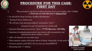
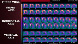
1
MYOCARDIAL PERFUSION
IMAGING
Case Study – Nuclear Cardiology Center
PSMMC
Prepared by:
Al-waleed Mohammed Al-hussain
Supervised by:
Mohammed Abdullah Alshehri
OUTLINES
• Introduction
• Indication
• Instrumentation
• Materials
• Procedure
• Results
• Conclusion
• Reference
INTRODUCTION
MYOCARDIAL PERFUSION IMAGING
- This test is designed to evaluate regional myocardial perfusion under
Rest and Stress parts, to define regional myocardial perfusion reserve.
INDICATION
• Detection or evaluate coronary artery disease.
• Evaluation for coronary bypass surgery or angioplasty.
• Detection of myocardial Infarction or Ischemia.
• Shortness of Breath
• Chest Pain
• History/Family History of heart disease
INSTRUMENTATION
the scan was done on
- CardioMD Gamma Camera 
• 14*8 Rectangular FOV
• small footprint
• Low energy High resolution collimator.
• Window of 20% at 140 KeV.
• Nb. Of frames in RR: 8
• Nb. Projection: 32
• Start angle: -45 degrees
• Time for 1st projection (in seconds): 25
(a) Philips (ADAC) CardioMD Gamma Camera
MATERIALS
Materials for this case:
• Radioactive : Technetium (99mTc) // t1⁄2 : 6hours // energy: 140Kev
• Pharmaceutical (Kit) : Myoview // Expired: 8 hours after use
• Radiopharmaceutical was studied: for MPI [Technetium (99mTc)-Myoview]
• Stress Room (Pharmacological) : Persantine
CASE STUDY
• Full history:
Male patient,
54 years old,
130kg weight,
known HT,
Morbid obesity,
Renal impairment (Kidney failure),
Nearly diagnosed heart failure.
PROCEDURE FOR THIS CASE:
FIRST DAY
1. The procedure was done in 2days because he is over weight 120+ (130kg)
in First day we will take him in (Stress Part)
• he should be Stop 24 hours (Caffine/Tea/Smoke)
• fasting 6 hours before test
• We will give him pharmacological ( persantine, why? )
because he can’t do exercise in a tread mill.
• Injection Radiopharmaceutical / Dose : (99mTc)-Myoview / 600 MBq
• Injection of radiopharmaceutical two minute after persantine end
(persantine given within 4 mins).
• After 15 minutes we can start scan
• Place patient supine and arms above of head. (especially in left arm) 
• Connect patient with ECG ( So we can use GatedSPECT)
• Scanning time : 7.12mins (b) Philips CardioMD
(patient position)
PROCEDURE FOR THIS CASE:
SECONDE DAY
2. Second day we will take him in (Rest Part)
• Fasting 2 hours before test
• Injection Radiopharmaceutical / Dose: (99mTc)-Myoview / 600 MBq
• After 40 minutes we can start scan
• Place patient supine and arms above of head (especially in left arm)
• Connect patient with ECG ( So we can use GatedSPECT)
• Scanning time : 7.12mins
RESULTS
THREE VIEW:
SHORT
AXIS
HORIZONTAL
AXIS
VERTICAL
AXIS
DIAGNOSIS/FINDINGS
• Diagnosis:
- Ejection Fraction:
20%
- Tomographic images at stress showed:
patchy uptake, reduced perfusion in apex, inferolateral segment.
- Tomographic images at rest showed:
slight improvement at above area.
• Findings(conclusion):
Dilated LV (left ventricle)
No significant ischemia
No infarction
Picture of DCM (Dilated cardiomyopathy)
CONCLUSION
- In Cardiac Perfusion Studies we identify defect in the myocardium by the intensity
and the perfusion which is reflected as the colours the brightness colouring the most
perfusion part will be.
REFRENCE
1. PSMMC, www.psmmc.med.sa (results images)
2. Philips (ADAC), www.ais-nuclear.com/nuclear-medicine-cameras (a)
3. Philips CardioMD, www.maiedge.com/product (b)
THANK YOU

Nuclear Medicine - MPI - Case Study

  • 1.
    1 MYOCARDIAL PERFUSION IMAGING Case Study– Nuclear Cardiology Center PSMMC Prepared by: Al-waleed Mohammed Al-hussain Supervised by: Mohammed Abdullah Alshehri
  • 2.
    OUTLINES • Introduction • Indication •Instrumentation • Materials • Procedure • Results • Conclusion • Reference
  • 3.
    INTRODUCTION MYOCARDIAL PERFUSION IMAGING -This test is designed to evaluate regional myocardial perfusion under Rest and Stress parts, to define regional myocardial perfusion reserve.
  • 4.
    INDICATION • Detection orevaluate coronary artery disease. • Evaluation for coronary bypass surgery or angioplasty. • Detection of myocardial Infarction or Ischemia. • Shortness of Breath • Chest Pain • History/Family History of heart disease
  • 5.
    INSTRUMENTATION the scan wasdone on - CardioMD Gamma Camera  • 14*8 Rectangular FOV • small footprint • Low energy High resolution collimator. • Window of 20% at 140 KeV. • Nb. Of frames in RR: 8 • Nb. Projection: 32 • Start angle: -45 degrees • Time for 1st projection (in seconds): 25 (a) Philips (ADAC) CardioMD Gamma Camera
  • 6.
    MATERIALS Materials for thiscase: • Radioactive : Technetium (99mTc) // t1⁄2 : 6hours // energy: 140Kev • Pharmaceutical (Kit) : Myoview // Expired: 8 hours after use • Radiopharmaceutical was studied: for MPI [Technetium (99mTc)-Myoview] • Stress Room (Pharmacological) : Persantine
  • 7.
    CASE STUDY • Fullhistory: Male patient, 54 years old, 130kg weight, known HT, Morbid obesity, Renal impairment (Kidney failure), Nearly diagnosed heart failure.
  • 8.
    PROCEDURE FOR THISCASE: FIRST DAY 1. The procedure was done in 2days because he is over weight 120+ (130kg) in First day we will take him in (Stress Part) • he should be Stop 24 hours (Caffine/Tea/Smoke) • fasting 6 hours before test • We will give him pharmacological ( persantine, why? ) because he can’t do exercise in a tread mill. • Injection Radiopharmaceutical / Dose : (99mTc)-Myoview / 600 MBq • Injection of radiopharmaceutical two minute after persantine end (persantine given within 4 mins). • After 15 minutes we can start scan • Place patient supine and arms above of head. (especially in left arm)  • Connect patient with ECG ( So we can use GatedSPECT) • Scanning time : 7.12mins (b) Philips CardioMD (patient position)
  • 9.
    PROCEDURE FOR THISCASE: SECONDE DAY 2. Second day we will take him in (Rest Part) • Fasting 2 hours before test • Injection Radiopharmaceutical / Dose: (99mTc)-Myoview / 600 MBq • After 40 minutes we can start scan • Place patient supine and arms above of head (especially in left arm) • Connect patient with ECG ( So we can use GatedSPECT) • Scanning time : 7.12mins
  • 10.
  • 11.
  • 13.
    DIAGNOSIS/FINDINGS • Diagnosis: - EjectionFraction: 20% - Tomographic images at stress showed: patchy uptake, reduced perfusion in apex, inferolateral segment. - Tomographic images at rest showed: slight improvement at above area. • Findings(conclusion): Dilated LV (left ventricle) No significant ischemia No infarction Picture of DCM (Dilated cardiomyopathy)
  • 14.
    CONCLUSION - In CardiacPerfusion Studies we identify defect in the myocardium by the intensity and the perfusion which is reflected as the colours the brightness colouring the most perfusion part will be.
  • 15.
    REFRENCE 1. PSMMC, www.psmmc.med.sa(results images) 2. Philips (ADAC), www.ais-nuclear.com/nuclear-medicine-cameras (a) 3. Philips CardioMD, www.maiedge.com/product (b)
  • 16.