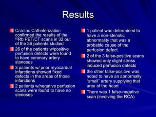
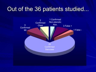
Comparison of  82 Rb PET/CT Myocardial Perfusion Imaging with Cardiac Catheterization by H. Backhaus, C. Johnson St. Joseph’s Hospital, Marshfield, WI
Background St. Joseph’s Hospital is a 500+ bed facility located in north central Wisconsin.  It serves the primarily rural population of Northern and Central Wisconsin plus Upper Michigan St. Joseph’s has one of the largest Nuclear Medicine departments in the region It is also the only facility in the State of Wisconsin to utilize  82 Rb PET/CT imaging
 
Myocardial Perfusion Imaging The primary modality used for myocardial perfusion studies at St. Joseph’s Hospital is  99m Tc-sestamibi SPECT/CT imaging However, for all patients weighing over 250 lbs or with a BMI  ≥ 35  82 Rb PET/CT imaging is preferred by our physicians
Why? 82 Rb emits a higher energy gamma than  99m Tc The higher energy gamma is more able to penetrate excess tissue This results in more counts reaching the camera Ultimately, higher quality diagnostic images are obtained
Plus… 82 Rb PET/CT imaging takes only about 1 hour to complete ( 99m Tc-sestamibi is a 2-day protocol for obese patients) This results in less time spent at the hospital by the patients – making them much happier And, this allows more patients to be seen in a shorter amount of time – resulting in higher throughput
Objectives The purpose of this study was to determine the diagnostic quality of  82 Rb PET/CT myocardial perfusion imaging on obese patients  This was done by comparing the Nuclear Medicine physician’s interpretations of these images to the results of cardiac catheterizations performed on the same patients
Methods Out of all patients seen between January 2008 & March 2009 thirty-six  (25 male, 11 female) underwent both  82 Rb PET/CT MPI & cardiac catheterization  All patients were imaged on a Philips Gemini GXL PET/CT system
Methods (cont.) Surview Topogram  CT Transmission Scan Injected 30-50 mCi  82 Rb over 30-60 sec 6 min of Gated Rest Images Adenosine Stress (over 7 min) Injected 30-50 mCi  82 Rb over 30-60 sec (2 min after start of adenosine injection)  6 min of Gated Stress Images CT Transmission Scan
Methods (cont.) Images were processed by technologist & read by either a Nuclear Medicine physician or Nuclear Cardiologist All 36 patients in this study were referred to & received Cardiac Catheterization following their  82 Rb PET/CT scans Physicians’ reports from the Nuclear Medicine procedures were compared to the physicians’ reports from the Catheterizations in order to determine any correlation between the findings
Results Cardiac Catheterization confirmed the results of the  82 Rb PET/CT scans in 32 out of the 36 patients studied 26 of the patients w/positive perfusion defects were found to have coronary artery stenoses 3 patients w/ prior myocardial infarctions showed fixed defects in the areas of those infarctions  2 patients w/negative perfusion scans were found to have no stenoses 1 patient was determined to have a non-stenotic abnormality that was a probable cause of the perfusion defect 2 of the 3 false-positive scans showed only slight stress induced perfusion defects  the other false-positive was noted to have an abnormally “small” artery supplying that area of the heart There was 1 false-negative scan (involving the RCA)
Results…
Out of the 36 patients studied... 26 Confirmed Stenoses 3  Confirmed MI 2 Confirmed Clear 1 Confirmed Non-stenotic Abn. 3 False + 1 False -
Example #1 (attenuation corrected images) - No definitive fixed defects  - Moderate size, mild, stress-induced defect of mid lateral wall
Example #2 (attenuation corrected images) - Confirmation of known fixed defect - Prior MI to apex
Example #3 (attenuation corrected images) - No ischemia
Example #4 Attenuation corrected vs. non attenuation corrected - Corrected images show more realistic perfusion - Uncorrected images show fixed defect in the inferior wall (attenuation corrected)  (uncorrected)
Results (cont.) 100% of the perfusion defects to locations in the myocardium supplied by the LCX artery (lateral wall) were verified by catheterization 89% of the perfusion defects involving areas of the heart typically supplied by the LAD artery (anterior wall & apex) were confirmed to be LAD occlusions by cardiac catheterization 79% of the perfusion defects to areas of the heart supplied by the RCA (inferior wall) were confirmed by cardiac catheterization
Conclusions Based on these results, it seems more difficult to make an accurate diagnosis of a small perfusion defect to an area of the myocardium that is partially blocked by a solid organ such as the liver (count attenuation) Attenuation correction software does help improve the ability to differentiate true perfusion defects from organ attenuation However, smaller perfusion defects can sometimes be masked Nevertheless, 89% of the perfusion defect diagnoses made using the  82 Rb PET/CT scans correlated completely with the findings of cardiac catheterization
Conclusions (cont.) 82 Rb MPI can also be a valuable tool for detecting non-stenotic abnormalities that have a deleterious effect on the blood flow to a patient’s heart In one case an RCA that originated from the circumflex system instead of the aorta was found In another, an uncharacteristically small RCA supplying the patient’s inferior wall was found
Conclusions (cont.) So, overall,  82 Rb PET/CT myocardial perfusion imaging is an accurate and efficient diagnostic tool for detecting life-threatening pathologies in obese patients exhibiting symptoms of CAD And, this makes everyone happy!
 
Questions?

Snm 09 Presentation Hkb

  • 1.
    Comparison of 82 Rb PET/CT Myocardial Perfusion Imaging with Cardiac Catheterization by H. Backhaus, C. Johnson St. Joseph’s Hospital, Marshfield, WI
  • 2.
    Background St. Joseph’sHospital is a 500+ bed facility located in north central Wisconsin. It serves the primarily rural population of Northern and Central Wisconsin plus Upper Michigan St. Joseph’s has one of the largest Nuclear Medicine departments in the region It is also the only facility in the State of Wisconsin to utilize 82 Rb PET/CT imaging
  • 3.
  • 4.
    Myocardial Perfusion ImagingThe primary modality used for myocardial perfusion studies at St. Joseph’s Hospital is 99m Tc-sestamibi SPECT/CT imaging However, for all patients weighing over 250 lbs or with a BMI ≥ 35 82 Rb PET/CT imaging is preferred by our physicians
  • 5.
    Why? 82 Rbemits a higher energy gamma than 99m Tc The higher energy gamma is more able to penetrate excess tissue This results in more counts reaching the camera Ultimately, higher quality diagnostic images are obtained
  • 6.
    Plus… 82 RbPET/CT imaging takes only about 1 hour to complete ( 99m Tc-sestamibi is a 2-day protocol for obese patients) This results in less time spent at the hospital by the patients – making them much happier And, this allows more patients to be seen in a shorter amount of time – resulting in higher throughput
  • 7.
    Objectives The purposeof this study was to determine the diagnostic quality of 82 Rb PET/CT myocardial perfusion imaging on obese patients This was done by comparing the Nuclear Medicine physician’s interpretations of these images to the results of cardiac catheterizations performed on the same patients
  • 8.
    Methods Out ofall patients seen between January 2008 & March 2009 thirty-six (25 male, 11 female) underwent both 82 Rb PET/CT MPI & cardiac catheterization All patients were imaged on a Philips Gemini GXL PET/CT system
  • 9.
    Methods (cont.) SurviewTopogram CT Transmission Scan Injected 30-50 mCi 82 Rb over 30-60 sec 6 min of Gated Rest Images Adenosine Stress (over 7 min) Injected 30-50 mCi 82 Rb over 30-60 sec (2 min after start of adenosine injection) 6 min of Gated Stress Images CT Transmission Scan
  • 10.
    Methods (cont.) Imageswere processed by technologist & read by either a Nuclear Medicine physician or Nuclear Cardiologist All 36 patients in this study were referred to & received Cardiac Catheterization following their 82 Rb PET/CT scans Physicians’ reports from the Nuclear Medicine procedures were compared to the physicians’ reports from the Catheterizations in order to determine any correlation between the findings
  • 11.
    Results Cardiac Catheterizationconfirmed the results of the 82 Rb PET/CT scans in 32 out of the 36 patients studied 26 of the patients w/positive perfusion defects were found to have coronary artery stenoses 3 patients w/ prior myocardial infarctions showed fixed defects in the areas of those infarctions 2 patients w/negative perfusion scans were found to have no stenoses 1 patient was determined to have a non-stenotic abnormality that was a probable cause of the perfusion defect 2 of the 3 false-positive scans showed only slight stress induced perfusion defects the other false-positive was noted to have an abnormally “small” artery supplying that area of the heart There was 1 false-negative scan (involving the RCA)
  • 12.
  • 13.
    Out of the36 patients studied... 26 Confirmed Stenoses 3 Confirmed MI 2 Confirmed Clear 1 Confirmed Non-stenotic Abn. 3 False + 1 False -
  • 14.
    Example #1 (attenuationcorrected images) - No definitive fixed defects - Moderate size, mild, stress-induced defect of mid lateral wall
  • 15.
    Example #2 (attenuationcorrected images) - Confirmation of known fixed defect - Prior MI to apex
  • 16.
    Example #3 (attenuationcorrected images) - No ischemia
  • 17.
    Example #4 Attenuationcorrected vs. non attenuation corrected - Corrected images show more realistic perfusion - Uncorrected images show fixed defect in the inferior wall (attenuation corrected) (uncorrected)
  • 18.
    Results (cont.) 100%of the perfusion defects to locations in the myocardium supplied by the LCX artery (lateral wall) were verified by catheterization 89% of the perfusion defects involving areas of the heart typically supplied by the LAD artery (anterior wall & apex) were confirmed to be LAD occlusions by cardiac catheterization 79% of the perfusion defects to areas of the heart supplied by the RCA (inferior wall) were confirmed by cardiac catheterization
  • 19.
    Conclusions Based onthese results, it seems more difficult to make an accurate diagnosis of a small perfusion defect to an area of the myocardium that is partially blocked by a solid organ such as the liver (count attenuation) Attenuation correction software does help improve the ability to differentiate true perfusion defects from organ attenuation However, smaller perfusion defects can sometimes be masked Nevertheless, 89% of the perfusion defect diagnoses made using the 82 Rb PET/CT scans correlated completely with the findings of cardiac catheterization
  • 20.
    Conclusions (cont.) 82Rb MPI can also be a valuable tool for detecting non-stenotic abnormalities that have a deleterious effect on the blood flow to a patient’s heart In one case an RCA that originated from the circumflex system instead of the aorta was found In another, an uncharacteristically small RCA supplying the patient’s inferior wall was found
  • 21.
    Conclusions (cont.) So,overall, 82 Rb PET/CT myocardial perfusion imaging is an accurate and efficient diagnostic tool for detecting life-threatening pathologies in obese patients exhibiting symptoms of CAD And, this makes everyone happy!
  • 22.
  • 23.