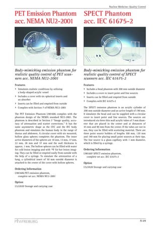
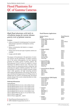
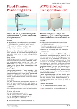
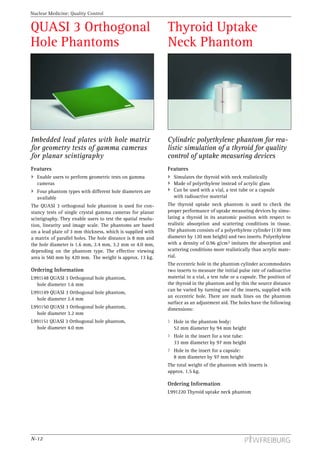
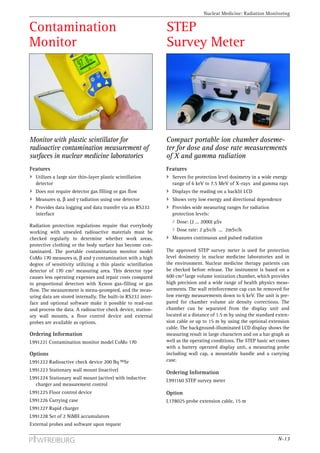
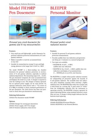
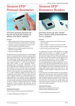
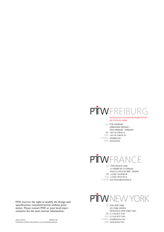
2. Nuclear Medicine: Introduction
Let us introduce ourselves
PTW-Freiburg is an internationally operating company, manufacturing and marketing specialized dosimetry and quality
control equipment for the medical radiology market. Founded in 1922, the company is located in Freiburg on the western
side of the famous Black Forest mountains in southwestern Germany.
Our History
In 1922, twenty-seven years after Roentgen discovered the X-rays, Professor Hammer from the Physics Institute of
Freiburg University founded PTW to produce and market his development of an X-ray dosemeter based on the electrostatic
relais, a revolutionary new electromechanical component for measuring very small electrical charges.
In 1927, Dr. Herbert Pychlau took over the company and developed it during four decades into an internationally recognized
manufacturer of quality dosemeters for medical radiology.
PTW has developed and manufactured many generations of up-to-date products over the years, based on the newest
technology. The company has grown continuously.
Today, 140 employees achieve an annual turnover of 16 Million EUR. A new building has been constructed just recently,
which enlarges the total production area to 3,500 m2.
Our Operations
PTW-Freiburg designs, develops, manufactures and distributes high quality dosimetry and QC equipment mainly for use in
the medical field, especially in radiation therapy, diagnostic radiology and nuclear medicine. The development and
production of mechanical, electronic and software components are all done in house. We have a well-equipped and modern
workshop with 6 CNC machines. Our products, especially the PTW ionization chambers, are well known throughout the
world and are recognized for their workmanship and high level of quality. PTW-Freiburg is the market leader in its major
product lines.
The PTW distribution is organized internationally. A number of exclusive PTW representations are established in many coun-
tries around the world. In 1995, the PTW New York Corporation was established to improve our service to customers in America
and to distribute and service PTW products all over the American continent. In January 2002, PTW-France was established.
PTW-Freiburg runs an accredited secondary standard dosimetry laboratory. We perform radiological calibrations for
dosemeters used in radiation therapy, diagnostic radiology and health physics, which are directly traceable to the primary
standard. Our calibration lab provides a complete range of radiological calibrations from low X-ray energies up to 60Co
and from low to high dose rates.
We cooperate closely with official public agencies worldwide, and we participate actively in national and international work
groups for the standardization of devices and procedures for dose measurement and quality control in medical
radiology.
Our Company Philosophy
From the very beginning, PTW-Freiburg’s products have been optimized for practicability, precision and quality. All
dosemeters are thoroughly tested in radiation beams, and calibration certificates are attached to every dosemeter.
Thanks to the constant improvement of our quality standards and internal procedures, our quality assurance system was cer-
tified in 1995 as compliant with the standards ISO 9001 and EN 46001. Our medical products are CE marked according to the
European Medical Device Directive and they are FDA 510(k) approved. The calibration laboratories are part of the quality
management system of PTW-Freiburg.
We at PTW-Freiburg are proud to work with our customers as a competent and reliable partner. We provide you with prod-
ucts, support and service in line with good manufacturing practice, with the added touch of personal attention from our
motivated staff. We constantly strive to improve the international presence and reputation of PTW-Freiburg.
We intend to continue our efforts in the future as we did in the past to develop the most useful, most reliable and highest
quality dosimetry products possible. Our commitment to maximum customer satisfaction will remain unchanged.
For more details please visit our websites at http://www.ptw.de (PTW-Freiburg) and http://www.ptwny.com (PTW New York).
N-02
3. Nuclear Medicine: Content
Content
Radiation Measurement and
Quality Control in Nuclear Medicine N-04
Introduction
Isotope Calibrators N-05
Activity Measurement
CURIEMENTOR 3 Isotope Calibrator N-05
CURIEMENTOR 4 Isotope Calibrator N-05
CURIEMENTOR Printer N-06
Ionization Chamber Lead Shielding N-06
Quality Control N-07
QC Phantoms
PET Cylinder Phantom
acc. NEMA NU2-2001 N-07
PET Sensitivity Phantom
acc. NEMA NU2-2001 N-07
PET Emission Phantom acc. CEC Project N-08
PET Emission Phantom acc. IEC 61675-1 N-08
PET Emission Phantom
acc. NEMA NU2-2001 N-09
SPECT Phantom acc. IEC 61675-2 N-09
Flood Phantoms for QC of Gamma Cameras N-10
Flood Phantom Applications N-10
Flood Phantom Positioning Carts N-11
ATW3 Shielded Transportation Cart N-11
QUASI 3 Orthogonal Hole Phantoms N-12
Thyroid Uptake Neck Phantom N-12
Radiation Monitoring Equipment N-13
Radiation Monitoring Equipment
Contamination Monitor N-13
STEP Survey Meter N-13
Personal Dosemeters N-14
N-03
4. Nuclear Medicine: Introduction
Radiation Measurement
and Quality Control in
Nuclear Medicine
Introduction
General Radiation Monitoring
Since the discovery of X-rays in 1895 by Roentgen and Radiation Monitoring is another important measuring
uranium radioactivity in 1896 by Becquerel, the use of task in nuclear medicine laboratories:
ionizing radiation in medicine has undergone fascinating
Surfaces must be checked regularly with respect to
development. Nuclear medicine started in 1937 with the
radioactive contamination using a contamination
first clinical use of “artificial radioactivity” for treating a
monitor.
patient with leukemia. Today there are nearly one hundred
different nuclear medicine procedures that provide infor- Radiation levels, for example behind shielding or in
mation about virtually every major organ system within the vicinity of therapy patients before release, must
the body. Modern computer based techniques such as PET be measured using a survey meter.
(Positron Emission Tomography) and SPECT (Single The radiation exposition to the staff must be
Photon Emission Computed Tomography) offer methods measured using personal radiation monitors.
for diagnostic imaging that are unique by documenting
dynamic organ function and structure. Quality Control
PET is rapidly becoming a major diagnostic imaging The complex techniques of modern nuclear medicine
modality. PET images demonstrate the chemistry of organs imaging systems require sophisticated test equipment for
and other tissues such as tumors. A radiopharmaceutical, regular quality control. The test tools must be adapted to
especially FDG (fluorodeoxyglucose), marked with the the different imaging techniques according to interna-
positron emitting isotope 18F, is injected into the patient’s tional standards. Special phantoms for quality control
body and its emissions are measured by a PET scanner. procedures of PET and SPECT scanners, gamma cameras
and uptake measuring equipment are available.
SPECT, like PET, acquires information on the concentration
of radionuclides introduced to patient’s body by rotating a Delivery
photon detector array around the body. SPECT scanners The products in this catalog comply with international
offer less imaging resolution compared to PET, but the standards and regulations. Medical and electric products
availability of radiopharmaceuticals, particularly for the are supplied with the CE mark. All deliveries come with a
brain and head and the practical and economic aspects manual in English.
make this mode of emission tomography attractive for
clinical studies of the brain. International Standards and Regulations in Nuclear Medicine
Scintigraphy with planar gamma cameras and with thyroid IEC 60789 Characteristics and test conditions of radionuclide imaging
uptake measuring equipment are further helpful methods devices – Anger type gamma camera
in nuclear medicine. IEC 61145 Calibration and usage of ionization chamber systems for assay
Besides the diagnostic procedures more and more therapy of radionuclides
methods are used in nuclear medicine. From the beginning, IEC 61675-1 Radionuclide imaging devices – Characteristics and test
patients with thyroid cancer were treated with 131I. Quite a conditions – Part 1: Positron emission tomographs
number of further therapy procedures are available today. IEC 61675-2 Radionuclide imaging devices – Characteristics and test
conditions – Part 2: Single photon emission computed tomographs
Nuclear medicine uses very small amounts of radioactive
materials or radiopharmaceuticals to diagnose and treat dis- IEC 61675-3 Radionuclide imaging devices – Characteristics and test
conditions – Part 3: Gamma camera-based whole-body imaging systems
eases. Nevertheless the radiopharmaceuticals emit ionizing
radiation to which patients and operating staff can be National Electrical Manufacturers Association, USA
NEMA Standards Publication NU2-2001
exposed unintentionally. To avoid unnecessary irradiation Performance Measurements of Positron Emission Tomographs
and to fulfill international regulations, radiation measure-
CEC Concerted Action Project,
ment and quality control procedures for determination, Methodologies for Performance Evaluation of Positron Emission
recording and evaluation are mandatory in nuclear medicine. Tomographs, J Nucl Biol Med; 35: 141-157
European Council Directive 97/43/EURATOM of 30 June 1997 on health
Radiation Measurement protection of individuals against the danger of ionizing radiation in rela-
Activity Measurement tion to medical exposure
Nuclear medicine procedures use many different radioac- ICRP 60, International Commission on Radiation Protection, Report No. 60
tive isotopes for radiation diagnostics and for therapy. The
amount of radioactivity has to be determined before it is
applied to a patient. The isotope calibrators have precisely
to measure the radioactivity of gamma and beta emitting
isotopes with different energies for high quality imaging
and for applying the right amount of radiation to treat dis-
ease. The isotope calibrators should allow easy and fast
operation in routine work as well as quick and effective
cleaning in case of contamination.
N-04
5. Nuclear Medicine: Isotope Calibrators
CURIEMENTOR® 3 CURIEMENTOR® 4
Isotope Calibrator Isotope Calibrator
Standard isotope calibrator for radio- Deluxe isotope calibrator for radionuclide
nuclide activity measurement in nuclear activity measurement in nuclear medicine
medicine and intravascular brachytherapy and intravascular brachytherapy
Features Features
Combines modern technology with user friendly, State-of-the-art development of an isotope calibrator
straight-forward operation Stores calibration factors for all isotopes used in
Stores calibration factors for all isotopes used in nuclear medicine with direct access to thirty of them
nuclear medicine with direct access to five of them Displays measuring results continuously for isotopes
Features auto-start, auto-print and auto-reset functions with very short half-value time
Complies with IEC 61145 Provides data logging and data transfer via an RS232
interface
The CURIEMENTOR 3 isotope calibrator enables users to Complies with IEC 61145
measure activity of radioactive isotopes as used for all
medical applications in diagnostic and therapeutic nuclear The CURIEMENTOR 4 isotope calibrator is a highly
medicine and in intravascular brachytherapy. Great impor- sophisticated instrument for activity measurements in
tance has been attached to the design of the instrument to nuclear medicine including PET. It includes and exceeds
focus on the needs of operators. The display and keyboard the features of the CURIEMENTOR 3 isotope calibrator.
panel with its flat keys are specially designed for intuitive The user has direct access to 30 different isotope calibra-
handling and easy cleaning in case of contamination. The tions. When measuring isotopes with rapid decay, the dis-
measuring results can either be displayed in Bq, Ci or in A. played measuring values are updated each half second,
The pressurized well-type ionization chamber has nearly which provides continuous display. 18F activities of up to
4π measuring geometry and connects to the display unit 25 GBq for PET applications can be measured. If higher
by a 1.5 m connection cable, which makes it possible to activities should be measured, a special ionization cham-
shield the chamber separately. A radioactive check device ber can be supplied. Up to 140 measuring results includ-
is available to check the proper performance of the ing administrative data can be stored internally. The fea-
device regularly. The display unit is supplied with an tures with respect to the chamber design, check device,
RS232 interface. A printer can be connected optionally. RS232 interface and printer option are identical with the
The printing mode can be set for labels or endless-paper CURIEMENTOR 3 features.
on the CURIEMENTOR display unit.
Ordering Information
L981129 CURIEMENTOR 4 isotope calibrator 115/230 V
L981962 CURIEMENTOR 3 isotope calibrator 115/230 V incl. display unit, chamber, liner and syringe holder
L981101 CURIEMENTOR 3 isotope calibrator 115/230 V L981130 CURIEMENTOR 4 isotope calibrator 115/230 V
with wall mount display unit with wall mount display unit
L971001 Radioactive check device 3.7 MBq 137Cs, incl. L971001 Radioactive check device 3.7 MBq 137Cs, incl.
shielded container (comply with national regulations!) shielded container (comply with national regulations!)
Option Option
T23365/50 Lead absorber to detect a Mo break-through T23365/50 Lead absorber to detect a Mo break-through
CURIEMENTOR Printer N-06 CURIEMENTOR Printer N-06
Ionization Chamber Lead Shielding N-06 Ionization Chamber Lead Shielding N-06
N-05
6. Nuclear Medicine: Isotope Calibrators
CURIEMENTOR® Ionization Chamber
Printer Lead Shielding
Thermal printer for printing Sectional lead rings for radiation shiel-
CURIEMENTOR measuring results ding of the CURIEMENTOR ionization
on labels chamber
Features Features
Prints CURIEMENTOR results on label paper Makes it possible to shield the CURIEMENTOR cham-
Printing is automatically controlled by the ber separately
CURIEMENTOR isotope calibrator Protects measurements against falsification by exter-
Connects to the CURIEMENTOR 3 or nal radioactive sources
CURIEMENTOR 4 display units Protects operating staff against irradiation from
radioactive sources inside the ion chamber
To print the measuring data of the CURIEMENTOR 3 and
CURIEMENTOR 4 isotope calibrators on paper labels, the The separation of the CURIEMENTOR ionization chamber
suitable CITIZEN type printer can be connected to the dis- and the display unit makes it possible to fit a lead shield-
play unit via a serial interface. The user has the possibility ing around the chamber. This concept ensures optimum
to print the measuring results including administrative radiation protection. The operating staff are not exposed
data on self-adhesive stickers of a thermal paper roll. to radiation during the measurement of activity placed in
The width of the paper roll is 58 mm. The label size is the chamber well, and other radioactive sources in the
50 mm x 25 mm. The printer is connected to the mains via vicinity of the chamber cannot falsify the measurement.
a power supply, which is included in the delivery.
The lead shielding model T23365/60-65 consists of a steel
Ordering Information carrier ring, 15 mm high with four feet and three sectional
L991056 CITIZEN Thermal printer lead rings. The dimensions of the assembled shielding are:
including power supply (100 ... 230) V Outside diameter: 250 mm
L178048 Connection cable between printer and Height: 240 mm
CURIEMENTOR isotope calibrator Lead wall thickness: 20 mm
L502076 Roll with 750 self-adhesive stickers Weight: approx. 31 kg
CURIEMENTOR 3 Isotope Calibrator N-05 An additional sectional lead ring model T23365/60 of
CURIEMENTOR 4 Isotope Calibrator N-05 70 mm height can be used to extend the shielding to
a height of 310 mm.
Ordering Information
T23365/60-62 CURIEMENTOR lead shield for the
shielding of the well-type ion chamber
Option
T23365/60 CURIEMENTOR lead shield, ring
CURIEMENTOR 3 Isotope Calibrator N-05
CURIEMENTOR 4 Isotope Calibrator N-05
N-06
7. Nuclear Medicine: Quality Control
PET Cylinder Phantom PET Sensitivity Phantom
acc. NEMA NU2-2001 acc. NEMA NU2-2001
Cylinder phantom with line source tube Sensitivity tube phantom with line source
for quality control of positron emission tube for quality control of positron
tomographs (PET scanners) emission tomographs (PET scanners)
Features Features
Enables users to perform scatter fraction, count loss Enables users to measure sensitivity on PET scanners
and random measurements on PET scanners Consists of five metal sleeves with different diameters
Consists of a long polyethylene cylinder with an and a line source tube
eccentric line source tube Complies with Section 5 of NEMA standard NU2-2001
Complies with Section 4 of NEMA standard NU2-2001
The purpose of the test procedure based on the PET sensi-
One purpose of the test procedure based on the PET cylin- tivity phantom is to measure the sensitivity or ability of
der phantom is to measure the relative system sensitivity positron emission tomographs to detect positrons. The
of positron emission tomographs to scattered radiation. phantom used for this purpose is a set of five metal tubes
Another purpose is to measure the effects of system dead- of 700 mm length and 2.5 mm wall thickness each.
time and the generation of random events at several levels The outside diameters are 9.5 mm, 12.7 mm, 15.9 mm,
of source activity. The phantom used for these purposes is 19.1 mm and 22.2 mm. A plastic tube homogeneously
a polyethylene cylinder of 700 mm length and 200 mm filled with 18F liquid is inserted for the measurement. This
outside diameter. A plastic tube homogeneously filled line source has an outside diameter of approx. 5 mm and a
with 18F liquid is inserted for the measurement at a dis- length of approx. 800 mm. Successive measurements are
tance of 45 mm parallel to the central axis. This line made by accumulating the sleeve wall thickness with the
source has an outside diameter of approx. 5 mm and a uniform line source surrounded by known absorbers.
length of approx. 800 mm. The measurement setup, data From these measurements, the sensitivity without
collection and analysis are described in Section 4 of the absorbers can be extrapolated to arrive at an attenuation
NEMA standard NU2-2001. free measurement. The measurement setup, data collec-
tion and analysis are described in section 5 of the
Ordering Information NEMA standard NU2-2001.
L991204 PET Cylinder phantom acc. NEMA NU2-2001,
Section 4 Ordering Information
L991205 PET Sensitivity phantom acc. NEMA NU2-2001,
Section 5
N-07
8. Nuclear Medicine: Quality Control
PET Emission Phantom PET Emission Phantom
acc. CEC Project acc. IEC 61675-1
Body-mimicking emission phantom for Body-mimicking emission phantom for
realistic quality control of PET scanners realistic quality control of PET scan-
acc. CEC Concerted Action Project ners acc. IEC 61675-1
Features Features
Simulates realistic conditions by utilizing Simulates realistic conditions by utilizing body-
body-shaped acrylic vessels shaped acrylic vessels
Includes covers with various inserts Includes covers with various inserts
Inserts can be filled and emptied from outside Inserts can be filled and emptied from outside
Complies with the European PET Instrumentation Program Complies with IEC 61675-1
The PET emission phantom L981602 was published by a The international standard IEC 61675-1 adopted the
task group of the Commission of the European Community proven concept of the PET emission phantom according to
(CEC Concerted Action Project). The phantom has been the European PET program. The major components are
developed in line with the philosophy that assessment of a identical with the PET phantom based on the European
clinical measuring system should always be made under standard. They realistically simulate a patient during a
the most realistic conditions possible to imitate the PET examination in the same manner and fulfill the
patient’s influence as an absorbing and scattering medium. requirements of the IEC standard.
One of the circular covers with different inserts may be The package includes:
mounted to the head phantom H or to the body phantom B,
Body phantom B with two arms attached
with a water tight seal in all cases. The possibility of filling
and emptying the inserts and phantom from outside is one Head phantom H
of the main advantages of this phantom. Cover D with three cylinders, each 50 mm in diameter
The package includes: for different absorbing material
Body phantom B with two arms attached Cover E with six spheres of 10 mm, 13 mm, 17 mm,
22 mm, 28 mm and 37 mm inner active diameter
Head phantom H
Blind cover G without inserts
Cover D with three cylinders, each 50 mm in diameter
for different absorbing material Cover M with point, line and scatter sources
Cover E with six spheres of 10 mm, 13 mm, 17 mm, Carrying and storage case
22 mm, 28 mm and 37 mm inner active diameter
Ordering Information
Blind cover G without inserts,
L981605 PET emission phantom,
Cover M with point, line and scatter sources
complete set acc. IEC 61675-1
Cover R with a rod source of 21 mm inner diameter
Heart phantom C
Carrying and storage case
Ordering Information
L981602 PET emission phantom,
complete set acc. CEC project
N-08
9. Nuclear Medicine: Quality Control
PET Emission Phantom SPECT Phantom
acc. NEMA NU2-2001 acc. IEC 61675-2
Body-mimicking emission phantom for Body-mimicking emission phantom for
realistic quality control of PET scan- realistic quality control of SPECT
ners acc. NEMA NU2-2001 scanners acc. IEC 61675-2
Features Features
Simulates realistic conditions by utilizing Includes a head phantom with 200 mm outside diameter
a body-shaped acrylic vessel Includes a cover to insert point and line sources
Includes a cover with six spherical inserts and Inserts can be filled and emptied from outside
an absorber
Complies with IEC 61675-2
Inserts can be filled and emptied from outside
Complies with Section 7 of NEMA NU2-2001 The SPECT emission phantom is an acrylic cylinder of
200 mm outside diameter and an active length of 190 mm.
The PET Emission Phantom L981606 complies with the It simulates the head and can be supplied with a circular
phantom design of the NEMA standard NU2-2001. The cover to insert point and line sources. The sources are
phantom is described in Section 7 “Image quality, accu- introduced via three thin wall acrylic tubes of 7 mm diam-
racy of attenuation and scatter corrections.” It has the eter that are placed in the center and at distances of
same asymmetric shape as the CEC and the IEC body 45 mm and 90 mm from the center. If the tubes are not in
phantom and simulates the human body in the range of use, they can be filled with scattering material. There are
thorax and abdomen. A circular cover with six mounted, three point source holders of lengths 100 mm, 120 mm
hollow glass spheres completes the phantom. The inner and 140 mm for placing small point sources at their tips.
active diameters of the spheres are 10 mm, 13 mm, 17 mm, The line source is a glass capillary with 1 mm diameter,
22 mm, 28 mm and 37 mm and the wall thickness is which is filled by a syringe.
approx. 1 mm. The hollow spheres can be filled with water
for cold lesion imaging and with 18F for hot lesion imag- Ordering Information
ing. They can be filled or emptied easily from outside with L981607 SPECT emission phantom,
the help of a syringe. To simulate the attenuation of a complete set acc. IEC 61675-2
lung, a cylindrical insert of 50 mm outside diameter is
attached to the center of the cover with hollow spheres. Option
L522020 Storage and carrying case
Ordering Information
L981606 PET emission phantom,
complete set acc. NEMA NU2-2001
Option
L522020 Storage and carrying case
N-09
10. Nuclear Medicine: Quality Control
Flood Phantoms for
QC of Gamma Cameras
Rigid flood phantoms with built-in Flood Phantom Applications
circulation pump for system inhomo- Gamma Camera Flood Phantom
geneity tests of gamma cameras Model Model
Philips (Picker/Marconi)
Features Prism 1000 FP65
Utilize an integrated circulating pump for automatic Prism 1500 FP65/ FP67S
Prism 2000 FP65
fluid mixing to guarantee a homogenous activity Prism 3000 FP47
distribution AXIS FP67S
Reduce contamination risk thanks to a compact, IRIX FP67S
closed system
Philips (ADAC)
Various sizes for the most types of gamma cameras are Forte FP67S
available Skylight FP67S
Vertex FP65
Comply with IEC 60789 Argus Epic FP65
Cardio Epic FP65
The purpose of the phantoms is the generation of correc- Genesys Epic FP65
tion matrices and the performance of regular constancy Solus Epic FP65
tests of the system inhomogeneity, the most important
General Electric
parameter for quality control of gamma cameras. The Millennium MPR FP65
flood phantoms of the FP series are available in various Millennium MPS FP57
dimensions for different detector sizes. They have some Optima-NX FP47
unique features for measuring precisely, saving time and Hawk Eye FP67S
protecting users against radiation exposition: The cuvette
General Electric (Elscint)
wall thickness of 20 mm and a defined extension volume Vari Cam FP67S
ensure stable geometry and high measuring precision SPX Helix FP67S
when using the filled phantom. The activity of the test iso- SPX Cardial FP50
tope is distributed homogeneously in the filling volume of SPX 6 FP67S
distilled water by a built-in circulating pump. The special SPX 4 FP67S
chamber design of the cuvette volume provides for a bub- General Electric (SMV/Sopha)
ble-free field of view. Vision DST XL FP67S
Vision DSX FP67S
Ordering Information Vision FX FP67S
Vision FX 40 FP65
Flood phantoms Vision FX 80 FP65
L991206 Model FP47 for 24 cm x 40 cm field of view Vision DS7 FP57
L991207 Model FP50 for 30 cm x 40 cm field of view
L991208 Model FP57 for 40 cm x 40 cm field of view Siemens
L991209 Model FP65 for 39 cm x 52 cm field of view E.Cam FP67S
Multispect 2 FP67S
L991210 Model FP67 for 40 cm x 54 cm field of view Multispect 3 FP65
L991211 Model FP67S for 40 cm x 54 cm field of view Diacam FP67S
L991212 Power supply 230 V Orbiter FP57
L991213 Power supply (90 ... 264) V
Mediso
X-Ring R FP67S
Flood Phantom Positioning Carts N-11 X-Ring C FP57
ATW3 Shielded Transportation Cart N-11
N-10
11. Nuclear Medicine: Quality Control
Flood Phantom ATW3 Shielded
Positioning Carts Transportation Cart
Mobile trestles to position flood phan- Shielded cart for the storage and
toms in relation to gamma cameras for transport of up to two flood phantoms,
homogeneity tests filled with radionuclide activated water
Features Features
Ensure precise, stable and reproducible positioning of Stores flood phantoms with radioactive filling for
FP flood phantoms in relation to gamma cameras shielded decay after use
The trestles are mobile and handling is easy Includes two compartments for simultaneous storage
Three standard solutions are available of up to two flood phantoms of any type
Charging and discharging can be done from top and
Because of their substantial weight including water from one side.
filling, especially the large flood phantoms require an
appropriate mobile device for precise and reproducible The shielded transportation cart ATW3 is used for easy
positioning. transportation and radiation-protected storage of up to
Model PW67S: two flood phantoms of the FP flood phantom series.
The PW67S positioning cart is a mobile trestle with frame Phantoms up to the size of the FP67S flood phantom can
for positioning the flood phantom model FP67S in rela- be stored after use. This keeps the filling in the flood
tion to the Philips gamma cameras types PRISM 1500, phantoms and the radioactive test isotope can decay over
AXIS, IRIX, Forte and Skylight, the Siemens gamma cam- time. The emptying and refilling of the flood phantoms
era type E.Cam and the Mediso gamma cameras types are avoided. The interior space of the cart is divided into
X-Ring R and X-Ring C. two compartments by a dividing wall, ensuring that the
flood phantoms cannot damage each other in case of dou-
Model APW1:
ble load. Charging and discharging can be done from top
The APW1 positioning cart is a mobile trestle with 3 mm
after having opened the cover flap as well as laterally after
lead shielding for storage and positioning of the FP65
having opened the side door.
flood phantom in relation to Philips gamma cameras types
PRISM 1000 and PRISM 2000. The GR65 holding frame is The cart is equipped with a 3 mm lead shielding in all
required to operate the phantom in combination with the directions. This is destined for safe storage of flood phan-
model APW1. toms with radioactive fillings that have not decayed yet.
Large castors make the cart easy to move. The weight is
Model HR65:
approx. 95 kg.
The HR65 holding frame is an economic alternative to
the APW1 model to position the FP65 flood phantom in Ordering Information
relation to Philips gamma cameras types PRISM 1000 and
L991214 Shielded transportation cart model ATW3
PRISM 2000.
Other types of gamma cameras require individual solutions. Flood Phantoms for QC of Gamma Cameras N-10
Ordering Information
L991215 Positioning cart model PW67S
L991216 Shielded positioning cart model APW1
L991217 Holding frame model GR65 for APW1
L991218 Holding frame model HR65
Flood Phantoms for QC of Gamma Cameras N-10
N-11
12. Nuclear Medicine: Quality Control
QUASI 3 Orthogonal Thyroid Uptake
Hole Phantoms Neck Phantom
Imbedded lead plates with hole matrix Cylindric polyethylene phantom for rea-
for geometry tests of gamma cameras listic simulation of a thyroid for quality
for planar scintigraphy control of uptake measuring devices
Features Features
Enable users to perform geometric tests on gamma Simulates the thyroid with neck realistically
cameras Made of polyethylene instead of acrylic glass
Four phantom types with different hole diameters are Can be used with a vial, a test tube or a capsule
available with radioactive material
The QUASI 3 orthogonal hole phantom is used for con- The thyroid uptake neck phantom is used to check the
stancy tests of single crystal gamma cameras for planar proper performance of uptake measuring devices by simu-
scintigraphy. They enable users to test the spatial resolu- lating a thyroid in its anatomic position with respect to
tion, linearity and image scale. The phantoms are based realistic absorption and scattering conditions in tissue.
on a lead plate of 3 mm thickness, which is supplied with The phantom consists of a polyethylene cylinder (130 mm
a matrix of parallel holes. The hole distance is 8 mm and diameter by 120 mm height) and two inserts. Polyethylene
the hole diameter is 1.6 mm, 2.4 mm, 3.2 mm or 4.0 mm, with a density of 0.96 g/cm3 imitates the absorption and
depending on the phantom type. The effective viewing scattering conditions more realistically than acrylic mate-
area is 560 mm by 420 mm. The weight is approx. 13 kg. rial.
The eccentric hole in the phantom cylinder accommodates
Ordering Information two inserts to measure the initial pulse rate of radioactive
L991148 QUASI 3 Orthogonal hole phantom, material in a vial, a test tube or a capsule. The position of
hole diameter 1.6 mm the thyroid in the phantom and by this the source distance
can be varied by turning one of the inserts, supplied with
L991149 QUASI 3 Orthogonal hole phantom,
an eccentric hole. There are mark lines on the phantom
hole diameter 2.4 mm
surface as an adjustment aid. The holes have the following
L991150 QUASI 3 Orthogonal hole phantom, dimensions:
hole diameter 3.2 mm
L991151 QUASI 3 Orthogonal hole phantom, Hole in the phantom body:
hole diameter 4.0 mm 52 mm diameter by 94 mm height
Hole in the insert for a test tube:
33 mm diameter by 97 mm height
Hole in the insert for a capsule:
8 mm diameter by 97 mm height
The total weight of the phantom with inserts is
approx. 1.5 kg.
Ordering Information
L991220 Thyroid uptake neck phantom
N-12
13. Nuclear Medicine: Radiation Monitoring
Contamination STEP
Monitor Survey Meter
Monitor with plastic scintillator for Compact portable ion chamber doseme-
radioactive contamination measurement of ter for dose and dose rate measurements
surfaces in nuclear medicine laboratories of X and gamma radiation
Features Features
Utilizes a large size thin-layer plastic scintillation Serves for protection level dosimetry in a wide energy
detector range of 6 keV to 7.5 MeV of X-rays and gamma rays
Does not require detector gas filling or gas flow Displays the reading on a backlit LCD
Measures α, β and γ radiation using one detector Shows very low energy and directional dependence
Provides data logging and data transfer via an RS232 Provides wide measuring ranges for radiation
interface protection levels:
Dose: (2 ... 2000) µSv
Radiation protection regulations require that everybody
working with unsealed radioactive materials must be Dose rate: 2 µSv/h ... 2mSv/h
checked regularly to determine whether work areas, Measures continuous and pulsed radiation
protective clothing or the body surface has become con-
taminated. The portable contamination monitor model The approved STEP survey meter is used for protection
CoMo 170 measures α, β and γ contamination with a high level dosimetry in nuclear medicine laboratories and in
degree of sensitivity utilizing a thin plastic scintillation the environment. Nuclear medicine therapy patients can
detector of 170 cm2 measuring area. This detector type be checked before release. The instrument is based on a
causes less operating expenses and repair costs compared 600 cm3 large volume ionization chamber, which provides
to proportional detectors with Xenon gas-filling or gas high precision and a wide range of health physics meas-
flow. The measurement is menu-prompted, and the meas- urements. The wall reinforcement cap can be removed for
uring data are stored internally. The built-in RS232 inter- low energy measurements down to 6 keV. The unit is pre-
face and optional software make it possible to read-out pared for chamber volume air density corrections. The
and process the data. A radioactive check device, station- chamber can be separated from the display unit and
ary wall mounts, a floor control device and external located at a distance of 1.5 m by using the standard exten-
probes are available as options. sion cable or up to 15 m by using the optional extension
cable. The background-illuminated LCD display shows the
Ordering Information measuring result in large characters and on a bar graph as
L991221 Contamination monitor model CoMo 170 well as the operating conditions. The STEP basic set comes
with a battery operated display unit, a measuring probe
Options including wall cap, a mountable handle and a carrying
L991222 Radioactive check device 200 Bq 90Sr case.
L991223 Stationary wall mount (inactive)
Ordering Information
L991224 Stationary wall mount (active) with inductive
L991160 STEP survey meter
charger and measurement control
L991225 Floor control device Option
L991226 Carrying case L178025 probe extension cable, 15 m
L991227 Rapid charger
L991228 Set of 2 NiMH accumulators
External probes and software upon request
N-13
14. Nuclear Medicine: Radiation Monitoring
Model FH39RP BLEEPER
Pen Dosemeter Personal Monitor
Personal pen-sized dosemeter for Personal pocket-sized
gamma and X-ray measurements radiation monitor
Features Features
Very small size and lightweight pocket dosemeter for Suitable for personal X and gamma radiation
personal radiation protection measurements of X and protection measurements
gamma radiation Provides audible dose rate indication and generates
Makes it possible to read the accumulated dose one bleep per 15 minutes in a natural background
at any time Two types are available:
Suitable for measuring low energy X-rays and high BLEEPER III, 45 keV ... 6 MeV,
energy photons in the range from 18 keV to 3 MeV audible dose rate indication
BLEEPER Sv Sw, 45 keV ... 6 MeV, audible dose rate
The model FH39RP is a direct-reading ion chamber
indication and display of accumulated dose
dosemeter for personal radiation protection in environ-
(1 ... 999999) µSv on an LCD, reset function.
ments of X and gamma radiation, especially in medical
radiation departments. The measuring range is (0 ... 2) mSv.
The BLEEPER is a simple robust pocket radiation monitor
The pen dosemeter integrates the total accumulated dose
for safeguarding all personnel against the hazards of
and displays the result on a scale. A charger is required to
X and gamma radiation. The available dose rate indica-
reset the reading to zero. The charger is battery operated,
tion gives immediate warning when entering a higher
but an optional power supply can be used instead. An
radiation level. The BLEEPER slips easily into a pocket and
optional radioactive check device with a 137Cs source of
is held firmly by a strong clip. A quiet series of "clicks"
0.33 MBq is available to check consistent performance of
from the loudspeaker indicates that the instrument is
the pen dosemeter. The check device has three test posi-
operating correctly. The BLEEPER is battery operated. Its
tions to check three pen dosemeters simultaneously.
typical operating lifetime using standard batteries is more
than one year. A change in bleep length indicates that the
Ordering Information
batteries need replacing.
L991025 Pen dosemeter model FH39RP
Ordering Information
Options
L991005 BLEEPER III Personal Monitor
L991003 Charger for zeroing pen dosemeters L991032 BLEEPER Sv Sw Personal Monitor
L991021 Power supply 230 V for charger
L991147 Dositest radioactive check device
N-14
15. Nuclear Medicine: Radiation Monitoring
Siemens EPD® Siemens EPD®
Personal Dosemeter Dosemeter Readers
Electronic personal dosemeter for Electronic devices for data transfer
Hp(10) and Hp(0.07) readouts of from a Siemens EPD personal doseme-
gamma, beta and X-radiation ter to a computer
Features Features
Highly sophisticated multi-functional personal radia- Establish communication between EPD dosemeters
tion monitor based upon modern detector technology and a computer
Combines unequalled radiological performance with Make it possible to read dose values from the EPD
advanced software and hardware features Make it possible to program special features of an EPD
Measures the new radiation quantities Hp(10) and Does not need external power supply
Hp(0.07) according to ICRP
Small size and light weight To complement the Siemens EPD dosemeter, two digital
readers for data transfer to a computer are available.
The EPD electronic personal dosemeter is a modern digital These readers connect directly to the PC's serial port.
monitor for personal radiation protection measurements, The entry level reader is the basic IrDA reader, which reads
based upon diode detectors and with multiple functions. the data from the EPD dosemeter via the infrared inter-
The nominal useful energy range for gamma radiation is face and transfers the data to the PC COMport by a
15 keV to 10 MeV, and the mean energy of beta particles self-contained serial RS232 cable of 1.8 m length.
may vary from 250 keV to 1.5 MeV. The dosemeter pro- For more demanding environments and where it is neces-
vides direct readout of dose equivalents Hp(10) - sary to ensure that only one specific EPD can be 'seen' by
deep/whole body - and Hp(0.07) - shallow/skin - accord- the reader, the desktop model may be used. It connects to
ing to ICRP definitions, displayed in Sievert (Sv) or rem. the PC COMport via a built-in serial RS232 cable of 1.8 m
The resolution is 1 µSv or 0.1 mrem. The easy-to-read dis- length. Both readers obtain power from the PC. The
play can show dose and dose rate values. Alarm thresh- EasyEPD2 software package is included with each of the
olds for the dose and dose rate measuring modes can be readers.
set. A variety of different audible and visual alarms are Certain functions of the EPD dosemeter are only program-
configurable via IR interface. The measurement, the alarm mable by the external readers, such as the presets of alarm
and the display functions are easily programmed by a thresholds, timer functions and personal ID numbers.
single pushbutton. The dosemeter features independent Special memory functions as 10 year data retention, pass-
memories for short-term and long-term data logging, dose word protected advanced dosimetry service, peak dose
rate peak storage, password protected personal dosimetry rates with time of occurrence, dose profile history and
as well as a number of further storage functions. The EPD event log for incidence assessments can also be activated
dosemeter is battery operated. The small size and the only by the readers connected.
attached clip make it possible to keep the dosemeter in a
pocket. Ordering Information
L991018 Siemens EPD Basic IrDA reader
Ordering Information L991019 Siemens EPD Desktop reader
L991017 Siemens EPD Electronic personal dosemeter
Siemens EPD Personal Dosemeter N-15
Siemens EPD Dosemeter Readers N-15
N-15
16. PHYSIKALISCH-TECHNISCHE WERKSTÄTTEN
DR. PYCHLAU GMBH
PTW-FREIBURG
LÖRRACHER STRASSE 7
791 FREIBURG · GERMANY
15
☎ +49 7 61 4 90 55-0
FAX +49 7 61 4 90 55-70
E-MAIL info@ptw.de
WEB www.ptw.de
PTW-FRANCE SARL
41 CHEMIN DE LA CERISAIE
91620 LA VILLE DU BOIS · FRANCE
☎ +33 (0) 1 64 49 98 58
FAX +33 (0) 1 69 01 59 32
E-MAIL ptw-france@wanadoo.fr
PTW reserves the right to modify the design and
specifications contained herein without prior PTW-NEW YORK
notice. Please contact PTW or your local repre- 201 PARK AVENUE
sentative for the most current information. HICKSVILLE, NEW YORK 11801
☎ (1-516) 827 3181
FAX (1-516) 827 3184
D702.229.0/1 200209-2B E-MAIL ptw@ptwny.com
Printed on cellulose bleached by a non-polluting method. WEB www.ptwny.com