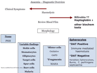
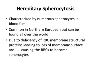
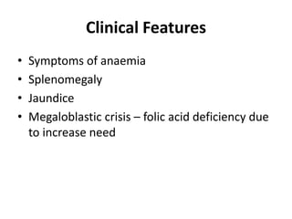
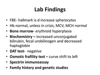
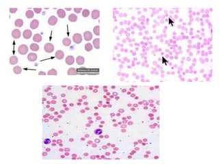
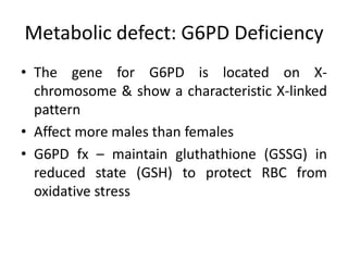
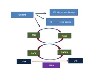
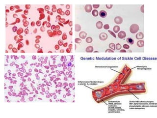
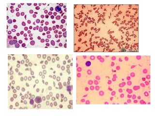
CLASSIFICATION OF ANAEMIA:
NORMOCHROMIC NORMOCYTIC
          ANAEMIA
Chapter outline
• Membrane defects: Hereditary spherocytosis
• Metabolic defect: pyruvate kinase deficiency,
  G6PD deficiency
• Haemoglobinopathies: sickle cell disease
• Extrinsic abnormalities: antibody mediated
  transfusion reaction
• Mechanical destruction: MAHA, infections
• Underlying causes:
1. Loss of RBC:Haemorrhage
2. Increased destruction of RBC
3. Reduced erythropoieisis
4. Reduced RBC’s survival

Haemolytic anaemia can be classified into:
1. Congenital and acquired
2. Intrinsic or extrinsic
3. Intravascular or extravascular haemolysis
Morphologic classification of Anaemia – MCV based
                                                                         N or h RETIC
               Normocytic - MCV 80-100 fl                                Bone marrow
                                                                         Acellular
                                                                         Hypercellular
                       h RETIC                                           Normocellular


                        Haemolytic
                         anaemia


       Intrinsic                                     Extrinsic

                              PNH

     Membrane
                                                                 Chemical &    Mechanical -
                                     Antibody   Infection                      MAHA
                                                                 Physical
     Enzyme                          mediated
                                                                 damage

    Haeme/globin
Anaemia - Diagnostic Overview

       Clinical Symptoms
                                                          Haemolysis
                                                                                   Bilirubin hh
                                                                                   Haptoglobin i
                                                       Review Blood Film
                                                                                   other biochem
                                                                                   tests
                                                          Morphology

      None
       PNH                                                                     Spherocytes
                               Variable findings                               *DAT Positive
                                                              *Blister cells
                                        Sickle cells                            Immune mediated
                                   Stomatocytes                 Oxidative         haemolysis
                                    Elliptocytes                  G6PD         *DAT Negative
                                        Target cells          *Fragments       Hereditary Spherocytosis,
                                                                               Burns, C. perfringens
                                        Spur cells               MAHA          septicaemia
Source; modified from AH Turner, 2004   Burr cells
                                         Malaria
Hereditary Spherocytosis
• Characterized by numerous spherocytes in
  blood film
• Common in Northern European but can be
  found all over the world
• Due to deficiency of RBC membrane structural
  proteins leading to loss of membrane surface
  are----- causing the RBCs to become
  spherocytes
Pathophysiology
• Defect in membrane skeletal proteins that
  connect the membrane skeleton to the lipid
  bilayer ( spectrin, ankyrin, protein 4.2, band 3)
• Cause RBCs progressively to lose unsupported
  lipid membrane because of the local
  disconnection
• RCs become rigid and their survival in the
  spleen decrease
Clinical Features
•   Symptoms of anaemia
•   Splenomegaly
•   Jaundice
•   Megaloblastic crisis – folic acid deficiency due
    to increase need
Lab Findings
•   FBE- hallmark is d increase spherocytes
•   Hb normal, unless in crisis, MCV, MCH normal
•   Bone marrow- erythroid hyperplasia
•   Biochemistry – increased unconjugated
    bilirubin, fecal urobilinogen and decreased
    haptoglobin
•   DAT test- negative
•   Osmotic frafility test – curve shift to left
•   Spectrin immunoassay
•   Family history and genetic studies
Treatment & differential diagnosis
• Splenectomy – post splenectomy cause the
  appearance of target cells, howell jolly bodies,
  pappenheimer bodies
• Differential diagnosis – other causes of
  spherocytes must be ruled out AIHA,
  PNH,sepsis, pyropoikilocytosis
Metabolic defect: G6PD Deficiency
• The gene for G6PD is located on X-
  chromosome & show a characteristic X-linked
  pattern
• Affect more males than females
• G6PD fx – maintain gluthathione (GSSG) in
  reduced state (GSH) to protect RBC from
  oxidative stress
RBC Membrane damage
Oxidant


                 Hb        Heinz bodies


          GSH                 GSSG


          NADP               NADPH


   G-6P                                   6PG
                 G6PD
Clinical Features
• G6PD deficiency is usually asymptomatic but:
 1.Acute haemolytic anaemia in response to
 oxidative stress such as drugs, fava beans,
 infections, etc.
 2.Neonatal jaundice
 3.Congenital     non-spherocytic   haemolytic
 anaemia
Lab Findings
• FBE – depends on severity – normochromic
  normocytic,         marked          anisocytosis,
  poikilocytosis with bite cells and blister cells
• Biochemical - haptoglobin severely               ,
  unconjugated bilirubin and plasma Hb
• G6PD screening test – G6PD fluoresence spot
  test, G6PD enzyme detection kit, PCR analysis
  of mutations.
Differential Diagnosis
• Drug induced HA
• Other enzymopathies eg Pyruvate kinase
  deficiency
• Haemoglobinopathies due to oxidative stress
Haem defects: Sickle Cell Disease
• Hb S is defined by structural formula α2β2Glu-val
  which indictaes that on the B chain at 6th
  position, glutamic acid is replaced by valine
• resulting in a structural change in RBC shape
  relates to the amount of O2 bound to the Hb
  molecules
Pathophysiology
• When HbS is fully oxygenated it remains soluble in the
  erythrocyte similar to Hb A, maintaining its normal
  shape
• On deoxygenation, Hb S becomes less soluble and
  causing sickling
• The blood becomes more viscous when sickle cells are
  created. Results in reduced blood flow which prolongs
  the exposure of HbS containing erythrocytes to a
  hypoxic environment which further promotes sickling,
• The end result is occlusion of capillaries and arterioles
  by sickled RBCs and infarction of surrounding tissues
Clinical Features of SCD
• Hallmark feature is vaso occlusive
• Gradual loss of splenic fx
• Splenic sequestration

Laboratory diagnosis of SCD
 •Peripheral    blood       smear    –      poikilocytosis
 &anisocytosis,         sickle       cells,         target
 cells, nRBCs, spherocytes, basohilic stippling, Howell-
 jolly bodies
 •Moderate leukocytosis & neutrophilia
 •thrombocytosis
Lab Diagnosis (Cont)
•   BM- erythroid hyperplasia
•   Elevated Indirect & direct bilirubin
•   Solubility & sickling test
•   Hb Electrophoresis

TREATMENT
 1. BM transplantation
 2. Transfusion
 3. Hydroxyurea therapy
Microangiopathic Haemolytic
            Anaemia (MAHA)
• Due to mechanical destruction
• It is a group of clinical disorders characterized by
  RBC fragmentation in the circulation, resulting
  from IV haemolysis
• The fragmentation occurs as RBCs passed through
  fibrin deposits inside the lumen of arterioles &
  capillaries or through damaged epithelium &
  vessel walls
• RBCs being forced through a fibrin clot, attaching
  to fibrin, folding around the strands &
  fragmenting by the force of the flowing blood
• Disorders include
 1) Thrombocytic thrombocytopenic purpurea
    (TTP)
 2) Haemolyric uraemic syndrome (HUS)
 3) Disseminated intravascular coagulation (DIC)
 4) Haemolysis Elevated Liver enzymes & Low
    Platelet (HELLP)
Normochromic normocytic anaemia

Normochromic normocytic anaemia

  • 1.
  • 2.
    Chapter outline • Membranedefects: Hereditary spherocytosis • Metabolic defect: pyruvate kinase deficiency, G6PD deficiency • Haemoglobinopathies: sickle cell disease • Extrinsic abnormalities: antibody mediated transfusion reaction • Mechanical destruction: MAHA, infections
  • 3.
    • Underlying causes: 1.Loss of RBC:Haemorrhage 2. Increased destruction of RBC 3. Reduced erythropoieisis 4. Reduced RBC’s survival Haemolytic anaemia can be classified into: 1. Congenital and acquired 2. Intrinsic or extrinsic 3. Intravascular or extravascular haemolysis
  • 4.
    Morphologic classification ofAnaemia – MCV based N or h RETIC Normocytic - MCV 80-100 fl Bone marrow Acellular Hypercellular h RETIC Normocellular Haemolytic anaemia Intrinsic Extrinsic PNH Membrane Chemical & Mechanical - Antibody Infection MAHA Physical Enzyme mediated damage Haeme/globin
  • 5.
    Anaemia - DiagnosticOverview Clinical Symptoms Haemolysis Bilirubin hh Haptoglobin i Review Blood Film other biochem tests Morphology None PNH Spherocytes Variable findings *DAT Positive *Blister cells Sickle cells Immune mediated Stomatocytes Oxidative haemolysis Elliptocytes G6PD *DAT Negative Target cells *Fragments Hereditary Spherocytosis, Burns, C. perfringens Spur cells MAHA septicaemia Source; modified from AH Turner, 2004 Burr cells Malaria
  • 6.
    Hereditary Spherocytosis • Characterizedby numerous spherocytes in blood film • Common in Northern European but can be found all over the world • Due to deficiency of RBC membrane structural proteins leading to loss of membrane surface are----- causing the RBCs to become spherocytes
  • 7.
    Pathophysiology • Defect inmembrane skeletal proteins that connect the membrane skeleton to the lipid bilayer ( spectrin, ankyrin, protein 4.2, band 3) • Cause RBCs progressively to lose unsupported lipid membrane because of the local disconnection • RCs become rigid and their survival in the spleen decrease
  • 8.
    Clinical Features • Symptoms of anaemia • Splenomegaly • Jaundice • Megaloblastic crisis – folic acid deficiency due to increase need
  • 9.
    Lab Findings • FBE- hallmark is d increase spherocytes • Hb normal, unless in crisis, MCV, MCH normal • Bone marrow- erythroid hyperplasia • Biochemistry – increased unconjugated bilirubin, fecal urobilinogen and decreased haptoglobin • DAT test- negative • Osmotic frafility test – curve shift to left • Spectrin immunoassay • Family history and genetic studies
  • 10.
    Treatment & differentialdiagnosis • Splenectomy – post splenectomy cause the appearance of target cells, howell jolly bodies, pappenheimer bodies • Differential diagnosis – other causes of spherocytes must be ruled out AIHA, PNH,sepsis, pyropoikilocytosis
  • 12.
    Metabolic defect: G6PDDeficiency • The gene for G6PD is located on X- chromosome & show a characteristic X-linked pattern • Affect more males than females • G6PD fx – maintain gluthathione (GSSG) in reduced state (GSH) to protect RBC from oxidative stress
  • 13.
    RBC Membrane damage Oxidant Hb Heinz bodies GSH GSSG NADP NADPH G-6P 6PG G6PD
  • 14.
    Clinical Features • G6PDdeficiency is usually asymptomatic but: 1.Acute haemolytic anaemia in response to oxidative stress such as drugs, fava beans, infections, etc. 2.Neonatal jaundice 3.Congenital non-spherocytic haemolytic anaemia
  • 15.
    Lab Findings • FBE– depends on severity – normochromic normocytic, marked anisocytosis, poikilocytosis with bite cells and blister cells • Biochemical - haptoglobin severely , unconjugated bilirubin and plasma Hb • G6PD screening test – G6PD fluoresence spot test, G6PD enzyme detection kit, PCR analysis of mutations.
  • 16.
    Differential Diagnosis • Druginduced HA • Other enzymopathies eg Pyruvate kinase deficiency • Haemoglobinopathies due to oxidative stress
  • 18.
    Haem defects: SickleCell Disease • Hb S is defined by structural formula α2β2Glu-val which indictaes that on the B chain at 6th position, glutamic acid is replaced by valine • resulting in a structural change in RBC shape relates to the amount of O2 bound to the Hb molecules
  • 19.
    Pathophysiology • When HbSis fully oxygenated it remains soluble in the erythrocyte similar to Hb A, maintaining its normal shape • On deoxygenation, Hb S becomes less soluble and causing sickling • The blood becomes more viscous when sickle cells are created. Results in reduced blood flow which prolongs the exposure of HbS containing erythrocytes to a hypoxic environment which further promotes sickling, • The end result is occlusion of capillaries and arterioles by sickled RBCs and infarction of surrounding tissues
  • 20.
    Clinical Features ofSCD • Hallmark feature is vaso occlusive • Gradual loss of splenic fx • Splenic sequestration Laboratory diagnosis of SCD •Peripheral blood smear – poikilocytosis &anisocytosis, sickle cells, target cells, nRBCs, spherocytes, basohilic stippling, Howell- jolly bodies •Moderate leukocytosis & neutrophilia •thrombocytosis
  • 21.
    Lab Diagnosis (Cont) • BM- erythroid hyperplasia • Elevated Indirect & direct bilirubin • Solubility & sickling test • Hb Electrophoresis TREATMENT 1. BM transplantation 2. Transfusion 3. Hydroxyurea therapy
  • 23.
    Microangiopathic Haemolytic Anaemia (MAHA) • Due to mechanical destruction • It is a group of clinical disorders characterized by RBC fragmentation in the circulation, resulting from IV haemolysis • The fragmentation occurs as RBCs passed through fibrin deposits inside the lumen of arterioles & capillaries or through damaged epithelium & vessel walls • RBCs being forced through a fibrin clot, attaching to fibrin, folding around the strands & fragmenting by the force of the flowing blood
  • 24.
    • Disorders include 1) Thrombocytic thrombocytopenic purpurea (TTP) 2) Haemolyric uraemic syndrome (HUS) 3) Disseminated intravascular coagulation (DIC) 4) Haemolysis Elevated Liver enzymes & Low Platelet (HELLP)