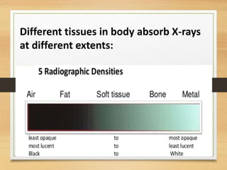
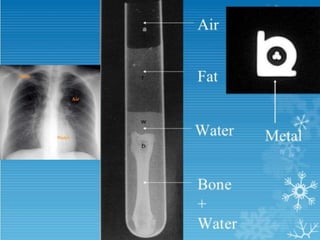
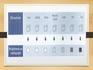
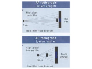
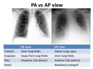
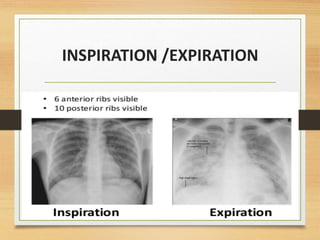
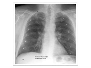
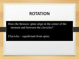
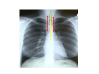
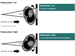
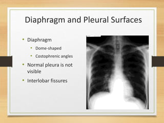
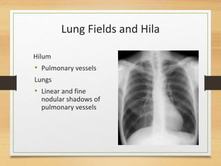
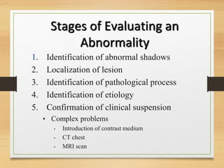
GOOD MORNING..!!
NORMAL X-RAY
INTERPRETATION
BY
GEETHIKHA B
Recognition
of abnormal
first requires
knowledge of
normal.
Different tissues in body absorb X-rays
at different extents:
DATA BASE
7
Date
Position
markers
Type of film
Patient’s
position
Technical
quality
TECHNICAL ASPECTS
Orientation
Inspiration
Penetration
Rotation
Angulation
Factors to be considered on all x-rays include:
ORIENTATION
• PA- the x-rays penetrate through the back of the
patient on to the film
• AP-the x-rays penetrate through the front of the
patient on to the film
INSPIRATION /EXPIRATION
PENETRATION
ROTATION
LOOK INTO
• Bony Framework
• Soft Tissues
• Lung Fields and Hila
• Diaphragm and Pleural
Spaces
• Mediastinum and
Heart
• Abdomen and Neck
Bony Fragments
• Ribs
• Spine
• Shoulder
girdle
• Clavicles
Soft Tissues
• Breast shadows
• Supraclavicular areas
• Axillae
• Tissues along side of
breasts
Mediastinum and Heart
• Heart size on PA
• Right side
• Inferior vena cava
• Right atrium
• Superior vena cava
Mediastinum and Heart
• Left side
• Left ventricle
• Pulmonary artery
• Aortic knuckle
Diaphragm and Pleural Surfaces
• Diaphragm
• Dome-shaped
• Costophrenic angles
• Normal pleura is not
visible
• Interlobar fissures
Lung Fields and Hila
Hilum
• Pulmonary vessels
Lungs
• Linear and fine
nodular shadows of
pulmonary vessels
Abdomen and Neck
• Abdomen
• Gastric bubble
• Neck
Stages of Evaluating an
Abnormality
1. Identification of abnormal shadows
2. Localization of lesion
3. Identification of pathological process
4. Identification of etiology
5. Confirmation of clinical suspension
• Complex problems
• Introduction of contrast medium
• CT chest
• MRI scan
And finally..!!
• Practice and repetition.
• Plain XR is the starting point, findings on the
XR will direct you towards further imaging.
• Some diseases have certain XR patterns, but
this is not written in stone!
• Always keep the patient clinical presenting
features in mind when coming up with your
differential diagnosis.
Normal chest x ray

Normal chest x ray