Neurological manifestations are common in COVID-19, occurring in around 80% of hospitalized patients. The document discusses several neurological effects such as smell and taste disorders, encephalopathy, cerebrovascular disease, neuromuscular disorders, seizures, sleep disorders, and various acute neurological manifestations. The pathogenesis is multifactorial, involving direct viral invasion, immune dysfunction, coagulopathies, and the renin-angiotensin system.
Scott Letendre, MD
Professor in Residence
Division of Infectious Diseases & Global Public Health
Departments of Medicine and Psychiatry
University of California, San Diego
Neurological Manifestations of COVID-19 InfectionSudhir Kumar
COVID-19 primarily affects respiratory system, however, it can affect other systems too, including nervous system. This presentation offers details about neurological symptoms and disorders seen in patients with COVID-19.
Neuropsychiatric manifestations of endocrine disordersDheeraj kumar
This is a subject seminar of neuropsychiatric manifesations of endocrine disorders.It took a lot of time to prepare,it helps fellow residents of Gen medicine to download and present as it is.
Neuropsychiatric manifestations of endocrine disordersDheeraj kumar
This is a subject seminar of neuropsychiatric manifesations of endocrine disorders.It took a lot of time to prepare,it helps fellow residents of Gen medicine to download and present as it is.
Transverse Myelitis in a Patient with COVID-19: A Case Reportkomalicarol
There has been growing evidence of COVID-19
potentially causing a wide range of neurological abnormalities
from as mild as anosmia to as serious as stroke. It is important to
recognize that amid this pandemic, we have been seeing different
manifestations and associations of COVID-19
TheNeuroSurgeons sponsored the presentation to the Zimbabwe Association of Neurological Surgeons.
we are learning more about the neurological manifestations of the novel coronavirus as we are frantically looking for solution to this formidable pandemic.
Guillain Barre Syndrome & Covid-19: A Case Reportclinicsoncology
Besides respiratory symptoms, coronavirus disease 2019 (COVID-19), like the severe acute respiratory syndrome coronavirus (SARS-CoV) and Middle East respiratory syndrome coronavirus (MERS-CoV), has neurological signs. Symptoms like myalgia, headaches, dizziness, anosmia, ageusia and disorder of consciousness confirms that the nervous system is involved in COVID-19 infection.
Guillain Barre Syndrome & Covid-19: A Case Reportpateldrona
Besides respiratory symptoms, coronavirus disease 2019 (COVID-19), like the severe acute respiratory syndrome coronavirus (SARS-CoV) and Middle East respiratory syndrome coronavirus (MERS-CoV), has neurological signs
Guillain Barre Syndrome & Covid-19: A Case Reportkomalicarol
Besides respiratory symptoms, coronavirus disease 2019
(COVID-19), like the severe acute respiratory syndrome coronavirus (SARS-CoV) and Middle East respiratory syndrome coronavirus (MERS-CoV), has neurological signs. Symptoms like myalgia,
headaches, dizziness, anosmia, ageusia and disorder of consciousness confirms that the nervous system is involved in COVID-19 infection. Guillain barre syndrome (GBS) is a neurological disorder
that usually follows a viral infection, it is possible that COVID-19
infection and GBS are closely related. In this case report, we try to
elucidate the relation between SARS-CoV-2 and GBS.
The coronavirus infection coronavirus disease 2019 (COVID-19) first presented as an outbreak of atypical pneumonia in Wuhan, China, on December 12, 2019.1,2 Since then, it has spread globally to infect >1 963 943 individuals and killed >123 635 in >200 countries as of April 14, 2020. This infection has affected health and the economy worldwide on an unprecedented scale.
The coronavirus disease outbreak has proven to be a major health crisis affecting virtually every facets of our lives.
Coronavirus disease is an ongoing pandemic disease. The disease which is caused by a new type of virus, known as severe
acute respiratory syndrome coronavirus 2 (SARS-CoV-2). Many patients hospitalized with COVID-19 will develop muscle
weakness particularly those admitted in intensive care unit (ICU). Studies have shown that muscle weakness is one of the
direct consequences of critical illness. We systematically reviewed literature that quantified changes in muscle strength and it
relationship with COVID- 19 in Intensive care unit in humans.
COVID-19 Associated Large Vessel Thrombosis and Ischemic Stroke: A Case Seriesmahendrareddychirra
The novel severe acute respiratory syndrome coronavirus (SARS-COV-2) affects different people in different ways. Most infected people will develop mild to moderate respiratory flu-like illness and recover without the need for hospitalization.
Una nuova ricerca spiega le possibilità delle terapie di neuromodulazione di intervenire nella cura dei problemi neurologici derivati dalla pandemia da COVID-19
CONCEPT OF NODOPATHIES AND PARANODOPATHIES.pptxNeurologyKota
emergence of autoimmune neuropathies and role of nodal and paranodal regions in their pathophysiology.
Peripheral neuropathies are traditionally categorized into demyelinating or axonal.
dysfunction at nodal/paranodal region key for better understanding of patients with immune mediated neuropathies.
antibodies targeting node and paranode of myelinated nerves have been increasingly detected in patients with immune mediated neuropathies.
have clinical phenotype similar common inflammatory neuropathies like Guillain Barre syndrome and chronic inflammatory demyelinating polyradiculoneuropathy
they respond poorly to conventional first line immunotherapies like IVIG
This presentation briefs out the approach of dementia assessment in line with consideration of recent advances. Now the pattern of assessment has evolved towards examining each individual domain rather than lobar assessment.
This presentation contains information about Dementia in Young onset. Also it describes the etiologies, clinical feature of common YOD & their management.
Entrapment Syndromes of Lower Limb.pptxNeurologyKota
This presentation contains information about the various Entrapment syndromes of Lower limb in descending order of topography. It also contains information about etiology, clinical features and management of each of these entrapment syndromes with special emphasis on electrodiagnostic confirmation.
Report Back from SGO 2024: What’s the Latest in Cervical Cancer?bkling
Are you curious about what’s new in cervical cancer research or unsure what the findings mean? Join Dr. Emily Ko, a gynecologic oncologist at Penn Medicine, to learn about the latest updates from the Society of Gynecologic Oncology (SGO) 2024 Annual Meeting on Women’s Cancer. Dr. Ko will discuss what the research presented at the conference means for you and answer your questions about the new developments.
Lung Cancer: Artificial Intelligence, Synergetics, Complex System Analysis, S...Oleg Kshivets
RESULTS: Overall life span (LS) was 2252.1±1742.5 days and cumulative 5-year survival (5YS) reached 73.2%, 10 years – 64.8%, 20 years – 42.5%. 513 LCP lived more than 5 years (LS=3124.6±1525.6 days), 148 LCP – more than 10 years (LS=5054.4±1504.1 days).199 LCP died because of LC (LS=562.7±374.5 days). 5YS of LCP after bi/lobectomies was significantly superior in comparison with LCP after pneumonectomies (78.1% vs.63.7%, P=0.00001 by log-rank test). AT significantly improved 5YS (66.3% vs. 34.8%) (P=0.00000 by log-rank test) only for LCP with N1-2. Cox modeling displayed that 5YS of LCP significantly depended on: phase transition (PT) early-invasive LC in terms of synergetics, PT N0—N12, cell ratio factors (ratio between cancer cells- CC and blood cells subpopulations), G1-3, histology, glucose, AT, blood cell circuit, prothrombin index, heparin tolerance, recalcification time (P=0.000-0.038). Neural networks, genetic algorithm selection and bootstrap simulation revealed relationships between 5YS and PT early-invasive LC (rank=1), PT N0—N12 (rank=2), thrombocytes/CC (3), erythrocytes/CC (4), eosinophils/CC (5), healthy cells/CC (6), lymphocytes/CC (7), segmented neutrophils/CC (8), stick neutrophils/CC (9), monocytes/CC (10); leucocytes/CC (11). Correct prediction of 5YS was 100% by neural networks computing (area under ROC curve=1.0; error=0.0).
CONCLUSIONS: 5YS of LCP after radical procedures significantly depended on: 1) PT early-invasive cancer; 2) PT N0--N12; 3) cell ratio factors; 4) blood cell circuit; 5) biochemical factors; 6) hemostasis system; 7) AT; 8) LC characteristics; 9) LC cell dynamics; 10) surgery type: lobectomy/pneumonectomy; 11) anthropometric data. Optimal diagnosis and treatment strategies for LC are: 1) screening and early detection of LC; 2) availability of experienced thoracic surgeons because of complexity of radical procedures; 3) aggressive en block surgery and adequate lymph node dissection for completeness; 4) precise prediction; 5) adjuvant chemoimmunoradiotherapy for LCP with unfavorable prognosis.
Title: Sense of Taste
Presenter: Dr. Faiza, Assistant Professor of Physiology
Qualifications:
MBBS (Best Graduate, AIMC Lahore)
FCPS Physiology
ICMT, CHPE, DHPE (STMU)
MPH (GC University, Faisalabad)
MBA (Virtual University of Pakistan)
Learning Objectives:
Describe the structure and function of taste buds.
Describe the relationship between the taste threshold and taste index of common substances.
Explain the chemical basis and signal transduction of taste perception for each type of primary taste sensation.
Recognize different abnormalities of taste perception and their causes.
Key Topics:
Significance of Taste Sensation:
Differentiation between pleasant and harmful food
Influence on behavior
Selection of food based on metabolic needs
Receptors of Taste:
Taste buds on the tongue
Influence of sense of smell, texture of food, and pain stimulation (e.g., by pepper)
Primary and Secondary Taste Sensations:
Primary taste sensations: Sweet, Sour, Salty, Bitter, Umami
Chemical basis and signal transduction mechanisms for each taste
Taste Threshold and Index:
Taste threshold values for Sweet (sucrose), Salty (NaCl), Sour (HCl), and Bitter (Quinine)
Taste index relationship: Inversely proportional to taste threshold
Taste Blindness:
Inability to taste certain substances, particularly thiourea compounds
Example: Phenylthiocarbamide
Structure and Function of Taste Buds:
Composition: Epithelial cells, Sustentacular/Supporting cells, Taste cells, Basal cells
Features: Taste pores, Taste hairs/microvilli, and Taste nerve fibers
Location of Taste Buds:
Found in papillae of the tongue (Fungiform, Circumvallate, Foliate)
Also present on the palate, tonsillar pillars, epiglottis, and proximal esophagus
Mechanism of Taste Stimulation:
Interaction of taste substances with receptors on microvilli
Signal transduction pathways for Umami, Sweet, Bitter, Sour, and Salty tastes
Taste Sensitivity and Adaptation:
Decrease in sensitivity with age
Rapid adaptation of taste sensation
Role of Saliva in Taste:
Dissolution of tastants to reach receptors
Washing away the stimulus
Taste Preferences and Aversions:
Mechanisms behind taste preference and aversion
Influence of receptors and neural pathways
Impact of Sensory Nerve Damage:
Degeneration of taste buds if the sensory nerve fiber is cut
Abnormalities of Taste Detection:
Conditions: Ageusia, Hypogeusia, Dysgeusia (parageusia)
Causes: Nerve damage, neurological disorders, infections, poor oral hygiene, adverse drug effects, deficiencies, aging, tobacco use, altered neurotransmitter levels
Neurotransmitters and Taste Threshold:
Effects of serotonin (5-HT) and norepinephrine (NE) on taste sensitivity
Supertasters:
25% of the population with heightened sensitivity to taste, especially bitterness
Increased number of fungiform papillae
The prostate is an exocrine gland of the male mammalian reproductive system
It is a walnut-sized gland that forms part of the male reproductive system and is located in front of the rectum and just below the urinary bladder
Function is to store and secrete a clear, slightly alkaline fluid that constitutes 10-30% of the volume of the seminal fluid that along with the spermatozoa, constitutes semen
A healthy human prostate measures (4cm-vertical, by 3cm-horizontal, 2cm ant-post ).
It surrounds the urethra just below the urinary bladder. It has anterior, median, posterior and two lateral lobes
It’s work is regulated by androgens which are responsible for male sex characteristics
Generalised disease of the prostate due to hormonal derangement which leads to non malignant enlargement of the gland (increase in the number of epithelial cells and stromal tissue)to cause compression of the urethra leading to symptoms (LUTS
Ozempic: Preoperative Management of Patients on GLP-1 Receptor Agonists Saeid Safari
Preoperative Management of Patients on GLP-1 Receptor Agonists like Ozempic and Semiglutide
ASA GUIDELINE
NYSORA Guideline
2 Case Reports of Gastric Ultrasound
Ethanol (CH3CH2OH), or beverage alcohol, is a two-carbon alcohol
that is rapidly distributed in the body and brain. Ethanol alters many
neurochemical systems and has rewarding and addictive properties. It
is the oldest recreational drug and likely contributes to more morbidity,
mortality, and public health costs than all illicit drugs combined. The
5th edition of the Diagnostic and Statistical Manual of Mental Disorders
(DSM-5) integrates alcohol abuse and alcohol dependence into a single
disorder called alcohol use disorder (AUD), with mild, moderate,
and severe subclassifications (American Psychiatric Association, 2013).
In the DSM-5, all types of substance abuse and dependence have been
combined into a single substance use disorder (SUD) on a continuum
from mild to severe. A diagnosis of AUD requires that at least two of
the 11 DSM-5 behaviors be present within a 12-month period (mild
AUD: 2–3 criteria; moderate AUD: 4–5 criteria; severe AUD: 6–11 criteria).
The four main behavioral effects of AUD are impaired control over
drinking, negative social consequences, risky use, and altered physiological
effects (tolerance, withdrawal). This chapter presents an overview
of the prevalence and harmful consequences of AUD in the U.S.,
the systemic nature of the disease, neurocircuitry and stages of AUD,
comorbidities, fetal alcohol spectrum disorders, genetic risk factors, and
pharmacotherapies for AUD.
HOT NEW PRODUCT! BIG SALES FAST SHIPPING NOW FROM CHINA!! EU KU DB BK substit...GL Anaacs
Contact us if you are interested:
Email / Skype : kefaya1771@gmail.com
Threema: PXHY5PDH
New BATCH Ku !!! MUCH IN DEMAND FAST SALE EVERY BATCH HAPPY GOOD EFFECT BIG BATCH !
Contact me on Threema or skype to start big business!!
Hot-sale products:
NEW HOT EUTYLONE WHITE CRYSTAL!!
5cl-adba precursor (semi finished )
5cl-adba raw materials
ADBB precursor (semi finished )
ADBB raw materials
APVP powder
5fadb/4f-adb
Jwh018 / Jwh210
Eutylone crystal
Protonitazene (hydrochloride) CAS: 119276-01-6
Flubrotizolam CAS: 57801-95-3
Metonitazene CAS: 14680-51-4
Payment terms: Western Union,MoneyGram,Bitcoin or USDT.
Deliver Time: Usually 7-15days
Shipping method: FedEx, TNT, DHL,UPS etc.Our deliveries are 100% safe, fast, reliable and discreet.
Samples will be sent for your evaluation!If you are interested in, please contact me, let's talk details.
We specializes in exporting high quality Research chemical, medical intermediate, Pharmaceutical chemicals and so on. Products are exported to USA, Canada, France, Korea, Japan,Russia, Southeast Asia and other countries.
MANAGEMENT OF ATRIOVENTRICULAR CONDUCTION BLOCK.pdfJim Jacob Roy
Cardiac conduction defects can occur due to various causes.
Atrioventricular conduction blocks ( AV blocks ) are classified into 3 types.
This document describes the acute management of AV block.
Title: Sense of Smell
Presenter: Dr. Faiza, Assistant Professor of Physiology
Qualifications:
MBBS (Best Graduate, AIMC Lahore)
FCPS Physiology
ICMT, CHPE, DHPE (STMU)
MPH (GC University, Faisalabad)
MBA (Virtual University of Pakistan)
Learning Objectives:
Describe the primary categories of smells and the concept of odor blindness.
Explain the structure and location of the olfactory membrane and mucosa, including the types and roles of cells involved in olfaction.
Describe the pathway and mechanisms of olfactory signal transmission from the olfactory receptors to the brain.
Illustrate the biochemical cascade triggered by odorant binding to olfactory receptors, including the role of G-proteins and second messengers in generating an action potential.
Identify different types of olfactory disorders such as anosmia, hyposmia, hyperosmia, and dysosmia, including their potential causes.
Key Topics:
Olfactory Genes:
3% of the human genome accounts for olfactory genes.
400 genes for odorant receptors.
Olfactory Membrane:
Located in the superior part of the nasal cavity.
Medially: Folds downward along the superior septum.
Laterally: Folds over the superior turbinate and upper surface of the middle turbinate.
Total surface area: 5-10 square centimeters.
Olfactory Mucosa:
Olfactory Cells: Bipolar nerve cells derived from the CNS (100 million), with 4-25 olfactory cilia per cell.
Sustentacular Cells: Produce mucus and maintain ionic and molecular environment.
Basal Cells: Replace worn-out olfactory cells with an average lifespan of 1-2 months.
Bowman’s Gland: Secretes mucus.
Stimulation of Olfactory Cells:
Odorant dissolves in mucus and attaches to receptors on olfactory cilia.
Involves a cascade effect through G-proteins and second messengers, leading to depolarization and action potential generation in the olfactory nerve.
Quality of a Good Odorant:
Small (3-20 Carbon atoms), volatile, water-soluble, and lipid-soluble.
Facilitated by odorant-binding proteins in mucus.
Membrane Potential and Action Potential:
Resting membrane potential: -55mV.
Action potential frequency in the olfactory nerve increases with odorant strength.
Adaptation Towards the Sense of Smell:
Rapid adaptation within the first second, with further slow adaptation.
Psychological adaptation greater than receptor adaptation, involving feedback inhibition from the central nervous system.
Primary Sensations of Smell:
Camphoraceous, Musky, Floral, Pepperminty, Ethereal, Pungent, Putrid.
Odor Detection Threshold:
Examples: Hydrogen sulfide (0.0005 ppm), Methyl-mercaptan (0.002 ppm).
Some toxic substances are odorless at lethal concentrations.
Characteristics of Smell:
Odor blindness for single substances due to lack of appropriate receptor protein.
Behavioral and emotional influences of smell.
Transmission of Olfactory Signals:
From olfactory cells to glomeruli in the olfactory bulb, involving lateral inhibition.
Primitive, less old, and new olfactory systems with different path
Pulmonary Thromboembolism - etilogy, types, medical- Surgical and nursing man...VarunMahajani
Disruption of blood supply to lung alveoli due to blockage of one or more pulmonary blood vessels is called as Pulmonary thromboembolism. In this presentation we will discuss its causes, types and its management in depth.
Couples presenting to the infertility clinic- Do they really have infertility...Sujoy Dasgupta
Dr Sujoy Dasgupta presented the study on "Couples presenting to the infertility clinic- Do they really have infertility? – The unexplored stories of non-consummation" in the 13th Congress of the Asia Pacific Initiative on Reproduction (ASPIRE 2024) at Manila on 24 May, 2024.
The POPPY STUDY (Preconception to post-partum cardiovascular function in prim...
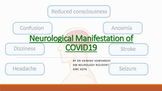
Neurological manifestation of covid19
1. BY DR VAIBHAV SOMVANSHI
DM NEUROLOGY RESIDENT
GMC KOTA
Neurological Manifestation of
COVID19
2. INTRODUCTION
SARS-COV-2 SS mRNA (30kb) virus belongs to Beta corona family
At the end of 2019, a novel coronavirus identified as cause of a cluster of pneumonia cases in
Wuhan, a city in the Hubei Province of China.
Acronym – CO(corona) VI(virus) D(diasease) 19(year of identification)
Neurologic implication common – around 80% in hospitalized patients at some point.
Liotta EM, Batra A, Clark JR, Shlobin NA, Hoffman SC, Orban ZS, Koralnik IJ. Frequent neurologic manifestations and encephalopathy-associated morbidity in
Covid-19 patients. Ann Clin Transl Neurol. 2020 Nov;7(11):2221-2230. doi: 10.1002/acn3.51210. Epub 2020 Oct 5. PMID: 33016619; PMCID: PMC7664279.
4. Pathophysiology
Viral entry receptor :
ACE-2
Entry dependent on
interaction between
spike protein,
TMPRSS2 and ACE2.
Portal of entry:
Respiratory tract
mucosa
5.
6.
7.
8. Neuropathology
of COVID-19 –
Schematic
Representaion
Olfactory
neurons, neural
stem cells, and
Sustentacular
cells
Virus bind
to ACE2
Cytokine
storm in
vasculature
ACE2 on
respiratory
epithelium
BBB and ACE2
expressing
pericytes
cerebral microinfarcts from
vascular plugging and
vasoconstriction
Transsynaptic
spread
9. NEUROPATHOGENESIS
Mechanisms of neurologic complications in patients with COVID-19 - Diverse and multifactorial
Neurologic injury from
systemic dysfunction
Renin-angiotensin system
dysfunction
Immune dysfunction
Proinflammatory
state
Parainfectious and
postinfectious
triggers
Direct viral invasion of the
nervous system
Post-hypoxic
leukoencephalopathy
Encephalopathy due to
metabolic
derangements Cytokine storm
Capillary
endothelitis
coagulopathy
ADEM
GBS
Olfactory
Vagus nerve
Neurotropis
m
Transcytosis
Endocytosis
Transynaptic
transmission
14. Risk factors for more severe COVID-19 illness in
patients with pre-existing neurological
disorders
Age >=60 years
Pre-existing neurological disorders
Long-term facility or nursing home residents
Swallowing or breathing difficulty
Immunosuppressants-drugs
Comorbidities
Stroke
Chronic lung disease
Asthma
Serious heart diseases
Obesity
Diabetes
Hypertension
Renal disease
Liver disease
Immunocompromised
15. Prevalence of
Neurological
Manifestations
in COVID-19
Patients
Saniasiaya J, Islam MA, Abdullah B. Prevalence of Olfactory Dysfunction in Coronavirus Disease 2019 (COVID-19): A Meta-analysis of 27,492 Patients. Laryngoscope.
2021 Apr;131(4):865-878. doi: 10.1002/lary.29286. Epub 2020 Dec 5. PMID: 33219539; PMCID: PMC7753439
18. SMELL AND TASTE DISORDERS
Anosmia and dysgeusia - common early symptoms in patients with COVID-19.
meta-analysis of 83 studies involving more than 27,000 patients, olfactory dysfunction - 48 %
Transient anosmia - d/t inflammatory changes in sustentacular cells
In a survey of nonhospitalized patients with olfactory dysfunction from Italy,
83 % - complete recovery at a mean of 37 days after symptom onset.
Saniasiaya J, Islam MA, Abdullah B. Prevalence of Olfactory Dysfunction in Coronavirus Disease 2019 (COVID-19): A Meta-analysis of 27,492
Patients. Laryngoscope. 2021 Apr;131(4):865-878. doi: 10.1002/lary.29286. Epub 2020 Dec 5. PMID: 33219539; PMCID: PMC7753439.
19. ENCEPHALOPATHY
Encephalopathy is common in critically ill patients with COVID-19.
Delirium and agitation requiring sedation or may manifest somnolence and a decreased level
of consciousness.
Spectrum of neuroimaging abnormalities –
Hyperintensities in the medial temporal lobe, multifocal white matter lesions on FLAIR
isolated white matter micro-hemorrhages
Cytotoxic lesions in the splenium of the corpus callosum
Periventricular ischemic changes
EEG – non-specific
CSF- some case report shows + RTPCR for COVID19 otherwise WNL
Encephalopathy is a risk factor for poor outcome in COVID pts.
20. CEREBROVASCULAR DISEASE
incidence of ischemic stroke associated with COVID-19 in hospitalized patients- 0.4 to 2.7%
incidence of intracranial hemorrhage - 0.2 to 0.9%
incidence of CVT - 0.08%
Risk of stroke may vary according to the severity of COVID-19.
Due to the close association between COVID-19 and cerebrovascular disease, all patients
presenting with stroke need to be screened for SARS-CoV-2 infection,
COVID-19 is mainly associated with LVO strokes rather than the small vessel occlusion strokes.
Infection induced hypercoagulable state and embolisms d/t COVID-19-related myocarditis may
both result in strokes.
Mao L, Jin H, Wang M, Hu Y, Chen S, He Q, Chang J, Hong C, Zhou Y, Wang D, Miao X, Li Y, Hu B. Neurologic Manifestations of Hospitalized Patients With Coronavirus
Disease 2019 in Wuhan, China. JAMA Neurol. 2020 Jun 1;77(6):683-690. doi: 10.1001/jamaneurol.2020.1127. PMID: 32275288; PMCID: PMC7149362.
21. CEREBROVASCULAR DISEASE
Mostly stroke occurs 1 - 3weeks after onset of COVID-19 symptoms but can be initial
presentation.
Similar ischemic treatment protocol IV rTPA, aspirine, mechanical thrombectomy, with
unambiguous indication for full-dose anticoagulation.
Patients receiving ACE inhibitors or ARBs should continue treatment with these agents
PRES syndrome- Low availability of ACE 2 receptor leads to labile blood pressure results into
PRES like picture.
Widespread capillary endothelitis or a cytokine storm can also cause a diffuse
leukoencephalopathy with confluent posterior predominant white matter T2/FLAIR
hyperintensities and scattered microhemorrhage
Mao L, Jin H, Wang M, Hu Y, Chen S, He Q, Chang J, Hong C, Zhou Y, Wang D, Miao X, Li Y, Hu B. Neurologic Manifestations of Hospitalized Patients With Coronavirus
Disease 2019 in Wuhan, China. JAMA Neurol. 2020 Jun 1;77(6):683-690. doi: 10.1001/jamaneurol.2020.1127. PMID: 32275288; PMCID: PMC7149362.
22. NEUROMUSCULAR DISEASE
GBS associated with COVID 19 infection with low incidence.
Cases of GBS have been observed with the Ad26.COV2.S (Janssen/Johnson & Johnson) COVID-
19 vaccine.
AIDP more prevalent than other variants.
COVID-19 GBS seems to be more severe than non-COVID-19 GBS,
CSF and electrodiagnostic studies unremarkable.
Management remains the same.
Malik GR, Wolfe AR, Soriano R, Rydberg L, Wolfe LF, Deshmukh S, Ko JH, Nussbaum RP, Dreyer SD, Jayabalan P, Walter JM, Franz CK. Injury-prone: peripheral nerve injuries associated with prone
positioning for COVID-19-related acute respiratory distress syndrome. Br J Anaesth. 2020 Dec;125(6):e478-e480. doi: 10.1016/j.bja.2020.08.045. Epub 2020 Sep 4. PMID: 32948295; PMCID: PMC7473147.
23. NEUROMUSCULAR DISEASE
Myositis – prevalence 11% , myalgia and fatigue common symptoms.
HCQS, Azathioprine, dexamethasone induced iatrogenic myopathy and neuropathy
Focal and multifocal neuropathies - Ocular motor neuropathies , 7th , 10th , 11th, 12th, Tapia
syndrome, Neuralgic amyotrophy.
Critical illness neuropathy and myopathy
Peripheral nerve injuries after prone positioning- MC brachial plexus (lower trunk) – 14.5%
Malik GR, Wolfe AR, Soriano R, Rydberg L, Wolfe LF, Deshmukh S, Ko JH, Nussbaum RP, Dreyer SD, Jayabalan P, Walter JM, Franz CK. Injury-prone: peripheral nerve injuries associated with prone positioning for
COVID-19-related acute respiratory distress syndrome. Br J Anaesth. 2020 Dec;125(6):e478-e480. doi: 10.1016/j.bja.2020.08.045. Epub 2020 Sep 4. PMID: 32948295; PMCID: PMC7473147.
24. Seizure
Prevelence in COVID pts – 4.1%
Risk factors - metabolic alterations, and potential changes related to encephalitis.
Reported in patients with severe COVID-19 infection. Neuroimaging - abnormal in
approximately half of patients.
Saniasiaya J, Islam MA, Abdullah B. Prevalence of Olfactory Dysfunction in Coronavirus Disease 2019 (COVID-19): A Meta-analysis of 27,492 Patients. Laryngoscope. 2021 Apr;131(4):865-878. doi:
10.1002/lary.29286. Epub 2020 Dec 5. PMID: 33219539; PMCID: PMC7753439
25. Covidnosomnia
Risk factors -
Sleep disorders related to Anxiety, depression, poor sleep quality
Physical and social isolation, psychosocial disturbance
Quarantine, stress related to financial loss, loss of loved ones
Both insomnia and hypersomnia is prevalent.
26. Critical illness polyneuropathy/myopathy
Long stay admissions - post intensive care syndrome (PICS) - critical illness polyneuropathy and
myopathy (CIPNM)
SARS-CoV can infiltrate brainstem via trans-synaptic transfer - lead to dysfunction of the
cardiorespiratory centres of brainstem – need of long duration ventilation.
Risk factors for PICS - long durations of mechanical ventilation, hypoxia and sepsis,
Pro-inflammatory cytokines and free radicals - affect the microcirculation of both CNS & PNS by
reducing oxygen and nutrient delivery.
27. ACUTE NEUROLOGIC MANIFESTATIONS
Meningoencephalitis - Both viral (CSF RTPCR +ve for COVID19) and apparent
autoimmune(NMDA) meningoencephalitis have been reported in patients with COVID-19.
Rhombencephalitis - Brainstem encephalitis or isolated cerebellitis have been reported in adults
and children with COVID-19 infection. Patients with fulminant disease and brainstem
compression develop hydrocephalus.
ADEM and ANE - Hemorrhagic lesions in bilateral thalami, medial temporal lobes, and
subinsular lesions.
MS - might potentially experience both true and pseudorelapses.
28. ACUTE NEUROLOGIC MANIFESTATIONS
Multisystem inflammatory syndrome in children - illness similar to incomplete Kawasaki
disease- neurocognitive symptoms- headache, lethargy, confusion with MRI revealed signal
abnormality in the splenium of the corpus callosum .
Movement disorders reported in COVID-19 patients- tremors, myoclonus, and nonspecific
psychomotor agitation.
Posterior reversible encephalopathy syndrome (PRES) –due to hypertension and renal failure
29. Neurologic Manifestations of COVID-19 in
Children
Pathophysiology similar in pediatric population as well.
Pediatric patients shows prevalence of headache (4%), anosmia (2%), seizures (0.7%), and
cerebrovascular stroke(0.7%)
The pooled estimates frequency of seizure (3.1%) and encephalopathy (12.6% ) in children
with severe COVID 19.
Immune profiling suggests that autoantibodies in (MIS-C) shared sequence similarity between
SARS-CoV-2 and sialic acid residues on neural tissue.
Management - steroids and IV immunoglobulin in MIS-C, autoantibody depletion with
plasmapheresis.
Future potential immunologic interventions include blocking agents (against interleukin-1 and -
6), anti-CD20 monoclonal antibodies to ameliorate neurologic complications of COVID-19
Ranabothu S, Onteddu S, Nalleballe K, et al: Spectrum of COVID-19 in children. Acta Paediatr 2020; 109:1899–1900
30. PERSISTENT NEUROLOGIC SYMPTOMS
AFTER COVID-19 INFECTION
Constellation of persistent symptoms after acute COVID-19 infection described by various
terms- long COVID/post-COVID syndrome/post-COVID conditions.
Persists >6/6 weeks after SARS-CoV-2 infection, which have been reported to range between 5
and 80 percent of patients.
Umbrella term covers fatigue, dyspnea, brain fog" (81%), headache (68%), numbness/tingling
(60%), dysgeusia (59%), anosmia (55 %), and myalgias (55%)
Dysexecutive syndrome has been noted in around 30% of patients who have survived severe
life-threatening COVID-19 which encompasses emotional, motivational and behavioural
symptoms, as well as cognitive deficits.
It is unclear which, if any, part the neuroaxis or brain networks would be susceptible to
degeneration in the long-term, after acute COVID-19 illness. Long-term population follow-up
studies are needed to monitor for increased incidence of neurodegenerative problems
Graham EL, Clark JR, Orban ZS, Lim PH, Szymanski AL, Taylor C, DiBiase RM, Jia DT, Balabanov R, Ho SU, Batra A, Liotta EM, Koralnik IJ. Persistent neurologic symptoms and cognitive dysfunction in non-
hospitalized Covid-19 "long haulers". Ann Clin Transl Neurol. 2021 May;8(5):1073-1085. doi: 10.1002/acn3.51350. Epub 2021 Mar 30. PMID: 33755344; PMCID: PMC8108421.
31. PERSISTENT NEUROLOGIC SYMPTOMS
AFTER COVID-19 INFECTION
Extensive use of steroids/monoclonal antibodies/broad-spectrum antibiotics may lead to the
development/exacerbation of a preexisting fungal disease .
Rhino-oculo-cerebral mucormycosis (ROCM) is a rare disorder in which the olfactory system and brain become
infected with aerosolized spores from the Mucorales genus of fungi, which
includes Rhizopus, Apophysomyces, Mucor, Cunninghamella and Lichtheimia.
Spores nasal cavity necrosis nasal stuffiness and discharge, which can contain blood. mucor fungus -
highly angioinvasive- spreads to maxillary and ethmoidal sinuses, the orbits, the cavernous sinuses, the meninges
and, finally, the brain
Large multicentre study, 87% COVID-19-associated ROCM had used corticosteroids as a standard regimen to
treat the viral infection, diabetes mellitus in 78% of cases , diabetic ketoacidosis in 15% of cases
When routed through the cavernous sinuses maxillary and mandibular nerve palsies and internal carotid
artery thrombosis, pseudoaneurysm carotid cavernous fistula. From the sphenoid sinus clivus to the basal
meninges and basilar artery thrombosis or rupture with subarachnoid haemorrhage. nasal cavity or the
paranasal sinuses brain parenchyma, leading to cerebritis and abscesses.
Graham EL, Clark JR, Orban ZS, Lim PH, Szymanski AL, Taylor C, DiBiase RM, Jia DT, Balabanov R, Ho SU, Batra A, Liotta EM, Koralnik IJ. Persistent neurologic symptoms and cognitive dysfunction in non-
hospitalized Covid-19 "long haulers". Ann Clin Transl Neurol. 2021 May;8(5):1073-1085. doi: 10.1002/acn3.51350. Epub 2021 Mar 30. PMID: 33755344; PMCID: PMC8108421.
38. MANAGEMENT OF PATIENTS WITH
NEUROLOGIC CONDITIONS
Vaccination against COVID-19
Managing immunosuppressive therapy - At present there is no evidence to suggest that patients
with neurologic disease who are treated with immunosuppressive therapy are at increased risk of
COVID-19
Infection is a trigger for myasthenic crisis but has not been reported to be particularly prevalent in
patients with COVID-19
Alternative treatments may also be considered for patients who develop COVID-19 while taking
immunosuppressive therapy. As examples, immunoglobulin therapy, complement inhibitor therapy,
and plasma exchange are not expected to increase the risk of COVID-19
Conditions with features of raised ICP - treatment protocol comprises decongestants like mannitol,
hypertonic saline and hyperventilation to maintain target PCO2 around 30mmHg.
In ischemic stroke, with a high D-Dimer level, preventive anticoagulation is recommended.
In patients of intracranial hemorrhage, optimal blood pressure control is targeted using calcium
channel blockers and diuretics.
39. COVID-19 vaccination
Thromboembolic events with thrombocytopenia – CVT with and without hemorrhage, reported
in patients immunized - adenovirus-vector ChAdOx1 nCoV-19/AZD122 (AstraZeneca COVID-19)
and Ad26.COV2.S (Janssen COVID-19) vaccines
DNA from adenovirus vectors may bind to platelet factor 4 and trigger the production of
autoantibodies
Autoimmune vaccine-induced thrombotic thrombocytopenia (VITT) syndrome likely occurs
between 5 and 30 days post-vaccination
Benefits of vaccination to prevent the morbidity and mortality associated with COVID-19
infection greatly outweigh the risk of VITT
Management- anticoagulation, IVIG, hydration, plasma Exchange in refractory cases
Furie KL, Cushman M, Elkind MSV, Lyden PD, Saposnik G; American Heart Association/American Stroke Association Stroke Council Leadership. Diagnosis and Management of Cerebral Venous Sinus
Thrombosis With Vaccine-Induced Immune Thrombotic Thrombocytopenia. Stroke. 2021 Jul;52(7):2478-2482. doi: 10.1161/STROKEAHA.121.035564. Epub 2021 Apr 29. PMID: 33914590.
40. COVID-19 in Patients with Pre-Existing
Neurological Disorders
A review of 17.5 million NHS patient records in England found that patients with strokes,
dementia, and neurological disorders had a hazard ratio of COVID-19 related hospital death 2 to
3, after adjusting for age, sex, ethnicity, and other co-morbidities.
Population with neurological disease - older, on immunomodulators, social-distancing, and
frequent hand washing are challenging for patients with significant physical or cognitive
impairment, or behavioural issues. So more frequent screening needed.
Apart from the direct effects of the virus itself, changes in daily-routine and the additional
personal, familial, and social stress that the pandemic has caused have led to increased
prevalence of anxiety, depression, and obsessive-compulsive disruptive behaviours in the
general population
41. Practice of Neurology
Out-patient services for non-COVID-19 patients have been Crippled, travel restrictions,
patients’ fear of venturing to medical centers, re-allocation of medical services to care for
COVID-19 patients.
Access and availability of medication has also suffered - Developing e-pharmacies would also
help ease patients’ access to medicines.
Global disruption of various routine new-born, early childhood and other population-based
vaccination programs - may result in re-emergence or surge in neurological illnesses such as
polio, diphtheria, and sub-acute-sclerosing-panencephalitis.
Vaccination status of all children should be checked when they visit a medical facility.
Telemedicine could be the suportive role for wide availability of experts without facing health
hazards and could be alternative to conventional learning.
42. Neuroimaging in COVID-19
Encephalitis including limbic encephalitis radiological acute disseminated encephalomyelitis,
cytotoxic lesions of the corpus callosum, and radiological acute hemorrhagic necrotizing
encephalopathy.
Detection of acute stroke can be a strong prognostic marker of the poor patient outcome as it is
the most common neuroimaging finding among the COVID-19 patients.
Magnetic resonance imaging of COVID-19 patients with GBS showed enhancement of caudal
nerve roots and enhancement of facial nerve bilaterally.
55. Take Home Message
SARS CoV-2 has potential to affect almost all organs but whether it does so, is still fully understood.
Extra-pulmonary manifestations of COVID-19 must be managed as per existing guidelines based on other
etiologies.
Telemedicine should be encouraged and infrastructure strengthening to concur future waves is best shot to
manage covid neurological complications as well as non-covid neurological diseases safely.
Vaccination against COVID-19 is now strong combatting tool available. Whenever possible mRNA based vaccine
specially those who already experienced any GBS episode.
Development of novel therapies- combatting RAS disequilibrium induced by ACE2 loss using MasR agonists. In
this regard, a clinical trial (NCT04452435) of the MasR agonist C21 for treating nonneurologic complications of
COVID-19 was recently completed.
Long term degenerative neurological complications should also be monitored as having similar basic
mechanisms of neural inflammation, demyelination, vascular or fibrinogen leakage, astrocytes modulations.
Successful treatment of rodent stroke after nasal delivery of C21, suggesting the potential for future trials for
treating or preventing neurologic complications of COVID-19
Bennion DM, Jones CH, Dang AN, et al: Protective effects of the angiotensin II AT2 receptor agonist compound 21 in ischemic stroke: A nose-to-brain delivery
approach. Clin Sci (Lond) 2018; 132:581–593
57. Q. 56-year-old woman with encephalitis,
experienced headache, confusion, facial
palsy, ophthalmoparesis, refractory status
epilepticus and pleocytosis. SARS-CoV-2
PCR results were positive in respiratory
sample but negative in CSF. Diagnosis?
A. Encephalopathy
B. Encephalitis
C. Ischemic Stroke
D. ADEM
58. Q. 76-year-old man, developed altered
mental status 14 days after severe
respiratory symptoms. SARS-CoV-2 PCR
results were positive in nasopharyngeal
sample and negative in CSF; no pleocytosis
was noted. Diagnosis?
A. Encephalopathy
B. Encephalitis
C. Ischemic Stroke
D. ADEM
59. Q. 60-year-old woman, experienced
sudden right haemiparesis 11 days after
severe respiratory symptoms. SARS-CoV-2
PCR results were positive in
nasopharyngeal sample. Diagnosis?
A. Encephalopathy
B. Encephalitis
C. Ischemic Stroke
D. ADEM
60. Q. 60-year-old woman experienced sudden
right haemiparesis and aphasia after
withdrawal of sedation. SARS-CoV-2 PCR
results were positive in nasopharyngeal
sample. Diagnosis?
A. Encephalopathy
B. Encephalitis
C. Hemorrhagic Stroke
D. ADEM
61. Q. 49-year-old man whose CT revealed
interstitial pneumonia and with real-time
polymerase chain reaction (RT-PCR) was
positive for SARS-CoV-2. Endotracheal
intubation and mechanical ventilation with
prolonged sedation were required because
of severe respiratory failure. He presented
with delayed recovery of consciousness
after protracted sedation. Diagnosis?
A. Encephalopathy
B. Encephalitis
C. Hemorrhagic Stroke
D. ADEM
62. Q. 59-year-old woman with a background
of transfusion-dependent aplastic anemia
presented with seizures and reduced level
of consciousness 10 days after the onset of
subjective fever, cough, and headache.
Nasopharyngeal swab testing for severe
acute respiratory syndrome coronavirus
(SARS-CoV-2) was positive. Diagnosis?
A. Encephalopathy
B. Encephalitis
C. Hemorrhagic Stroke
D. ANE
63.
64. References
Dhamne, et al.: Guillian--Barre Syndrome and COVID-19 - http://www.annalsofian.org
UpToDate
Bradley and Daroff's Neurology in Clinical Practice, 2-Volume Set, 8th Edition
Vitalakumar D, Ankita Sharma, Anoop Kumar,and S. J. S. Flora ; Neurological Manifestations in
COVID-19 Patients: A Meta-Analysis; https://doi.org/10.1021/acschemneuro.1c00353 ACS
Chem. Neurosci. 2021, 12, 2776−2797
Editor's Notes
COVID‑19 has a wide‑ranging and multimodal neurological impact.
First - several neurological symptoms and complications are commonly observed in COVID‑19 pts
Second - medications and vaccinations used to counter the disease have secondary neurological effects
Third - pts with pre‑existing neurological disorders increased health‑risk due to COVID‑19
pandemic has disrupted the delivery of neurological and vaccination services & associated educational and research programs
virus has club‑shaped spikes that are
visible under the electron microscope as a SOLAR CORONA;
hence the name‑ CORONAVIRUS
PATHOGENESIS of COVID-19 evolves in three phases.
Early infection phase- inflammatory response localized to mucosa of upper respiratory tract – transmit n infect others
Pulmonary phase- virus proliferates and invades the lungs- lung damage, hypoxemia, and cardiovascular dysfunction
Inflammatory response phase- cytokine storm- MODS including the nervous system.
ACE2 abundant in small intestine, kidney, lungs, and heart, brain
highest expression in PONS AND MEDULLA
ACE2 is also expressed in cerebral vasculature & blood-brain barrier (BBB) - endothelium, pericytes, and
contractile cells, Purkinje cells, cortical layer V neurons, astrocytes,
and microglia also express ACE2 and TMPRSS2
ACE2 & MasR pathways.
Angiotensin II exerts vasoconstrictive procoagulant, proinflammatory, and prooxidant effects.
Angiotensin II may instead be inactivated by the ACE2.
ACE2 also converts angiotensin I and angiotensin II into angiotensin 1–9 and angiotensin 1–7, respectively.
Angiotensin 1–7 activates the MasR to promote anti-inflammatory, anticoagulant, vasodilatory, and antioxidant effects
Hematogenous route: SARS-CoV-2 invasion of CNS from the bloodstream is mediated by three mechanisms;
Transcellular migration- binding of the virus to ACE2, basigin , or neuropilin-1 of endothelial cells then crossing endothelial cells via TRANSCYTOSIS,
2. Infecting immune cells then cross BBB Trojan Horse mechanism
3. Paracellular route- disrupting endothelial cells’ tight junctions.
(B) SARS-CoV-2 infects olfactory epithelium and reaches the CNS viathe olfactory neurons
A coronavirus binding ACE2 on sustentacular cell allow viral invasion into olfactory
neurons followed by transsynaptic spread via cranial nerves and olfactory pathways
to enter the brainstem, basal ganglia, and cortex.
B Inhaled viral particles easily bind to ACE2 on respiratory epithelium to replicate and enter the bloodstream.
C, “cytokine storm” and the ACE2-expressing vascular endothelium.
D, ACE2 loss decreases flow in the cerebral microcirculation, in part by pericyte action on cerebral vessels.
E cerebral microinfarcts from vascular plugging and vasoconstriction
Transmembrane protease, serine 2 (TMPRSS2) enzyme, is needed to activate the spike protein.
A serine protease enzyme inhibitor blocks viral entry into the host cell.
This phenomenon can be exploited for developing a treatment of COVID-19, in the future
Encephalopathy Altered sensorium, in severe COVID-19, ranges from confusion, delirium, stupor to coma.
Encephalitis The SARS-CoV-2 virus has the potential to enter the brain.
Subjective neurological symptoms - -
Sleep disturbances were the most common complaint. Other complaints were dysgeusia, headache, hyposmia, depression, dizziness, numbness/paraesthesia, daytime sleepiness, and muscle ache
Hydroxychloroquine myopathy/myositis
META-ANALYSIS
(PRISMA guidelines) preferred reporting items for systematic reviews and meta-analyses
Publication Bias. The funnel plot has indicated the
involvement of publication bias confirmed by Eggers’s test (p > 0.05).
arthralgia, sleep disorders, malaise, movement disorders, and
Guillain−Barre syndrome, the p-value was found to be more
than 0.05
reduced expression of ACE2 in patients of
hypertension. New infection by SARS‑CoV‑2, the binding of
the virus to ACE2, further reduces the ability to lower blood
pressure causing sustained hypertension and predisposing
to intracranial bleed. Additionally, thrombocytopenia is a
risk factor for hemorrhagic stroke. Ischemic stroke occurs
secondary to elevated D‑Dimer level causing microthrombi
and septic shock causing watershed infarcts
reduced expression of ACE2 in patients of
hypertension. New infection by SARS‑CoV‑2, the binding of
the virus to ACE2, further reduces the availabilty of ACE2 causing sustained hypertension and predisposing
to intracranial bleed.
Additionally, thrombocytopenia is a risk factor for hemorrhagic stroke.
Ischemic stroke occurs secondary to elevated D‑Dimer level causing microthrombi
and septic shock causing watershed infarcts
TAPIA SYNDROME is synchronous paresis or paralysis of the Vagus and Hypoglossal nerves (CN's X and XII) occurring after orotracheal intubation with the head maintained in a flexed position
acute hemorrhagic necrotizing encephalopathy
acute hemorrhagic necrotizing encephalopathy
multisystem inflammatory syndrome in children
The 1918 Spanish flu (Influenza A; H1N1 influenza) pandemic
was followed by increased incidence of encephalitis
lethargica (post‑infection Parkinsonism) that was characterized
by substantia nigral degeneration
MICROVASCULAR INJURY CAUSE OF DEATH
Panel A MR microscopy of the olfactory bulb- hyperintense signal
Panel B multiplex immunofluorescence imaging - fibrinogen leakage
Panel B1 shows diffuse leakage of fibrinogen in the parenchyma
Panel B2 collagen IV immunostaining- intact (arrowhead) and thinned (arrow) basal lamina with fibrinogen leakage into the parenchyma
Panel C MR microscopy of the pons – Hypointensity – vascular leakage
Panel D fibrinogen staining - areas of increased signal intensity corresponding to the vascular leakage WITH areas of fibrinogen leakage in blood vessels
Panel F shows magnetic resonance microscopy of the medulla – LINEAR HYPOINTENSITIES
Panel H shows perivascular astrocytosis
(a) Complete eyelid ptosis, restricted eye movements and no-light perception in left eye. (b) Palate eschar. (c) Brain MRI, T1-weighted image after gadolinium injection revealed left ethmoid sinus opacity with mucosal thickening. Enlargement of medial rectus muscle and orbital fat infiltrative pattern. (d) Haematoxylin and eosin (H&E) staining showing broad aseptate right angled hyphae of mucormycosis
Axial postcontrast T1WI showing (A) orbital infiltration with optic nerve sheath inflammatory changes (white arrow), the extra ocular muscles are hypoenhancing (black arrow) secondary to ischemic changes, and (B) cavernous sinus infiltration with lack of expected enhancement (solid white arrow). Perineural spread of the fungus is seen along the trigeminal nerve (dashed white arrow). (C) Axial non-contrast CT reveals destructive sinonasal soft tissue mass with fragmented bone and air loculi secondary to osteonecrosis (arrowhead)
(A, B) A 5-year-old boy (case 6) with acute COVID-19 presented with acute facial paralysis in conjunction with respiratory failure. He had marked enhancement and thickening of multiple cranial nerves, for example the 12th nerve on the left (A; arrowhead) and the seventh nerves bilaterally (B; arrowheads). (C–F) A 9-year-old boy (case 7), also with acute COVID-19, showed similar cranial nerve enhancement of his third nerves (C; arrowheads) as well as his seventh and eighth nerves (D; arrowhead [shown on patient's right side]) and his sixth nerves bilaterally (D; circles). This child also had enhancement of the cauda equina (E; arrowheads) as well as his cervical spine nerve roots (F; arrowheads). (G, H) A 13-year-old boy (case 33) with labyrinthitis with enhancement of the basal turn of the cochlea (G; arrowhead) and partial obliteration of his horizontal semicircular canal (H; arrowhead). All panels show T1 postcontrast images except for panel H (fast-spin echo T2 image).
Figure 1: A, Unenhanced CT scan of head demonstrates symmetric low attenuation within the bilateral medial thalami (arrows). B, Axial CT venogram demonstrates patency of the cerebral venous vasculature, including the internal cerebral veins (arrows). C, Coronal reformation of CT angiogram demonstrates normal appearance of the basilar artery and proximal posterior cerebral arteries.
Figure 2: A, B, E, F, T2-weighted fluid-attenuated inversion recovery MRI scans demonstrate hyperintensity within the bilateral medial temporal lobes and thalami (arrows), with evidence of hemorrhage indicated by, C, G, hypointense signal (arrows) on susceptibility-weighted images and, D, H, rim enhancement (arrows) on contrast material–enhanced images.
Figure 2(a–d) MRI images showing deep watershed white matter hyperintensities with microhaemorrhages. (a) DWI shows multiple foci of high diffusion signal in the white matter, and (b) ADC map shows corresponding low ADC. (c,d) SWI images show foci of microhaemorrhage in the pons, right parietal white matter, and right side of the splenium of corpus callosum. (e–h) MRI images from another patient. (e,f) DWI and ADC images showing multiple deep watershed white matter hyperintensities, suggestive of acute infarcts. (g,h) DWI images show white matter hyperintensities in the corpus callosum and dentate nuclei of the cerebellar hemispheres, likely to represent infarcts.
Figure 4MRI images showing acute haemorrhagic necrotising encephalopathy. (a) FLAIR, (b) DWI, (c) ADC, and (d) SWI images show bilateral symmetrical cortical and subcortical lesions in parieto-occipital lobes, with restricted diffusion and microhaemorrhages, giving a PRES-like appearance. In addition, there is a focus of microhaemorrhage in the splenium of corpus callosum on SWI (bold arrow). (e) FLAIR image showing multiple cerebral infarcts. (f) SWI image shows curvilinear susceptibility artefact, likely to represent microthrombi in the superficial veins. (g,h) FLAIR and SWI images showing focal infarct with microhaemorrhages in the right cerebellar hemisphere.
(A–C) 5-year-old girl (case 8) presented with fever, headache, and seizures. Initial MRI showed an acute small left frontal infarct on axial FLAIR imaging (A; arrow). 5 days later she had extensive leptomeningeal enhancement in the basilar cisterns and perisylvian regions on postcontrast T1-weighted imaging (B; arrows). Findings progressed and, 3 weeks after presentation, markedly reduced diffusion on diffusion trace imaging (C; arrows) and oedema of the brain parenchyma were observed. The patient's brain biopsy was positive for SARS-CoV-2 viral inclusions on electron microscopy and positive for tuberculosis granulomata despite no tuberculosis contact. (D–F) A 5-year-old girl (case 9) presented with encephalopathy and acute respiratory distress and became septic with meticillin-resistant Staphylococcus aureus and varicella zoster virus infections, both of which were culture positive in the blood and CSF. She had multiple small foci of reduced diffusion on axial diffusion trace imaging, in keeping with microinfarcts (D, E; arrows), some of which had associated microhaemorrhage (F; arrow). This patient died 15 days after symptom onset. (G–J) A 6-year-old boy (case 10) with no previous comorbidities presented with scattered T2 hyperintensities in the supratentorial white matter. He also had marked choroid plexitis (G; arrowheads) and foci of ring enhancement on T1 postcontrast imaging, in keeping with small abscesses (H; circles). There was minimal ependymal enhancement (H; arrowhead). Reduced diffusion was noted in the affected choroid and abscesses (not shown). 2 weeks later, axial T2-weighted imaging showed extensive and rapid progression involving multiple supratentorial and infratentorial compartments with ependymal invasion around the fourth ventricle (I; arrowhead), the basal ganglia, and the lateral ventricles (J; arrowhead). These areas also showed enhancement and reduced diffusion (not shown). Mycobacterium tuberculosis infection was confirmed at open biopsy. (K–O) A 16-year-old boy (case 11) presented with encephalopathy, fever, sinusitis, and meningismus, and Fusobacterium necrophorum and Streptococcus constellatus co-infections in the blood and CSF. At presentation he had multivessel stenoses on his magnetic resonance angiogram, most markedly involving the anterior and middle cerebral arteries (K; arrows), and there was associated vessel wall enhancement on postcontrast arterial wall imaging (L; arrow) and multiple vascular territory infarcts on the apparent diffusion coefficient map and diffusion-weighted trace image (M, N; arrows), features all in keeping with multivessel vasculitis. This condition progressed to multiple regional infarctions as shown on CT at day 5 (O; arrows). FLAIR=fluid-attenuated inversion recovery.
(A) Axial T2-weighted image in a 14-year-old boy (case 26) with the classic appearance of a splenial lesion (arrowhead) in post-COVID-19 MIS-C. This lesion showed reduced diffusion at presentation (arrowhead), as shown by the diffusion trace image in the same patient (B). Four patients with MIS-C were also noted to have myositis, which could be focal (C; oval), as seen in an 8-year-old boy (case 28), or diffuse (D; arrows), as seen in a 9-year-old boy (case 23) on axial T2-weighted fat-saturated images. MIS-C=multisystem inflammatory syndrome in children.
(A, B) A 15-year-old girl (case 12) presented 27 weeks pregnant with fever, seizures, and hypertension, and COVID-19 pneumonia. Her CT at presentation (A) showed low-density areas in multiple locations (arrows). MRI 7 days later (B) showed small focal infarcts and a larger left occipital infarct (arrows) on diffusion trace imaging, findings compatible with unusually severe posterior reversible encephalopathy syndrome. (C, D) A 15-year-old girl (case 20) with subacute COVID-19 and no classical respiratory symptoms presented with fever, confusion, and headache. Complete occlusive thrombosis of the superior sagittal sinus was shown by the large filling defect in the postcontrast sagittal T1-weighted image (C; arrowheads), with resultant bilateral haemorrhagic venous infarcts on axial FLAIR images (D; arrows). (E) A 15-year-old girl (case 27) with multisystem inflammatory syndrome in children who also developed multiple microthrombi, as shown on SWI. The microthrombi were relatively clinically silent and showed partial resolution at 3 weeks with full clinical resolution of symptoms at 3 months after presentation. (F–I) A 2-year-old girl (case 32, indeterminate category) presented with fever and pharyngeal pain with an acute left midbrain infarction (arrow) shown on the apparent diffusion coefficient map (F; arrow). She had a thrombus in the feeding anterior perforator vessel on SWI (G; arrow) and marked associated vessel wall enhancement on postcontrast T1 arterial wall imaging (H, arrow; I, circle). FLAIR=fluid-attenuated inversion recovery. SWI=susceptibility-weighted imaging.
C21 - angiotensin II AT2 receptor agonist compound 21
Axial T2WI (A–C) demonstrates multiple large hyperintense oval lesions predominantly affecting the subcortical WM of the cerebral hemispheres, the posterior arm of the right internal capsule, and the infratentorial fossa structures, particularly in the middle cerebellar peduncles. All lesions concurrently demonstrate diffusion restriction observed in the diffusion sequence (D–F) and gadolinium enhancement in the postcontrast T1 sequence (G–I). Most lesions have an open-ring enhancement pattern, best characterized in the right middle cerebellar peduncle (arrowhead in I).
MRI on day 6 demonstrated worsening brain stem swelling with symmetrical hemorrhagic lesions in the brain stem, amygdalae, putamina, and thalamic nuclei. Appearances were consistent with hemorrhagic ANE with early brain stem involvement