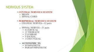
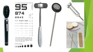
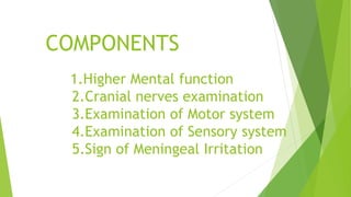
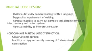
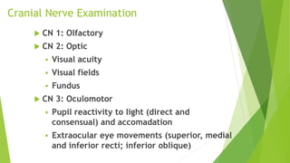
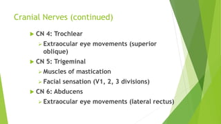
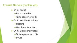
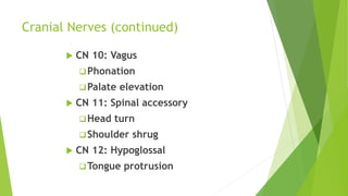
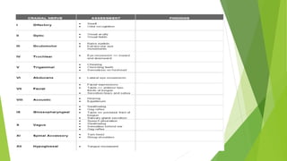
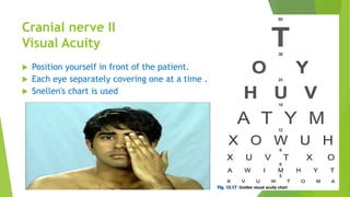
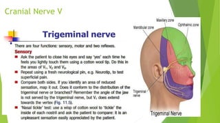
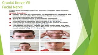
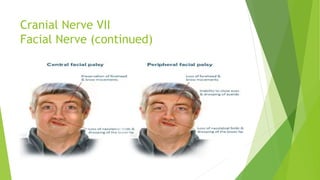
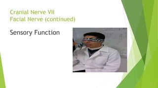
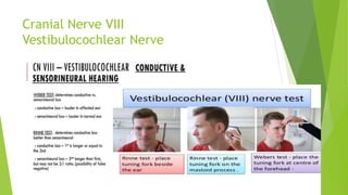
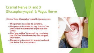
NERVOUS SYSTEM
EXAMINATION
Dr. Shahin Akter Nipa
MD (Phase A) Resident , Internal Medicine
Chittagong Medical College & Hospital
Nervous System Definition
The nervous system is the part
of body that coordinates its
action and transnmits signals
to and from different parts of
the body.
NERVOUS SYSTEM
 CENTRAL NERVOUS SYSTEM
 BRAIN
 SPINAL CORD
 PERIPHERAL NERVOUS SYSTEM
 CRANIAL NERVES---12 pairs
 SPINAL NERVES---31 pairs
• 8 CERVICAL
• 12 THORACIC
• 5 LUMBAR
• 5 SACRAL
• 1 COCCYGEAL
 AUTONOMIC NS
• SYMPATHETIC
• PARASYMPATHETIC
SYMPTOMS
(CANDIDATES FOR NS EXAMINATION)
 HEADACHE
 TRANSIENT LOSS OF CONCIOUSNESS (TLOC)
 TIA
 STROKE
 DIZZINESS
 VERTIGO
 FUNCTIONAL SYMTOMS
EQUIPMENTS for EXAMINATION
 pin
 Cotton
 Tunning fork
 Reflex hummer
 key
 Flashlight
 Ophthalmoscope
 Vision screeners
 Snellen chart
 Ishihara chart
 ophthalmoscope
 Gloves
 Coffee
COMPONENTS
1.Higher Mental function
2.Cranial nerves examination
3.Examination of Motor system
4.Examination of Sensory system
5.Sign of Meningeal Irritation
A.HIGHER
MENTAL
FUNCTION
1. Appearnce & Behavior
 General elements- eg:attire,signs of self neglect
 Disturbed / Agitated/ Confused/ Apathetic
 Neat & Tidy/ Untidy
 Silent/ Monosyllabic/ Over Talkative
 Reaction to greeting
 Presence of Facial Tics/ Inappropiate Behavior
2.Emotional state
Happy/ Distressed/Depressed/Irritable
Enjoy life/ Fed-up with life
3. DELUSION AND HALLUCINATION
Dellusion : It is a false belief in
something which is not a fact.
Hallucination: It is a false perception
of some special senses without any
external object or stimulus.
4. ORIENTATION IN PLACE AND TIME
5.LEVEL OF CONSCIOUSNESS
 Coma: Coma is state in which the patient makes no
psychological meaninngful response to external
stimulus or to inner need.
 Stupor: Show some response for instance to painful
stimuli
 Dementia: Patient awake and alert but muddled in
time,place,and person and has impaired memory
and mental processing
 Delirium: Patient confused but alertness is
impaired
6. Memory:
1. Recent memory:
 Day of the week
 Date in the month
2. Short term memory
 Memory for events of a few seconds or minutes past
 Test-repeat seven digits backwards
 Spell world backwards
3. Long Term Memory
7. INTELLIGENCE
 General knowledge
 Abstraction
 Judgment
 Insight
 Reasoning
8. SPEECH AND LANGUAGE
Listen to the patient’s spontaneous speech,
noting volume,rhythm and clarity.
Ask the patient to repeat phrases such as
‘yellow lorry’ to test lingual (tongue) sounds
and ‘baby hippopotamus’ for labial (lip)
sounds, then a tongue twister, e.g. ‘the Leith
police dismisseth us’.
Ask the patient to count steadily to 30 to
assess fatigue.
Ask the patient to cough and to say ‘Ah’;
observe the soft palate rising bilaterally.
SPEECH AND LANGUAGE (continued…)
 During spontaneous speech, listen to the fluency and appropriateness of the
content, particularly for paraphasias and neologisms.
 Show the patient a common object, e.g. coin or pen, and ask its name.
 Give a simple three-stage command, e.g. pick up this piece of paper, fold it in
half and place it under the book.
 Ask the patient to repeat a simple sentence, e.g. ‘Today is Tuesday’.
 Ask the patient to read a passage from a newspaper.
 Ask the patient to write a sentence; examine his handwriting.
SPEECH AND LANGUAGE (continued…)
 Dysarthria is a motor speech disorder. It results from impaired
movement of the muscles used for speech production, including
the lips, tongue, vocal folds etc.
 Aphasia is an impairment of language, affecting the production or
comprehension of speech and the ability to read or write.
 Dysphasia is loss of or deficiency in the power to use or
understand language as a result of injury to or disease of the
brain.
 Dysphonia is commonly referred to as hoarse voice, refers to
dysfunction in the ability to produce voice due to laryngeal
disorder.
TYPES OF DYSPHAISIA:
1.EXPRESSIVE (MOTOR) DYSPHASIA:
Damage to broca’s area(inferior frontal region)
Decrease verbal output
Non fluent speech
Errors of grammer and syntax
Comprehension is intact
2. RECEPTIVE (SENSORY) DYSPHASIA:
 Dysfunction in Wernicke’s area
 Poor comprehension
 Speech is fluent
 Meaningless
 Paraphasias(incorrect word)
 Neologisms(nonsense new words)
PARIETAL LOBE LESION:
Dyslexia:difficulty comprehending written language
Dysgraphia:impairement of writing
Apraxia: inability to carry out complex task despite having an
intact sensory and motor system
Agnosia:inability to interpret sensation
NONDOMINANT PARIETAL LOBE DYSFUNCTION:
Constructional apraxia:
Inability to copy accurately drawing of 3 dimensional
construction
Cranial Nerve Examination
 CN 1: Olfactory
 CN 2: Optic
 Visual acuity
 Visual fields
 Fundus
 CN 3: Oculomotor
 Pupil reactivity to light (direct and
consensual) and accomadation
 Extraocular eye movements (superior, medial
and inferior recti; inferior oblique)
Cranial Nerves (continued)
 CN 4: Trochlear
 Extraocular eye movements (superior
oblique)
 CN 5: Trigeminal
 Muscles of mastication
 Facial sensation (V1, 2, 3 divisions)
 CN 6: Abducens
 Extraocular eye movements (lateral rectus)
Cranial Nerves (continued)
 CN 7: Facial
Facial muscles
Taste (anterior 2/3)
 CN 8: Vestibulocochlear
Hearing
Vestibular function
 CN 9: Glossopharyngeal
Taste (posterior 1/3)
Uvula
Cranial Nerves (continued)
 CN 10: Vagus
Phonation
Palate elevation
 CN 11: Spinal accessory
Head turn
Shoulder shrug
 CN 12: Hypoglossal
Tongue protrusion
Cranial Nerve I
The Olfactory Nerve
 Each nostril should first be evaluated for
potency by compressing one nostril and
having the patient breath through the
opposite.
 Each nostril should then be tested
separately with a volatile, non-irritating
substance such as cloves, coffee or vanilla.
The patient should close his eyes, occlude
one nostril and identify the substance
placed under the open nostril.
 Causes of anosmia:
-upper RTI
-smoking,increasing age
- ethmoid tumor
-basal skull fracture,frontal fracture
- congenital-Kallmann’s syndrome
-meningioma
-following meningitis
Cranial nerve II
Visual Acuity
 Position yourself in front of the patient.
 Each eye separately covering one at a time .
 Snellen's chart is used
Cranial nerve II (continued)
Colour Vision Test
Cranial nerve II (continued)
OPTHALMOSCOPIC EXAMINATION
Retinal abnormalities.
(A) Left optic atrophy. Note the lack of a pink
neuroretinal rim.
(B) Preretinal haemorrhage.
(C) Pale white swollen disc. This is highly suggestive of
giant cell arteritis, particularly if associated with visual
loss.
(D) Arteriolar occlusion of the horizontal nerve fibre
layer.
Multiple cotton-wool spots in human immunodeficiency
virus (HIV) retinopathy.
(E) Cytomegalovirus retinitis. Note the large superficial
retinal infiltrate associated with flame haemorrhage.
(F) Central retinal artery occlusion. Note the milky-white
pale infarcted retina surrounding healthy pink fovea
(‘cherry-red spot’).
(G) Central retinal vein occlusion. Note the widespread
retinal haemorrhages and swollen optic disc.
(H) Diabetic retinopathy with multiple dot and blot
haemorrhages, indicating widespread capillary
occlusion, a precursor of new vessel formation.
Cranial nerves II , III
Pupils: Reaction to Light
 Have the patient look at a distant object
 Look at size, shape and symmetry of
pupils.
 Shine a light into each eye and observe
constriction of pupil .
 Flash a light on one pupil and watch it
contract briskly .
 Flash the light again and watch the opposite
pupil constriction (consensual reflex)
 Repeat this procedure on the opposite eye.
PUPIL Abnormalities:
 DM:small pupil,responds poorly,due to
autonomic neuropathy
 Argyll Robertson pupil:
-in syphilis
-pinpoint,irregular pupil
-constrict only on convergence
 Holmes adie pupil:
mid dilated,bilateral
responds poorly to convergence
 Macus gunn pupil:
optic nerve damage result in afferent pupillary defect
both pupil contsrict to light
Cranial nerves III, IV and VI
Extraocular Muscles
Cranial nerves III, IV and VI (continued..)
Extraocular Muscles
 3rd nerve palsy
-unilateral ptosis(complete)
-pupil:large(loss of parasympathetic )
-eye look inferolaterally
cause:posterior communicating
artery aneurysm
 Horner’s syndrome:
-partial ptosis
-pupil:small(sympathetic loss)
-drooping eyelid
-decrease sweating
 Myasthenia Gravis:
bilateral ptosis
NYSTAGMUS:
1.Peripheral vestibular nystagmus:
-horizontal
-vertical
-rotatory
2.CENTRAL VESTIBULAR NYSTAGMUS:
unidirectional
cause:-multiple sclerosis
-CVD
3.VERTICAL NYSTAGMUS:
brain stem lesion
Upbeat :
upper brain stem lesion
multiple sclerosis
infarction
Wernicke’s encephalopathy
 Down beat:
Arnold chiari malformation
phenytoin /lithium intoxication
4.PERIODIC ALTERNATING NYSTAGMUS:
Congenital
Drug intoxication
5.ATAXIC NYSTAGMUS:
Marked in abduction
Demyelination of medial longitudinal bundle
within brainstem
6.CONGENITAL NYSTAGMUS
horizontal/pendular
7.ACQUIRED PENDULAR NYSTAGMUS
cerebellar/brainstem disease
multiple sclerosis
spinocerebellar degeneration
brainstem ischaemia
DIPLOPIA:
Pure horizontal: 6th CN palsy
Vertical diplopia: 4th CN palsy, Thyroid eye
disease
Cranial Nerve V
Cranial Nerve V (continued)
Sensory Examination
Cranial Nerve V (continued)
Jaw Jerk
 The mandible or lower jaw—is
tapped at a downward angle just
below the lips at the chin while the
mouth is held slightly open.
 In response, the masseter muscles
will jerk the mandible upwards.
 Normally this reflex is absent or
very slight.
 However in individuals with upper
motor neuron lesions the jaw jerk
reflex can be quite pronounced.
TRIGEMINAL NERVE EXAMINATION
 Unilateral loss of 5th nerve:
direct injury
facial fracture
local invasion by cancer
 Lesion in cavernous sinus:
loss of corneal reflex
V1 ,V2 sensory loss
3,4 ,6 CN also affected
 Trigeminal neuralgia
- due to neurovascular compression
-sevre lancinating pain in V2,V3
 Reactivation of VZV
affect any sensory nerve
 Brisk jaw jerk:
pseudobulbur palsy
Cranial Nerve VII
Facial Nerve
Cranial Nerve VII
Facial Nerve (continued)
Cranial Nerve VII
Facial Nerve (continued)
Sensory Function
Cause of LMN facial palsy
 cerebellopontine angle tumor
 Acoustic angle tumor
 Trauma
 Parotid tumour
Bilateral facial palsy:
 GBS
 Sarcoidosis
 Lyme disease
 HIV
Cranial Nerve VIII
Vestibulocochlear Nerve
Cranial Nerve IX and X
Glossopharyngeal & Vagus Nerve
Unilateral X nerve palsy(recurrent laryngeal)
 lung cancer
 post thyroid surgery
 mediastinal lymphoma
 aortic arch aneurysm
Bilateral X nerve lesion:
 Progressive bulbar palsy(MND)
 Bilateral supranuclear lesion(Pseudobulbur
palsy)
 CVD
 Multiple sclerosis
Unilateral IX and X lesion:
 Skull base tumor
 Skull base fracture
 Stroke(lateral medullary syndrome)
Cranial Nerve XI
Accessory Nerve
•Have patient shrug shoulder against resistance and evaluate strength
of Trapezius muscle.
•Have patient turn head to one side against resistance and evaluate
strength and observe contracting sternomastoid muscle
Cranial Nerve XII
Hypoglossal Nerve
 Ask the patient to move the tongue side to side in the mouth and feel the strength
 Ask the patient to open mouth and observe the tongue whether any atrophy or fasciculation
present or not.
 Ask the patient to protrude the tongue. Protruded tongue deviates to the side of lesion of 12th
nerve.
Nervous system exam part 1

Nervous system exam part 1

  • 1.
    NERVOUS SYSTEM EXAMINATION Dr. ShahinAkter Nipa MD (Phase A) Resident , Internal Medicine Chittagong Medical College & Hospital
  • 2.
    Nervous System Definition Thenervous system is the part of body that coordinates its action and transnmits signals to and from different parts of the body.
  • 3.
    NERVOUS SYSTEM  CENTRALNERVOUS SYSTEM  BRAIN  SPINAL CORD  PERIPHERAL NERVOUS SYSTEM  CRANIAL NERVES---12 pairs  SPINAL NERVES---31 pairs • 8 CERVICAL • 12 THORACIC • 5 LUMBAR • 5 SACRAL • 1 COCCYGEAL  AUTONOMIC NS • SYMPATHETIC • PARASYMPATHETIC
  • 5.
    SYMPTOMS (CANDIDATES FOR NSEXAMINATION)  HEADACHE  TRANSIENT LOSS OF CONCIOUSNESS (TLOC)  TIA  STROKE  DIZZINESS  VERTIGO  FUNCTIONAL SYMTOMS
  • 6.
    EQUIPMENTS for EXAMINATION pin  Cotton  Tunning fork  Reflex hummer  key  Flashlight  Ophthalmoscope  Vision screeners  Snellen chart  Ishihara chart  ophthalmoscope  Gloves  Coffee
  • 8.
    COMPONENTS 1.Higher Mental function 2.Cranialnerves examination 3.Examination of Motor system 4.Examination of Sensory system 5.Sign of Meningeal Irritation
  • 9.
  • 10.
    1. Appearnce &Behavior  General elements- eg:attire,signs of self neglect  Disturbed / Agitated/ Confused/ Apathetic  Neat & Tidy/ Untidy  Silent/ Monosyllabic/ Over Talkative  Reaction to greeting  Presence of Facial Tics/ Inappropiate Behavior
  • 11.
  • 12.
    3. DELUSION ANDHALLUCINATION Dellusion : It is a false belief in something which is not a fact. Hallucination: It is a false perception of some special senses without any external object or stimulus.
  • 13.
    4. ORIENTATION INPLACE AND TIME
  • 15.
  • 16.
     Coma: Comais state in which the patient makes no psychological meaninngful response to external stimulus or to inner need.  Stupor: Show some response for instance to painful stimuli  Dementia: Patient awake and alert but muddled in time,place,and person and has impaired memory and mental processing  Delirium: Patient confused but alertness is impaired
  • 17.
    6. Memory: 1. Recentmemory:  Day of the week  Date in the month 2. Short term memory  Memory for events of a few seconds or minutes past  Test-repeat seven digits backwards  Spell world backwards 3. Long Term Memory
  • 18.
    7. INTELLIGENCE  Generalknowledge  Abstraction  Judgment  Insight  Reasoning
  • 19.
    8. SPEECH ANDLANGUAGE Listen to the patient’s spontaneous speech, noting volume,rhythm and clarity. Ask the patient to repeat phrases such as ‘yellow lorry’ to test lingual (tongue) sounds and ‘baby hippopotamus’ for labial (lip) sounds, then a tongue twister, e.g. ‘the Leith police dismisseth us’. Ask the patient to count steadily to 30 to assess fatigue. Ask the patient to cough and to say ‘Ah’; observe the soft palate rising bilaterally.
  • 20.
    SPEECH AND LANGUAGE(continued…)  During spontaneous speech, listen to the fluency and appropriateness of the content, particularly for paraphasias and neologisms.  Show the patient a common object, e.g. coin or pen, and ask its name.  Give a simple three-stage command, e.g. pick up this piece of paper, fold it in half and place it under the book.  Ask the patient to repeat a simple sentence, e.g. ‘Today is Tuesday’.  Ask the patient to read a passage from a newspaper.  Ask the patient to write a sentence; examine his handwriting.
  • 21.
    SPEECH AND LANGUAGE(continued…)  Dysarthria is a motor speech disorder. It results from impaired movement of the muscles used for speech production, including the lips, tongue, vocal folds etc.  Aphasia is an impairment of language, affecting the production or comprehension of speech and the ability to read or write.  Dysphasia is loss of or deficiency in the power to use or understand language as a result of injury to or disease of the brain.  Dysphonia is commonly referred to as hoarse voice, refers to dysfunction in the ability to produce voice due to laryngeal disorder.
  • 22.
    TYPES OF DYSPHAISIA: 1.EXPRESSIVE(MOTOR) DYSPHASIA: Damage to broca’s area(inferior frontal region) Decrease verbal output Non fluent speech Errors of grammer and syntax Comprehension is intact
  • 23.
    2. RECEPTIVE (SENSORY)DYSPHASIA:  Dysfunction in Wernicke’s area  Poor comprehension  Speech is fluent  Meaningless  Paraphasias(incorrect word)  Neologisms(nonsense new words)
  • 24.
    PARIETAL LOBE LESION: Dyslexia:difficultycomprehending written language Dysgraphia:impairement of writing Apraxia: inability to carry out complex task despite having an intact sensory and motor system Agnosia:inability to interpret sensation NONDOMINANT PARIETAL LOBE DYSFUNCTION: Constructional apraxia: Inability to copy accurately drawing of 3 dimensional construction
  • 25.
    Cranial Nerve Examination CN 1: Olfactory  CN 2: Optic  Visual acuity  Visual fields  Fundus  CN 3: Oculomotor  Pupil reactivity to light (direct and consensual) and accomadation  Extraocular eye movements (superior, medial and inferior recti; inferior oblique)
  • 26.
    Cranial Nerves (continued) CN 4: Trochlear  Extraocular eye movements (superior oblique)  CN 5: Trigeminal  Muscles of mastication  Facial sensation (V1, 2, 3 divisions)  CN 6: Abducens  Extraocular eye movements (lateral rectus)
  • 27.
    Cranial Nerves (continued) CN 7: Facial Facial muscles Taste (anterior 2/3)  CN 8: Vestibulocochlear Hearing Vestibular function  CN 9: Glossopharyngeal Taste (posterior 1/3) Uvula
  • 28.
    Cranial Nerves (continued) CN 10: Vagus Phonation Palate elevation  CN 11: Spinal accessory Head turn Shoulder shrug  CN 12: Hypoglossal Tongue protrusion
  • 30.
    Cranial Nerve I TheOlfactory Nerve  Each nostril should first be evaluated for potency by compressing one nostril and having the patient breath through the opposite.  Each nostril should then be tested separately with a volatile, non-irritating substance such as cloves, coffee or vanilla. The patient should close his eyes, occlude one nostril and identify the substance placed under the open nostril.
  • 31.
     Causes ofanosmia: -upper RTI -smoking,increasing age - ethmoid tumor -basal skull fracture,frontal fracture - congenital-Kallmann’s syndrome -meningioma -following meningitis
  • 32.
    Cranial nerve II VisualAcuity  Position yourself in front of the patient.  Each eye separately covering one at a time .  Snellen's chart is used
  • 35.
    Cranial nerve II(continued)
  • 37.
    Cranial nerve II(continued) Colour Vision Test
  • 38.
    Cranial nerve II(continued) OPTHALMOSCOPIC EXAMINATION
  • 40.
    Retinal abnormalities. (A) Leftoptic atrophy. Note the lack of a pink neuroretinal rim. (B) Preretinal haemorrhage. (C) Pale white swollen disc. This is highly suggestive of giant cell arteritis, particularly if associated with visual loss. (D) Arteriolar occlusion of the horizontal nerve fibre layer. Multiple cotton-wool spots in human immunodeficiency virus (HIV) retinopathy. (E) Cytomegalovirus retinitis. Note the large superficial retinal infiltrate associated with flame haemorrhage. (F) Central retinal artery occlusion. Note the milky-white pale infarcted retina surrounding healthy pink fovea (‘cherry-red spot’). (G) Central retinal vein occlusion. Note the widespread retinal haemorrhages and swollen optic disc. (H) Diabetic retinopathy with multiple dot and blot haemorrhages, indicating widespread capillary occlusion, a precursor of new vessel formation.
  • 41.
    Cranial nerves II, III Pupils: Reaction to Light  Have the patient look at a distant object  Look at size, shape and symmetry of pupils.  Shine a light into each eye and observe constriction of pupil .  Flash a light on one pupil and watch it contract briskly .  Flash the light again and watch the opposite pupil constriction (consensual reflex)  Repeat this procedure on the opposite eye.
  • 42.
    PUPIL Abnormalities:  DM:smallpupil,responds poorly,due to autonomic neuropathy  Argyll Robertson pupil: -in syphilis -pinpoint,irregular pupil -constrict only on convergence
  • 43.
     Holmes adiepupil: mid dilated,bilateral responds poorly to convergence  Macus gunn pupil: optic nerve damage result in afferent pupillary defect both pupil contsrict to light
  • 44.
    Cranial nerves III,IV and VI Extraocular Muscles
  • 45.
    Cranial nerves III,IV and VI (continued..) Extraocular Muscles
  • 46.
     3rd nervepalsy -unilateral ptosis(complete) -pupil:large(loss of parasympathetic ) -eye look inferolaterally cause:posterior communicating artery aneurysm
  • 47.
     Horner’s syndrome: -partialptosis -pupil:small(sympathetic loss) -drooping eyelid -decrease sweating
  • 48.
  • 49.
  • 50.
  • 51.
    3.VERTICAL NYSTAGMUS: brain stemlesion Upbeat : upper brain stem lesion multiple sclerosis infarction Wernicke’s encephalopathy
  • 52.
     Down beat: Arnoldchiari malformation phenytoin /lithium intoxication
  • 53.
    4.PERIODIC ALTERNATING NYSTAGMUS: Congenital Drugintoxication 5.ATAXIC NYSTAGMUS: Marked in abduction Demyelination of medial longitudinal bundle within brainstem
  • 54.
    6.CONGENITAL NYSTAGMUS horizontal/pendular 7.ACQUIRED PENDULARNYSTAGMUS cerebellar/brainstem disease multiple sclerosis spinocerebellar degeneration brainstem ischaemia
  • 55.
    DIPLOPIA: Pure horizontal: 6thCN palsy Vertical diplopia: 4th CN palsy, Thyroid eye disease
  • 56.
  • 57.
    Cranial Nerve V(continued) Sensory Examination
  • 58.
    Cranial Nerve V(continued)
  • 59.
    Cranial Nerve V(continued)
  • 60.
    Cranial Nerve V(continued) Jaw Jerk  The mandible or lower jaw—is tapped at a downward angle just below the lips at the chin while the mouth is held slightly open.  In response, the masseter muscles will jerk the mandible upwards.  Normally this reflex is absent or very slight.  However in individuals with upper motor neuron lesions the jaw jerk reflex can be quite pronounced.
  • 61.
  • 62.
     Unilateral lossof 5th nerve: direct injury facial fracture local invasion by cancer
  • 63.
     Lesion incavernous sinus: loss of corneal reflex V1 ,V2 sensory loss 3,4 ,6 CN also affected
  • 64.
     Trigeminal neuralgia -due to neurovascular compression -sevre lancinating pain in V2,V3  Reactivation of VZV affect any sensory nerve  Brisk jaw jerk: pseudobulbur palsy
  • 65.
  • 66.
    Cranial Nerve VII FacialNerve (continued)
  • 67.
    Cranial Nerve VII FacialNerve (continued) Sensory Function
  • 68.
    Cause of LMNfacial palsy  cerebellopontine angle tumor  Acoustic angle tumor  Trauma  Parotid tumour
  • 69.
    Bilateral facial palsy: GBS  Sarcoidosis  Lyme disease  HIV
  • 70.
  • 71.
    Cranial Nerve IXand X Glossopharyngeal & Vagus Nerve
  • 72.
    Unilateral X nervepalsy(recurrent laryngeal)  lung cancer  post thyroid surgery  mediastinal lymphoma  aortic arch aneurysm
  • 73.
    Bilateral X nervelesion:  Progressive bulbar palsy(MND)  Bilateral supranuclear lesion(Pseudobulbur palsy)  CVD  Multiple sclerosis
  • 74.
    Unilateral IX andX lesion:  Skull base tumor  Skull base fracture  Stroke(lateral medullary syndrome)
  • 75.
    Cranial Nerve XI AccessoryNerve •Have patient shrug shoulder against resistance and evaluate strength of Trapezius muscle. •Have patient turn head to one side against resistance and evaluate strength and observe contracting sternomastoid muscle
  • 76.
    Cranial Nerve XII HypoglossalNerve  Ask the patient to move the tongue side to side in the mouth and feel the strength  Ask the patient to open mouth and observe the tongue whether any atrophy or fasciculation present or not.  Ask the patient to protrude the tongue. Protruded tongue deviates to the side of lesion of 12th nerve.