This document discusses the structure and function of the peripheral nervous system. It covers topics such as:
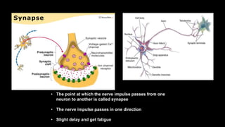
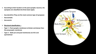
- The basic types of synapses based on their location in neurons.
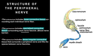
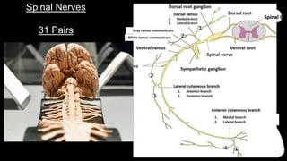
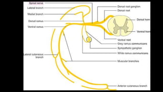
- The structure of peripheral nerves including the endoneurium, perineurium, and epineurium tissues.
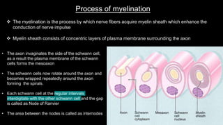
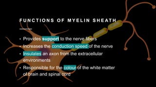
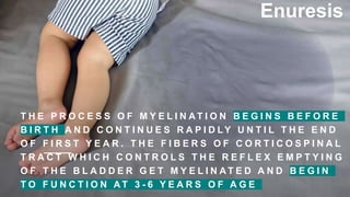
- Myelination including the process of how myelin sheaths form around axons and the functions they provide.
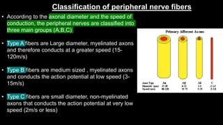
- Classification of peripheral nerve fibers based on axon diameter and conduction speed.
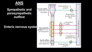
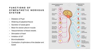
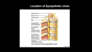
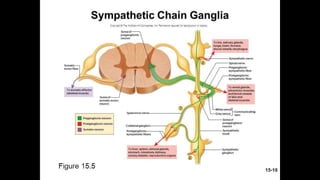
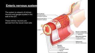
- Components of the autonomic nervous system including the sympathetic and parasympathetic systems.


![ONFH[AVN HIP] -TRIPLE REGIME -A NOVAL SURGICAL CONCEPT .pptx](https://cdn.slidesharecdn.com/ss_thumbnails/onfhavnhip2026koaconcalicutdrgokuldevdrmashraf-260210064517-213ec005-thumbnail.jpg?width=640&height=640&fit=bounds)


