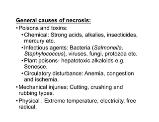
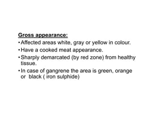
Necrosis
Necrosis
Necrosis
•Necrosis is death of cells and tissues in the
living animal.
•Focal/ Multifocal necrosis- terms used for one
or more, small, clearly defined areas of
necrosis.
•Diffuse necrosis- term used when necrosis
affects a large area or the entire tissue or
organ.
General causes of necrosis:
•Poisons and toxins:
•Chemical: Strong acids, alkalies, insecticides,
mercury etc.
•Infectious agents: Bacteria (Salmonella,
Staphylococcus), viruses, fungi, protozoa etc.
•Plant poisons- hepatotoxic alkaloids e.g.
Senesce.
•Circulatory disturbance: Anemia, congestion
and ischemia.
•Mechanical injuries: Cutting, crushing and
rubbing types.
•Physical : Extreme temperature, electricity, free
radical.
Gross appearance:
•Affected areas white, gray or yellow in colour.
•Have a cooked meat appearance.
•Sharply demarcated (by red zone) from healthy
tissue.
•In case of gangrene the area is green, orange
or black ( iron sulphide)
•Microscopic appearance: The microscopic
changes of necrosis vary with the type of
necrosis. Some general changes of necrosis in
the cytoplasm are:
•Eosinophilia: The cytoplasm stains darker red
in colour.
•Swelling and vacuolation: The cells are swollen
and contain different types of vacuoles.
•Changes in the nucleus: The nucleus may
show condensation (Pyknosis), fragmentation
(karyorrhexis) and may disappear (karyolysis).
Necrosis & Autolysis
•Autolysis:
•Autolysis is death of cells and tissues after the
death of the animal (somatic death) and it
should be distinguished from necrosis.
Cont0
•The enzymatic digestion of cells by enzymes
present within them. The cells most susceptible
to autolysis tend to be dying or dead cells
•1. No sharp line of demarcation between
affected and healthy tissue
•2. Circulatory changes like congestion and
haemorrhage are not present.
•AUTOLYSIS NECROSIS
1. No sharp line of demarcation
between affected and healthy
tissue
1. A line of demarcation is usually
present
2. Circulatory changes like
congestion and haemorrhage are
2. Circulatory changes are
present
congestion and haemorrhage are
not present.
present
3. Inflammatory changes are not
present.
3. Inflammatory changes like
infiltration of leukocytes are
present.
4. Growth of saprophytic
bacteria, bacillary rods in long
chains are often present.
4. Saprophytic growth not seen,
but pathogenic bacteria maybe
present.
Types of necrosis:
•Different types of necrosis are recognized
according to the causes, pathogenesis and the
tissue involved. These include Coagulative,
liquefactive, caseous and fat necrosis.
•Coagulative necrosis:
•Most common type of necrosis.
•Architectural outlines persist but cellular details
are lost.
•Type of tissue can be recognized.
•Denaturation (coagulation) of structural and
enzymic proteins blocks proteolysis.
• Causes:
1. Ischemia due to thrombosis/ embolism as in
infarcts.
2. Bacterial toxins e.g. Fusobacterium
necrophorum in livers in cattle.
3. Muscular dystrophy due to deficiency of
selenium and vit. E in cattle and sheep.
4. Necrosis of renal epithelium due to
poisoning from mercuric salts.
•Gross appearance:
•Necrotic area is firm, opaque with cooked meat
appearance.
•It is sharply demarcated from the healthy areas.
•Microscopic appearance: Architectural
outlines are present; cellular details are lacking.
•Result: Dead tissues remain in the body for a
long period, ultimately removed by
macrophages.
•Liquefactive necrosis: There is digestion and
liquefaction of necrotic tissue.
•Causes:
1. Pyogenic bacterial infections attract
neutrophils. Bacterial and leukocytic enzymes
liquefy dead cells and tissues.
2. Some chemicals like turpentine oil also attract
neutrophils and cause pus formation and
liquefactive necrosis.
3. The necrosis in the nervous tissue is mostly
liquefactive due to high content of lipids and
water.
•Gross appearance: The necrotic tissue is
liquefied and filled with semisolid creamy liquid
called pus.
•Pus:
•It is a thick, white or yellow, creamy liquid
consisting of exudate of leukocytes, tissue
debris and microorganisms.
•Proteolytic enzymes released from neutrophils
cause liquefaction of dead cells.
•Abscess:
•It is a localized collection of pus, surrounded by
fibrous capsule.
•Empyema: It is accumulation of pus in a body
cavity.
•Microscopic appearance:
No architectural or cellular details are visible in
the area of necrosis.
The necrotic area usually appears as a cavity
containing a mass of necrotic neutrophils,
bacteria and tissue debris.
The entire necrotic mass is surrounded by a
fibrous connective tissue capsule.
•Caseous necrosis: Dead tissue is converted
into a homogenous, granular mass resembling
cottage cheese.
•Cause: Associated with lesions of
Mycobacterium tuberculosis and
Arcanobacterium ovis, the cause of caseous
lymphadinitis.
Gross appearance:
• The area of necrosis is amorphous, granular,
friable, white-gray resembling cottage cheese.
•The caseous mass is enclosed within a
connective tissue capsule.
Microscopic appearance:
• The necrotic tissue is amorphous, granular
mass enclosed inside a zone of granulomatous
inflammation, containing macrophages. No
architectural or cellular details are seen.
Calcification commonly occurs in the necrotic
areas.
•Fat necrosis: Death of adipose tissue in a
living animal. There are three types of fat
necrosis:
•Pancreatic
•Traumatic and
•Nutritional
Pancreatic fat necrosis:
• Death of adipose tissue in and around
pancreas.
Causes:
• Pancreatitis and/or injury to pancreas and
its ducts release lipases which attack
adipose tissue in the peritoneum.
• Hydrolysis of triglycerides releases fatty
acids which combine with calcium to
produce chalky white areas (saponification).
.
• Gross appearance: Necrotic fat appears as
white or yellowish chalky masses. A zone of
inflammation appears around the necrotic
areas.
• Microscopic appearance: The necrotic
tissue is solid and homogenous and there
are numerous small needle-shaped clefts
occupied by fatty acid crystals
Traumatic fat necrosis:
• It occurs in subcutaneous adipose tissue
caused by mechanical injury.
Nutritional fat necrosis:
• This form of fat necrosis is seen in sheep
and cattle with extreme debility and
emaciation e.g. Tuberculosis and
Paratuberculosis
• Large, solid, chalky white masses occur in
the mesenteric adipose tissue. May interfere
with passage of ingesta.

necrosis and it's types and adaptation.

  • 1.
  • 2.
    Necrosis •Necrosis is deathof cells and tissues in the living animal. •Focal/ Multifocal necrosis- terms used for one or more, small, clearly defined areas of or more, small, clearly defined areas of necrosis. •Diffuse necrosis- term used when necrosis affects a large area or the entire tissue or organ.
  • 3.
    General causes ofnecrosis: •Poisons and toxins: •Chemical: Strong acids, alkalies, insecticides, mercury etc. •Infectious agents: Bacteria (Salmonella, Staphylococcus), viruses, fungi, protozoa etc. •Plant poisons- hepatotoxic alkaloids e.g. •Plant poisons- hepatotoxic alkaloids e.g. Senesce. •Circulatory disturbance: Anemia, congestion and ischemia. •Mechanical injuries: Cutting, crushing and rubbing types. •Physical : Extreme temperature, electricity, free radical.
  • 4.
    Gross appearance: •Affected areaswhite, gray or yellow in colour. •Have a cooked meat appearance. •Sharply demarcated (by red zone) from healthy •Sharply demarcated (by red zone) from healthy tissue. •In case of gangrene the area is green, orange or black ( iron sulphide)
  • 5.
    •Microscopic appearance: Themicroscopic changes of necrosis vary with the type of necrosis. Some general changes of necrosis in the cytoplasm are: •Eosinophilia: The cytoplasm stains darker red in colour. •Swelling and vacuolation: The cells are swollen •Swelling and vacuolation: The cells are swollen and contain different types of vacuoles. •Changes in the nucleus: The nucleus may show condensation (Pyknosis), fragmentation (karyorrhexis) and may disappear (karyolysis).
  • 6.
    Necrosis & Autolysis •Autolysis: •Autolysisis death of cells and tissues after the death of the animal (somatic death) and it should be distinguished from necrosis. should be distinguished from necrosis. Cont0
  • 7.
    •The enzymatic digestionof cells by enzymes present within them. The cells most susceptible to autolysis tend to be dying or dead cells •1. No sharp line of demarcation between affected and healthy tissue •2. Circulatory changes like congestion and •2. Circulatory changes like congestion and haemorrhage are not present.
  • 8.
    •AUTOLYSIS NECROSIS 1. Nosharp line of demarcation between affected and healthy tissue 1. A line of demarcation is usually present 2. Circulatory changes like congestion and haemorrhage are 2. Circulatory changes are present congestion and haemorrhage are not present. present 3. Inflammatory changes are not present. 3. Inflammatory changes like infiltration of leukocytes are present. 4. Growth of saprophytic bacteria, bacillary rods in long chains are often present. 4. Saprophytic growth not seen, but pathogenic bacteria maybe present.
  • 9.
    Types of necrosis: •Differenttypes of necrosis are recognized according to the causes, pathogenesis and the tissue involved. These include Coagulative, liquefactive, caseous and fat necrosis. •Coagulative necrosis: •Most common type of necrosis. •Most common type of necrosis. •Architectural outlines persist but cellular details are lost. •Type of tissue can be recognized. •Denaturation (coagulation) of structural and enzymic proteins blocks proteolysis.
  • 10.
    • Causes: 1. Ischemiadue to thrombosis/ embolism as in infarcts. 2. Bacterial toxins e.g. Fusobacterium necrophorum in livers in cattle. 3. Muscular dystrophy due to deficiency of selenium and vit. E in cattle and sheep. 4. Necrosis of renal epithelium due to poisoning from mercuric salts.
  • 11.
    •Gross appearance: •Necrotic areais firm, opaque with cooked meat appearance. •It is sharply demarcated from the healthy areas. •Microscopic appearance: Architectural outlines are present; cellular details are lacking. outlines are present; cellular details are lacking. •Result: Dead tissues remain in the body for a long period, ultimately removed by macrophages.
  • 12.
    •Liquefactive necrosis: Thereis digestion and liquefaction of necrotic tissue. •Causes: 1. Pyogenic bacterial infections attract neutrophils. Bacterial and leukocytic enzymes liquefy dead cells and tissues. 2. Some chemicals like turpentine oil also attract neutrophils and cause pus formation and liquefactive necrosis. 3. The necrosis in the nervous tissue is mostly liquefactive due to high content of lipids and water.
  • 13.
    •Gross appearance: Thenecrotic tissue is liquefied and filled with semisolid creamy liquid called pus. •Pus: •It is a thick, white or yellow, creamy liquid consisting of exudate of leukocytes, tissue debris and microorganisms. debris and microorganisms. •Proteolytic enzymes released from neutrophils cause liquefaction of dead cells. •Abscess: •It is a localized collection of pus, surrounded by fibrous capsule. •Empyema: It is accumulation of pus in a body cavity.
  • 14.
    •Microscopic appearance: No architecturalor cellular details are visible in the area of necrosis. The necrotic area usually appears as a cavity containing a mass of necrotic neutrophils, bacteria and tissue debris. bacteria and tissue debris. The entire necrotic mass is surrounded by a fibrous connective tissue capsule.
  • 15.
    •Caseous necrosis: Deadtissue is converted into a homogenous, granular mass resembling cottage cheese. •Cause: Associated with lesions of •Cause: Associated with lesions of Mycobacterium tuberculosis and Arcanobacterium ovis, the cause of caseous lymphadinitis.
  • 16.
    Gross appearance: • Thearea of necrosis is amorphous, granular, friable, white-gray resembling cottage cheese. •The caseous mass is enclosed within a connective tissue capsule. Microscopic appearance: Microscopic appearance: • The necrotic tissue is amorphous, granular mass enclosed inside a zone of granulomatous inflammation, containing macrophages. No architectural or cellular details are seen. Calcification commonly occurs in the necrotic areas.
  • 17.
    •Fat necrosis: Deathof adipose tissue in a living animal. There are three types of fat necrosis: •Pancreatic •Traumatic and •Nutritional
  • 18.
    Pancreatic fat necrosis: •Death of adipose tissue in and around pancreas. Causes: • Pancreatitis and/or injury to pancreas and • Pancreatitis and/or injury to pancreas and its ducts release lipases which attack adipose tissue in the peritoneum. • Hydrolysis of triglycerides releases fatty acids which combine with calcium to produce chalky white areas (saponification). .
  • 19.
    • Gross appearance:Necrotic fat appears as white or yellowish chalky masses. A zone of inflammation appears around the necrotic areas. • Microscopic appearance: The necrotic • Microscopic appearance: The necrotic tissue is solid and homogenous and there are numerous small needle-shaped clefts occupied by fatty acid crystals
  • 20.
    Traumatic fat necrosis: •It occurs in subcutaneous adipose tissue caused by mechanical injury. Nutritional fat necrosis: • This form of fat necrosis is seen in sheep and cattle with extreme debility and and cattle with extreme debility and emaciation e.g. Tuberculosis and Paratuberculosis • Large, solid, chalky white masses occur in the mesenteric adipose tissue. May interfere with passage of ingesta.