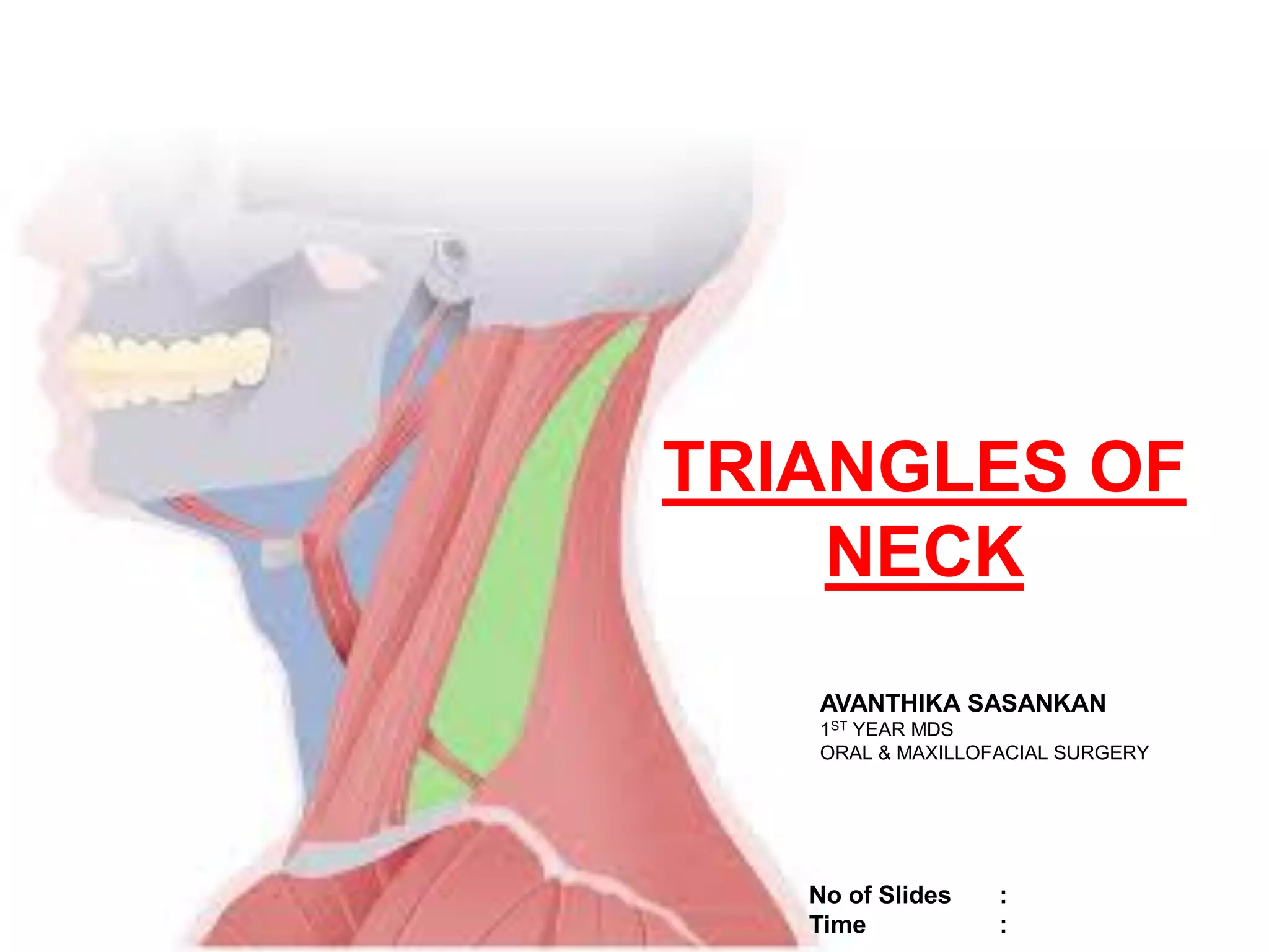
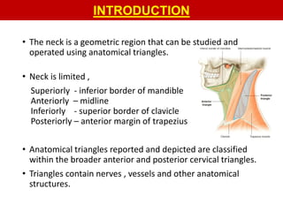
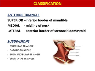
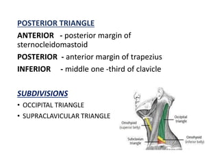
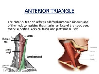
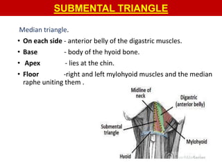
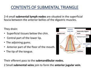
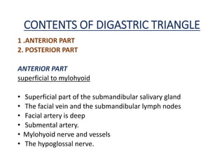
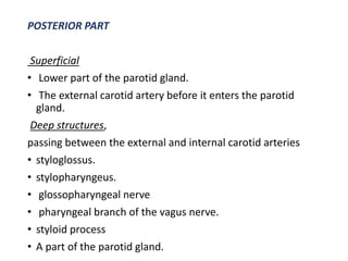
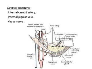
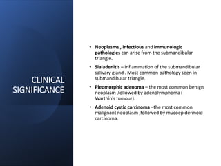
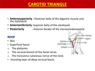
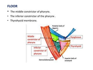
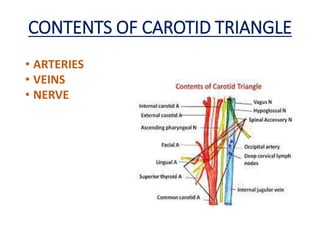
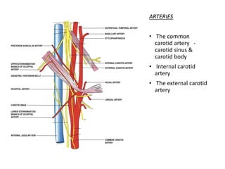
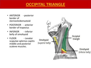
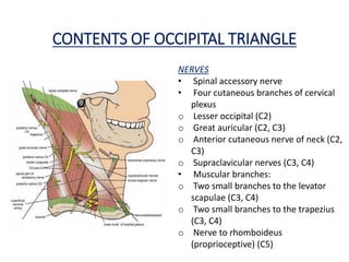
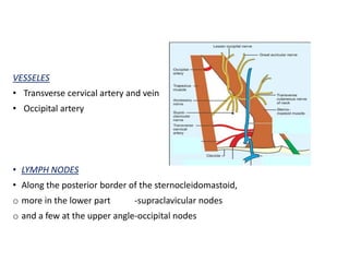
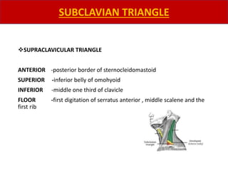
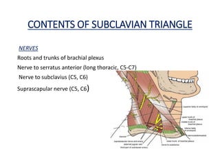
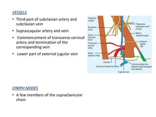
The document describes various anatomical triangles of the neck region. It discusses 11 triangles in detail, providing their boundaries, contents, and clinical significance. The triangles described include the anterior triangle, submental triangle, submandibular triangle, carotid triangle, muscular triangle, posterior triangle, occipital triangle, and supraclavicular triangle. Structures like nerves, vessels, muscles and lymph nodes contained within each triangle are outlined. Potential surgical and pathological implications are also mentioned.
Introduction
Suprahyoid muscle and its embryology
Relation of mylohyoid and digastric muscle
Submandibular gland and duct
Development and histology
Sublingual gland and duct ,it’s development and histology.
Submandibular ganglion and its relations
Clinical anatomy
Blood and nerve supply of submandibular and sublingual duct
Conclusion
References
hypoglossal nerve, origin course an termination of hypoglossal nerve, function of hypoglossal nerve, clinical examination of hypoglossal nerve, hypoglossal nerve palsy
Dr, Kathirvel Gopalakrishnan
M.D.S (OMFS)
Presentation on Anterior triangles of neck which helps for a quick refresh.
Applied aspects described well and all slides will be informative with lot of image based examples
scalp; is the soft tissue covering of cranial vault.
it extends anteriorly: supraorbital margin
posteriorly:external occipital protuberance and superior nuchal lines.
on each side: superior temporal lines.
introduction to skull, parts of skull, bones involved forming skull, different views of skull, norma basalis, anterio cranial middle cranial and posterior cranial fossa, clinical aspects of cranial fossa, foramens present in the cranial fossa
The parotid gland is a major salivary gland in many animals. In humans, the two parotid glands are present on either side of the mouth and in front of both ears. They are the largest of the salivary glands.
On every Wednesday The department of Otolaryngology & Head neck surgery of Sylhet MAG Osmani Medical College Does Case presentation session by Students and doctors.
this was done by the 5th year medical students (52nd MBBS,F batch 2013-14 session)
Recomendações da OMS sobre cuidados maternos e neonatais para uma experiência pós-natal positiva.
Em consonância com os ODS – Objetivos do Desenvolvimento Sustentável e a Estratégia Global para a Saúde das Mulheres, Crianças e Adolescentes, e aplicando uma abordagem baseada nos direitos humanos, os esforços de cuidados pós-natais devem expandir-se para além da cobertura e da simples sobrevivência, de modo a incluir cuidados de qualidade.
Estas diretrizes visam melhorar a qualidade dos cuidados pós-natais essenciais e de rotina prestados às mulheres e aos recém-nascidos, com o objetivo final de melhorar a saúde e o bem-estar materno e neonatal.
Uma “experiência pós-natal positiva” é um resultado importante para todas as mulheres que dão à luz e para os seus recém-nascidos, estabelecendo as bases para a melhoria da saúde e do bem-estar a curto e longo prazo. Uma experiência pós-natal positiva é definida como aquela em que as mulheres, pessoas que gestam, os recém-nascidos, os casais, os pais, os cuidadores e as famílias recebem informação consistente, garantia e apoio de profissionais de saúde motivados; e onde um sistema de saúde flexível e com recursos reconheça as necessidades das mulheres e dos bebês e respeite o seu contexto cultural.
Estas diretrizes consolidadas apresentam algumas recomendações novas e já bem fundamentadas sobre cuidados pós-natais de rotina para mulheres e neonatos que recebem cuidados no pós-parto em unidades de saúde ou na comunidade, independentemente dos recursos disponíveis.
É fornecido um conjunto abrangente de recomendações para cuidados durante o período puerperal, com ênfase nos cuidados essenciais que todas as mulheres e recém-nascidos devem receber, e com a devida atenção à qualidade dos cuidados; isto é, a entrega e a experiência do cuidado recebido. Estas diretrizes atualizam e ampliam as recomendações da OMS de 2014 sobre cuidados pós-natais da mãe e do recém-nascido e complementam as atuais diretrizes da OMS sobre a gestão de complicações pós-natais.
O estabelecimento da amamentação e o manejo das principais intercorrências é contemplada.
Recomendamos muito.
Vamos discutir essas recomendações no nosso curso de pós-graduação em Aleitamento no Instituto Ciclos.
Esta publicação só está disponível em inglês até o momento.
Prof. Marcus Renato de Carvalho
www.agostodourado.com
Flu Vaccine Alert in Bangalore Karnatakaaddon Scans
As flu season approaches, health officials in Bangalore, Karnataka, are urging residents to get their flu vaccinations. The seasonal flu, while common, can lead to severe health complications, particularly for vulnerable populations such as young children, the elderly, and those with underlying health conditions.
Dr. Vidisha Kumari, a leading epidemiologist in Bangalore, emphasizes the importance of getting vaccinated. "The flu vaccine is our best defense against the influenza virus. It not only protects individuals but also helps prevent the spread of the virus in our communities," he says.
This year, the flu season is expected to coincide with a potential increase in other respiratory illnesses. The Karnataka Health Department has launched an awareness campaign highlighting the significance of flu vaccinations. They have set up multiple vaccination centers across Bangalore, making it convenient for residents to receive their shots.
To encourage widespread vaccination, the government is also collaborating with local schools, workplaces, and community centers to facilitate vaccination drives. Special attention is being given to ensuring that the vaccine is accessible to all, including marginalized communities who may have limited access to healthcare.
Residents are reminded that the flu vaccine is safe and effective. Common side effects are mild and may include soreness at the injection site, mild fever, or muscle aches. These side effects are generally short-lived and far less severe than the flu itself.
Healthcare providers are also stressing the importance of continuing COVID-19 precautions. Wearing masks, practicing good hand hygiene, and maintaining social distancing are still crucial, especially in crowded places.
Protect yourself and your loved ones by getting vaccinated. Together, we can help keep Bangalore healthy and safe this flu season. For more information on vaccination centers and schedules, residents can visit the Karnataka Health Department’s official website or follow their social media pages.
Stay informed, stay safe, and get your flu shot today!
Pulmonary Thromboembolism - etilogy, types, medical- Surgical and nursing man...VarunMahajani
Disruption of blood supply to lung alveoli due to blockage of one or more pulmonary blood vessels is called as Pulmonary thromboembolism. In this presentation we will discuss its causes, types and its management in depth.
These simplified slides by Dr. Sidra Arshad present an overview of the non-respiratory functions of the respiratory tract.
Learning objectives:
1. Enlist the non-respiratory functions of the respiratory tract
2. Briefly explain how these functions are carried out
3. Discuss the significance of dead space
4. Differentiate between minute ventilation and alveolar ventilation
5. Describe the cough and sneeze reflexes
Study Resources:
1. Chapter 39, Guyton and Hall Textbook of Medical Physiology, 14th edition
2. Chapter 34, Ganong’s Review of Medical Physiology, 26th edition
3. Chapter 17, Human Physiology by Lauralee Sherwood, 9th edition
4. Non-respiratory functions of the lungs https://academic.oup.com/bjaed/article/13/3/98/278874
micro teaching on communication m.sc nursing.pdfAnurag Sharma
Microteaching is a unique model of practice teaching. It is a viable instrument for the. desired change in the teaching behavior or the behavior potential which, in specified types of real. classroom situations, tends to facilitate the achievement of specified types of objectives.
Tom Selleck Health: A Comprehensive Look at the Iconic Actor’s Wellness Journeygreendigital
Tom Selleck, an enduring figure in Hollywood. has captivated audiences for decades with his rugged charm, iconic moustache. and memorable roles in television and film. From his breakout role as Thomas Magnum in Magnum P.I. to his current portrayal of Frank Reagan in Blue Bloods. Selleck's career has spanned over 50 years. But beyond his professional achievements. fans have often been curious about Tom Selleck Health. especially as he has aged in the public eye.
Follow us on: Pinterest
Introduction
Many have been interested in Tom Selleck health. not only because of his enduring presence on screen but also because of the challenges. and lifestyle choices he has faced and made over the years. This article delves into the various aspects of Tom Selleck health. exploring his fitness regimen, diet, mental health. and the challenges he has encountered as he ages. We'll look at how he maintains his well-being. the health issues he has faced, and his approach to ageing .
Early Life and Career
Childhood and Athletic Beginnings
Tom Selleck was born on January 29, 1945, in Detroit, Michigan, and grew up in Sherman Oaks, California. From an early age, he was involved in sports, particularly basketball. which played a significant role in his physical development. His athletic pursuits continued into college. where he attended the University of Southern California (USC) on a basketball scholarship. This early involvement in sports laid a strong foundation for his physical health and disciplined lifestyle.
Transition to Acting
Selleck's transition from an athlete to an actor came with its physical demands. His first significant role in "Magnum P.I." required him to perform various stunts and maintain a fit appearance. This role, which he played from 1980 to 1988. necessitated a rigorous fitness routine to meet the show's demands. setting the stage for his long-term commitment to health and wellness.
Fitness Regimen
Workout Routine
Tom Selleck health and fitness regimen has evolved. adapting to his changing roles and age. During his "Magnum, P.I." days. Selleck's workouts were intense and focused on building and maintaining muscle mass. His routine included weightlifting, cardiovascular exercises. and specific training for the stunts he performed on the show.
Selleck adjusted his fitness routine as he aged to suit his body's needs. Today, his workouts focus on maintaining flexibility, strength, and cardiovascular health. He incorporates low-impact exercises such as swimming, walking, and light weightlifting. This balanced approach helps him stay fit without putting undue strain on his joints and muscles.
Importance of Flexibility and Mobility
In recent years, Selleck has emphasized the importance of flexibility and mobility in his fitness regimen. Understanding the natural decline in muscle mass and joint flexibility with age. he includes stretching and yoga in his routine. These practices help prevent injuries, improve posture, and maintain mobilit
Ethanol (CH3CH2OH), or beverage alcohol, is a two-carbon alcohol
that is rapidly distributed in the body and brain. Ethanol alters many
neurochemical systems and has rewarding and addictive properties. It
is the oldest recreational drug and likely contributes to more morbidity,
mortality, and public health costs than all illicit drugs combined. The
5th edition of the Diagnostic and Statistical Manual of Mental Disorders
(DSM-5) integrates alcohol abuse and alcohol dependence into a single
disorder called alcohol use disorder (AUD), with mild, moderate,
and severe subclassifications (American Psychiatric Association, 2013).
In the DSM-5, all types of substance abuse and dependence have been
combined into a single substance use disorder (SUD) on a continuum
from mild to severe. A diagnosis of AUD requires that at least two of
the 11 DSM-5 behaviors be present within a 12-month period (mild
AUD: 2–3 criteria; moderate AUD: 4–5 criteria; severe AUD: 6–11 criteria).
The four main behavioral effects of AUD are impaired control over
drinking, negative social consequences, risky use, and altered physiological
effects (tolerance, withdrawal). This chapter presents an overview
of the prevalence and harmful consequences of AUD in the U.S.,
the systemic nature of the disease, neurocircuitry and stages of AUD,
comorbidities, fetal alcohol spectrum disorders, genetic risk factors, and
pharmacotherapies for AUD.
Ozempic: Preoperative Management of Patients on GLP-1 Receptor Agonists Saeid Safari
Preoperative Management of Patients on GLP-1 Receptor Agonists like Ozempic and Semiglutide
ASA GUIDELINE
NYSORA Guideline
2 Case Reports of Gastric Ultrasound
263778731218 Abortion Clinic /Pills In Harare ,sisternakatoto
263778731218 Abortion Clinic /Pills In Harare ,ABORTION WOMEN’S CLINIC +27730423979 IN women clinic we believe that every woman should be able to make choices in her pregnancy. Our job is to provide compassionate care, safety,affordable and confidential services. That’s why we have won the trust from all generations of women all over the world. we use non surgical method(Abortion pills) to terminate…Dr.LISA +27730423979women Clinic is committed to providing the highest quality of obstetrical and gynecological care to women of all ages. Our dedicated staff aim to treat each patient and her health concerns with compassion and respect.Our dedicated group ABORTION WOMEN’S CLINIC +27730423979 IN women clinic we believe that every woman should be able to make choices in her pregnancy. Our job is to provide compassionate care, safety,affordable and confidential services. That’s why we have won the trust from all generations of women all over the world. we use non surgical method(Abortion pills) to terminate…Dr.LISA +27730423979women Clinic is committed to providing the highest quality of obstetrical and gynecological care to women of all ages. Our dedicated staff aim to treat each patient and her health concerns with compassion and respect.Our dedicated group of receptionists, nurses, and physicians have worked together as a teamof receptionists, nurses, and physicians have worked together as a team wwww.lisywomensclinic.co.za/
3. INTRODUCTION
• The neck is a geometric region that can be studied and
operated using anatomical triangles.
• Neck is limited ,
Superiorly - inferior border of mandible
Anteriorly – midline
Inferiorly - superior border of clavicle
Posteriorly – anterior margin of trapezius
• Anatomical triangles reported and depicted are classified
within the broader anterior and posterior cervical triangles.
• Triangles contain nerves , vessels and other anatomical
structures.
4. ANTERIOR TRIANGLE
SUPERIOR –inferior border of mandible
MEDIAL - midline of neck
LATERAL - anterior border of sternocleidomastoid
SUBDIVISIONS
• MUSCULAR TRIANGLE
• CAROTID TRIANGLE
• SUBMANDIBULAR TRIANGLE
• SUBMENTAL TRIANGLE
CLASSIFICATION
5. POSTERIOR TRIANGLE
ANTERIOR - posterior margin of
sternocleidomastoid
POSTERIOR - anterior margin of trapezius
INFERIOR - middle one -third of clavicle
SUBDIVISIONS
• OCCIPITAL TRIANGLE
• SUPRACLAVICULAR TRIANGLE
6. ANTERIOR TRIANGLE
The anterior triangle refer to bilateral anatomic subdivisions
of the neck comprising the anterior surface of the neck, deep
to the superficial cervical fascia and platysma muscle.
7. SUBMENTAL TRIANGLE
Median triangle.
• On each side - anterior belly of the digastric muscles.
• Base - body of the hyoid bone.
• Apex - lies at the chin.
• Floor -right and left mylohyoid muscles and the median
raphe uniting them .
8. CONTENTS OF SUBMENTAL TRIANGLE
2-4 small submental lymph nodes are situated in the superficial
fascia between the anterior bellies of the digastric muscles.
They drain:
• Superficial tissues below the chin.
• Central part of the lower lip.
• The adjoining gums.
• Anterior part of the floor of the mouth.
• The tip of the tongue.
Their efferent pass to the submandibular nodes.
2 Small submental veins join to form the anterior jugular vein.
9. CLINICAL
SIGNIFICANCE
• Tumors and cysts develop within its limits, and
abscesses from other regions extend to the area.
• Odontogenic infection from the lower central and
lateral incisor teeth can spread into the submental
space , may require external incision and drainage.
• For head and neck cancer staging , the neck is
divided into 6 anatomic lymph node levels . The
submental lymph nodes are classified as level Ia.
10. ❖ Submandibular triangle.
• Anteroinferiorly -Anterior belly of
digastric.
• Posteroinferiorly -Posterior belly of
digastric and the stylohyoid.
• Superiorly or base -Base of the mandible
and a line joining the angle of the
mandible to the mastoid process .
DIGASTRIC TRIANGLE
11. ROOF
• Skin.
• Superficial fascia,
a. The platysma.
b. The cervical branch of the facial nerve.
c. The ascending branch of the transverse or anterior
cutaneous nerve of the neck.
• Deep fascia, which splits to enclose the submandibular
salivary gland
FLOOR
• Anteriorly -Mylohyoid muscle
• Posteriorly -Hyoglossus.
• A small part of the middle constrictor muscle of the pharynx
12. CONTENTS OF DIGASTRIC TRIANGLE
1 .ANTERIOR PART
2. POSTERIOR PART
ANTERIOR PART
superficial to mylohyoid
• Superficial part of the submandibular salivary gland
• The facial vein and the submandibular lymph nodes
• Facial artery is deep
• Submental artery.
• Mylohyoid nerve and vessels
• The hypoglossal nerve.
13. POSTERIOR PART
Superficial
• Lower part of the parotid gland.
• The external carotid artery before it enters the parotid
gland.
Deep structures,
passing between the external and internal carotid arteries
• styloglossus.
• stylopharyngeus.
• glossopharyngeal nerve
• pharyngeal branch of the vagus nerve.
• styloid process
• A part of the parotid gland.
15. CLINICAL
SIGNIFICANCE
• Neoplasms , infectious and immunologic
pathologies can arise from the submandibular
triangle.
• Sialadenitis – inflammation of the submandibular
salivary gland . Most common pathology seen in
submandibular triangle.
• Pleomorphic adenoma – the most common benign
neoplasm ,followed by adenolymphoma (
Warthin’s tumour).
• Adenoid cystic carcinoma –the most common
malignant neoplasm ,followed by mucoepidermoid
carcinoma.
16. • Anterosuperiorly : Posterior belly of the digastric muscle and
the stylohyoid
• Anterioinferiorly: Superior belly of the omohyoid
• Posteriorly : Anterior border of the sternocleidomastoid
ROOF
• Skin.
• Superficial fascia
o The platysma.
o The cervical branch of the facial nerve.
o The transverse cutaneous nerve of the neck.
• Investing layer of deep cervical fascia.
CAROTID TRIANGLE
17. FLOOR
• The middle constrictor of pharynx.
• The inferior constrictor of the pharynx .
• Thyrohyoid membrane.
19. ARTERIES
• The common
carotid artery -
carotid sinus &
carotid body
• Internal carotid
artery
• The external carotid
artery
20. VEINS
• Internal jugular vein.
• The common facial vein internal jugular vein.
• A pharyngeal vein internal jugular vein.
• The lingual vein internal jugular vein.
21. NERVES
• Vagus
• Superior laryngeal branch of
the vagus,
o external laryngeal nerve
o internal laryngeal nerve
• Spinal accessory nerve
• Hypoglossal nerve .
o upper root of the Ansa
cervicalis or descendens
hyoglossi,
• Branch to the thyrohyoid
• Sympathetic chain
of 344
22. LYMPH NODES
• The deep cervical lymph nodes
• jugulodigastric node
• jugulo-omohyoid node
23. CLINICAL
SIGNIFICANCE
• Distention of the jugular vein can be palpated and
assessed.
• Internal jugular vein is usually utilized to establish
central venous access for parenteral nutrition ,
dialysis and administration of chemotherapy drugs.
• Carotid sinus is a baroreceptor that regulates
blood pressure . Supraventricular tachycardia may
be controlled by carotid sinus massage ,due to
inhibitory effects of vagus nerve on the heart.
• The carotid sinus is richly supplied by nerves. In
some persons ,the sinus may be hyper-sensitive. In
such persons , sudden rotation of the head may
cause slowing of heart . This condition is called
carotid sinus syndrome.
24. ANTERIORLY - Anterior median line of the neck from the
hyoid bone to the sternum.
POSTEROSUPERIORLY - Superior belly of the omohyoid muscle
POSTEROINFERIORLY -Lower part of anterior border of the
sternocleidomastoid muscle .
MUSCULAR TRIANGLE
26. • Superior thyroid artery
• Anterior jugular vein
• Inferior thyroid vein
• Ansa cervicalis
LYMPHATICS
• Anterior cervical
• Infrahyoid
• Pretracheal
• Paratracheal
MEDIAL PART
• Esophagus
• Trachea
• Thyroid gland
• Lower part of sternum
27. CLINICAL
SIGNIFICANCE
• Tracheostomy and thyroidectomy – surgically
accessed through the muscular triangle.
• A potential risk of both is damage to the recurrent
laryngeal nerve. Other than the cricothyroid
muscle, it supplies all the intrinsic muscles of the
larynx , including posterior cricoarytenoid ;the
only muscle that abducts the vocal cords. Injury
can cause hoarseness, aphonia and dyspnoea.
• Injury of the superior thyroid artery can result in
bleeding during surgery.
28. POSTERIOR TRIANGLE
The posterior triangle is a space on the side of the neck situated
behind the sternocleidomastoid muscle.
• ANTERIOR -Posterior border of sternocleidomastoid
• POSTERIOR -Anterior border of trapezius.
• INFERIOR OR BASE -Middle one-third of clavicle.
• APEX -Lies on the superior nuchal line where the
trapezius and sternocleidomastoid meet.
29. ROOF
• Investing layer of deep cervical fascia.
• The superficial fascia
o platysma.
o external jugular and posterior external jugular veins.
o Parts of the supraclavicular, great auricular, transverse cutaneous and
lesser occipital nerves .
o Unnamed arteries derived from the occipital, transverse cervical and
suprascapular arteries.
o Lymph vessels which pierce the deep fascia to end in the
supraclavicular nodes.
The external jugular vein: It lies deep to the platysma
30. FLOOR
• prevertebral layer of deep cervical fascia,
o Splenius capitis.
o Levator scapulae.
o Scalenus medius
o Semispinalis capitis
DIVISION OF POSTERIOR TRIANGLE
subdivided by the inferior belly of omohyoid into:
A larger upper part, called the occipital triangle.
A smaller lower part, called the supraclavicular or the subclavian triangle .
31. • ANTERIOR - posterior
border of
sternocleidomastoid
• POSTERIOR - anterior
border of trapezius
• INFERIOR - inferior
belly of omohyoid
• FLOOR - Levator
scapulae splenius capitis
middle and posterior
scalene muscles.
OCCIPITAL TRIANGLE
32. CONTENTS OF OCCIPITAL TRIANGLE
NERVES
• Spinal accessory nerve
• Four cutaneous branches of cervical
plexus
o Lesser occipital (C2)
o Great auricular (C2, C3)
o Anterior cutaneous nerve of neck (C2,
C3)
o Supraclavicular nerves (C3, C4)
• Muscular branches:
o Two small branches to the levator
scapulae (C3, C4)
o Two small branches to the trapezius
(C3, C4)
o Nerve to rhomboideus
(proprioceptive) (C5)
33. VESSELES
• Transverse cervical artery and vein
• Occipital artery
• LYMPH NODES
• Along the posterior border of the sternocleidomastoid,
o more in the lower part -supraclavicular nodes
o and a few at the upper angle-occipital nodes
34. CLINICAL
SIGNIFICANCE
• The most common swelling in the posterior
triangle is due to enlargement of the
supraclavicular lymph nodes .
• While doing biopsy of the lymph node ,one must
be careful in preserving the accessory nerve which
may get entangled amongst enlarged lymph nodes.
• Frequent surgical intervention include radical neck
dissection of lymph nodes along the
sternocleidomastoid after removal of head and
neck squamous cell carcinoma.
• Lymph node in this area can cause damage to the
accessory nerve and subsequently trapezius
innervation , leading to inability of the patient to
shrug their shoulder.
• Supraclavicular lymph nodes are commonly
enlarged in tuberculosis , Hodgkin’s disease and in
malignant growths of the breast ,arm or chest
• Torticollis or wry neck is a deformity in which the
head is bent to one side and the chin points to
other side . This is a result of spasm or contracture
of the muscles supplied by the spinal accessory
nerve , ie, sternocleidomastoid and trapezius.
35. ❖SUPRACLAVICULAR TRIANGLE
ANTERIOR -posterior border of sternocleidomastoid
SUPERIOR -inferior belly of omohyoid
INFERIOR -middle one third of clavicle
FLOOR -first digitation of serratus anterior , middle scalene and the
first rib
SUBCLAVIAN TRIANGLE
36. CONTENTS OF SUBCLAVIAN TRIANGLE
NERVES
Roots and trunks of brachial plexus
Nerve to serratus anterior (long thoracic, C5-C7)
Nerve to subclavius (C5, C6)
Suprascapular nerve (C5, C6)
37. VESSELS
• Third part of subclavian artery and
subclavian vein
• Suprascapular artery and vein
• Commencement of transverse cervical
artery and termination of the
corresponding vein
• Lower part of external jugular vein
LYMPH NODES
• A few members of the supraclavicular
chain
38. CLINICAL
SIGNIFICANCE
A cervical rib may compress the second part of
subclavian artery . In this cases, blood supply to
upper limb reaches via anastomoses around the
scapula .
Dysphagia caused by compression of the esophagus
by an abnormal subclavian artery is called dysphagia
lusoria.
In blalock’s operation for fallot’s tetralogy ,the right
subclavian artery is anastomosed end to side to short
circuit the pulmonary stenosis.
External jugular vein is clinically important . The
distention of this vein is visible in raised venous
pressure due to congestive heart failure. The height
of the column above the clavicle provides a rough
guide to the increase in the venous pressure. This is
called jugular venous pressure.
The left supraclavicular lymph node or Virchow’s
node is near the junction of the thoracic duct and the
left subclavian vein. Enlargement of this node can
indicate metastasis of gastrointestinal malignancy.
39. Anatomical landmarks can be useful during surgery of the neck.
The triangles of neck contain nerves ,arteries ,veins, lymph
vessels and other important structures.
Assessing neck lumps and understanding the surgical
approaches for pathological lesions requires a sound
understanding of neck anatomy
A better understanding of the triangles of the neck make
surgery more efficient and avoid intraoperative complications.
Anatomy of these triangles of the neck could help to minimize
surgical injuries and make surgical dissections more efficient.
Also , clinicians can easily identify some of these triangles and
their contents with palpation ,which provides valuable
assistance as surface landmarks for clinical examination.
The use of the divisions described as the triangles of the neck
permit the effective communication of the location of palpable
masses located in the neck between health care professionals.
CONCLUSION
40. • B.D Chaurasia - BD Chaurasia‘s Human Anatomy
• Frank H. Netter - Netter Atlas of Human Anatomy
REFERNCES