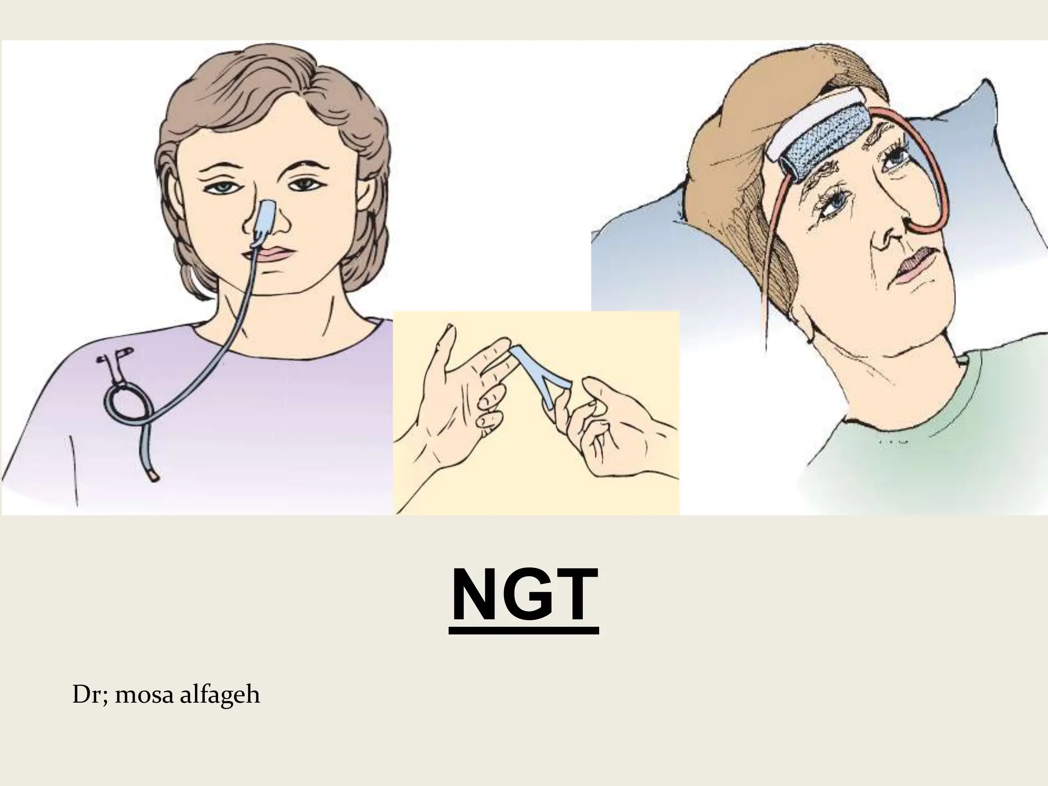
2. Definition
It is the insertion of a tube into the
oesophagus and stomach through the
nose
It is defined as the passage of tube
through the nose or mouth to stomach for
the purpose of the drainage, instillation,
decompression, lavage or performance of
diagnostic tests.
3. Purpose
• To feed the patient with fluids when oral
intake is not possible (Performing
gavage)
• To dilute and remove consumed poison
• To instill ice cold solution to control gastric
bleeding
• To prevent stress on operated site by
decompressing
• To relive vomiting and distension
4. Indications
• Patient who cannot eat (GIT functioning
normally)
– Comatose patient
– Mechanically ventilated
• Patient who will not eat
– Patients who refuse to eat
• Elderly
• Disoriented patients
– Patients who cannot maintain adequate oral
nutrition
• Patients with infection, trauma, cancer etc.
– Surgery
5. Contraindications
• Gastric surgery
• Ulcers
• Tracheoesophageal fistula
• Oesophageal surgery
• Recent nasal surgery, facial surgery
• Deviated nasal septum
• Patient on anticoagulant therapy
6. General instructions
• Remove the dentures ( to prevent it from
dislodging and blocking the respiratory tract)
• A rubber tube may be placed in a bowl of ice to
cool and stiffen
• Lubricate the tube
• While removing the tube, pinch the tube and pull it
out gently and quickly (so that the fluid may not
trickle down the pharynx )
• During introduction of the tube never use force (it
may cause injury to mucus membrane)
• Watch the complications (nausea, vomiting,
distension, diarrhea, aspiration, pneumonia etc.)
7. Articles Purposes
A try containing
Mackintosh with towel
To protect the bed and linen
A Ryle’s tube (8-12 Fr ) in a bowl
containing cold water
To make the tube hard and easy for
insertion
Cotton tipped applicator, Saline or
soda bicarbonate solution
To clean nostrils
Lubricant (liquid paraffin or
glycerine)
To prevent friction between mucus
membrane and tube
Adhesive plasters and scissor Fix the tube in position
A bowl of water, stethoscope,
syringe (10-20cc)
To test the location of tube
A kidney tray and a paper bag To collect waste
8. Ryle’s tube
• Flexible rubber or synthetic material
• Transparent or opaque
• Length : 36-50 inches
• Gastrostomy tube: 12-15 inches
• The lumen is measured by French (Fr)
11. Preliminary assessment
Check
– Doctors order for any specific instruction
– Patient ability to follow instructions
– General condition of the patient
– Articles available in the unit
12. Preparation of patient and unit
• Explain the sequence of procedure
• Arrange the articles at the bed side
• Provide privacy
• Provide comfortable position (fowler’s
position , comatose patient semi-fowlers
position)
• Place the mackintosh and towel across the
chest
• Remove the dentures
• Give mouth wash and help him to clean the
teeth
15. Method to confirm NG tube in
the stomach
• Aspirate: attach the syringe to the end of
NG tube and aspirate small amount of
gastric content
• Immerse distal end of tube into bowl of
water and check for air bubbles
• Auscultate : attach syringe to free end of
the tube, place diaphragm of stethescope
over left hypochondrium .inject 10 ml of air
and auscultate abdomen for gushing
sound.
16. After care
• Offer a mouth wash . Clean the face and
hands and dry them
• Remove the mackintosh and towel
• Make the patient comfortable in bed
• Take all articles to the utility room discard
the waste, clean it and replace it in a
proper place
• Wash hands
• Record the procedure
21. Definition
Nasogastric tube feeding is given
through tube which is inserted through
patient’s nose into stomach when patient
is unable to take food orally
It is a process of giving liquid nutrients
or medications through a tube into the
stomach when the oral intake is
inadequate or impossible
22. Gastric gavage
it is an artificial method of giving fluids
& nutrients through a tube, that has
passed into the oesophagus and stomach
through the nose, mouth or through the
opening made on the abdominal wall,
when oral intake is inadequate or
impossible
24. Advantages
• An adequate amount of all types of
nutrients including distasteful foods &
medications can be supplied
• Large amount of fluids can given with
safety
• It can be continued weeks with out any
danger
• The stomach may be aspirated at any
time of desired
• Overloading of the stomach can be
25. Principles
• A thorough knowledge of the anatomy
and physiology of digestive tract and
respiratory tract, ensures safe induction
of the tube
• Microorganisms enter the body through
food and drink
• Mental and physical preparation of the
patient facilitate introduction of the tube
• Systematic ways of working adds to the
comfort and safety of the client and help in
the economy of material, time and
26. Preliminary assessment
• Identify the correct patient
• Check the doctor’s order
• Check the level of consciousness
• Check whether feed is ready at hand
• Articles available in the unit
27. Preparation of patient and
environment
• Explain the sequence of procedure
• Provide adequate privacy
• Position (sitting or semi fowlers)
• Place mackintosh and towel around the
neck
• Arrange the articles at the bed side locker
• Clean the mouth by providing mouthwash
28. Articles
• A tray containing
– Mackintosh and towel
– 50cc syringe/ feeding tube and 5cc syringe
– Stethoscope
– Bowel with water
– Adhesive with scissors
– Feeds and water
– Ounce glass
– Kidney tray
29. Procedure
Wash hands
Place towel around the neck
Ensure the tube is in the stomach
Remove plunger from syringe…………..Pinch tube to
prevent air entry……….Remove spigot….connect
syringe to the tube.
Keep syringe about 12 inches above patients head.
Start feed with small measured amount of water and
allow feed to follow slowly and steadily through tube
in such a way, that air does not enter tube
Do not force fluid, allow to flow by gravity
At end of feed flush tube by pouring small measured
amount of water …..remove syringe and replace
spigot
30. After care
• Remove towel
• Place the patient in comfortable position
• Replace the articles to utility room, clean it
and replace it
• Record the procedure in nurses record
and intake out put chart.
33. Gastrostomy feeding :feeding is
given through an artificial opening made
surgically into the stomach through the
abdominal wall for feeding purpose when
the patient is not able to swallow through
oesophagus.
34. Jejunostomy feeding :
Jejunostomy is the surgical creation of
an opening (stoma) through the skin at the
front of the abdomen and the wall of
the jejunum(part of the small intestine)
• A jejunostomy is an alternative to
a gastrostomy .
• The advantage over a gastrostomy is its low
risk of aspiration due to its distal placement