Mid facial fractures ( Mid facial trauma )
•Download as PPTX, PDF•
3 likes•204 views

Content : Anatomy and fracture of maxilla nasal bone fracture Naso- ethmoidal-orbital fracture

Recommended
More Related Content
What's hot
What's hot (20)
Similar to Mid facial fractures ( Mid facial trauma )
Similar to Mid facial fractures ( Mid facial trauma ) (20)
Recently uploaded
Recently uploaded (20)
Mid facial fractures ( Mid facial trauma )
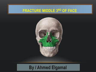
- 1. FRACTURE MIDDLE 3RD OF FACE
- 2. MIDDLE 3RD OF FACE Consists of : 2 maxillae 2 zygomatic bones 2 zygomatic process of temporal bones 2 palatine bones 2 nasal bones 2 lacrimal bones The vomer The ethmoid Pterygoid plates of sphenoid
- 3. ANATOMY OF THE MAXILLA Forms the largest part of the middle 3rd of face It is involved in the formation of the orbit, nose and palate, holds the upper teeth The body of each maxilla is hollowed by max sinus
- 4. ANATOMY OF MAXILLA Frontal process Palatine process Alveolar process Maxilla have 4 processes
- 5. ANATOMY OF MAXILLA Mid face skeleton is made up of thin segments of bone which supported by a more rigid frameworks of buttresses It is absorbs and transmits forces applied to facial skeleton It is : vertical buttresses : nasomaxillary & zygomaticomaxillary & pterygomaxillary Horizontal buttresses : frontal bar & orbital rims & zygomatic process of temporal bone maxillary alveolus
- 6. FRACTURE OF MAXILLA Radiographic examination CT scan with 3D ( more diagnosed ) Water’s view Caldwell view Lateral view Submentovertex view
- 7. CLASSIFICATION OF MAXILLARY FRACTURE Le fort I transverse fracture / floating maxilla Occurs transversally across the maxilla above the level of the teeth The fracture segment contains the alveolar process , portions of the wall of the maxillary sinus ,the palate and lower portion of the pterygoid plates
- 8. CLASSIFICATION OF MAXILLARY FRACTURE Le fort ll pyramidal fracture It involves the nasal bones and frontal process of maxilla and pass laterally through the lacrimal bones ,the inferior rim of the orbit and through the zygomaticomaxillay sutures Then continue backward along the lateral wall of maxilla through the pterygoid plates
- 9. CLASSIFICATION OF MAXILLARY FRACTURE Le fort lll craniofacial disjunction Complete separation of the facial bones from their cranial attachment The fracture occurs through the zygomatico-frontal , maxillo-frontal , naso-frontal sutures through orbital floor
- 10. CLASSIFICATION OF MAXILLARY FRACTURE Alveolar fracture of the maxilla This may involve bone containing one or more teeth Vertical split fracture of the maxilla Very rare that the maxilla splits along the sagittal plane
- 11. DIAGNOSIS OF MAXILLARY FRACTURE History of trauma Radiographic examination Clinical examination : >> Evidence of severe soft tissue injury in the facial region >> Bleeding from the nose,periorbital ,edema and ecchymosis >> Dish face deformity is a result of fractured and displaced middle third >> Malocclusion and open bite >> Diploplopia
- 16. RADIOGRAPHIC EXAMINATION Submentovertex viewWater view C.t scanPanoramaOcclusal
- 17. TREATMENT OF MAXILLARY FRACTURE Reduction Early reduction is mandatory to avoid complications Closed reduction manually is used in recent fracture when the fragment are not impacted, if fragment are impacted ,a row disimpaction forceps is used Closed reduction by traction is used in delayed fractures Open reduction is indicate when the traction is not effective
- 18. TREATMENT OF MAXILLARY FRACTURE
- 19. TREATMENT OF MAXILLARY FRACTURE Fixation Trans fixation wire used for fixation of comminuted unstable midfacial fractures suspension by wire adam wiring technique :A wire suspending the midfacial fracture to intact bone in higher level than the fracture fixation by miniplate Open fixation technique by wire ostepsynthesis
- 20. TREATMENT OF MAXILLARY FRACTURE
- 22. INTRODUCTION Nasal bone is the 3rd most commonly broken bone in the body If not properly managed >>> long term func & cosmetic problems (( foremost position Managed easily in children more than adults ( more cartilaginous )
- 23. ANATOMY
- 24. TYPES ACCORDING TO FORCE DIRECTION Frontal direction Sever flattening of nasal bones & septum Widening of nasal width
- 25. Lateral direction Depression of ipsilateral Nasal bone Out fracture of contralateral nasal bone ( sever ) Twisting of the nose
- 26. Below direction Fracture of septum Quadrangular cartilage dislocation
- 27. SIGNS & SYMPTOMS Pain Edema Nose deformation Epistaxis Rhinitis Airway blockage Crepitance Ecchymosis around the eyes Nasal septum deviation Step deformity ( palpated ) CSF rhinorrhea ( salty taste )
- 29. CLOSED REDUCTION Indications unilateral or bilateral fracture of nasal bone fracture of nasal septum with minimally deviated
- 30. CLOSED REDUCTION may be done under local or general anesthesia WALSHAM’S & ASCH’S septal forceps are used
- 32. OPEN REDUCTION Indication 1- extensive fractures 2- deviation of nasal pyramid greater than on half width of nasal bridge 3- persistent deformity after closed reduction
- 33. OPEN REDUCTION The septum may prevent proper reduction of nasal pyramid as the septal fragments are interlocked Be cautious when elevating the periosteum of the nasal bone because the the fractured segments may become unstable , deviated or lost so that conservative approach is better
- 34. CONTRAINDICATIONS FOR REDUCTION Some cases are contraindicated for TTT When : 1- The fragments are not displaced 2- Sever naso-ethmoid complex fractures ( precipitate or worsen CSF leak )
- 35. METHODS OF IMMOBILIZATION INTRANASAL METHODS ribbon gauze & stainless steel splint Ribbon gauze is the most standard 12 – 15 cm length is inserted into the nose in layers beginning from above downwards NOTE >>>> DON’T OVERPACK THE NOSE TO AVOID DISPLACING REDUCED FRACTURED SEGMENTS DISADVANTGES : 1- Inadequate anteroposterior support 2- Difficulty in breathing through nose 3- Potential source of infection
- 36. ERTRANASAL METHODES Plaster of Paris & lead splints Not all cases require intranasal splinting The plaster of Paris is most commonly used The plaster of Paris is applied while it is still wet and moulded to the shape of nose The splint shouldn’t be extent to the soft part of the nose This is left for about 1 week depending on the mobility of segments antibiotics may be placed in each nostril for 1 – 5 days
- 37. When the segments is very mobile and can’t be stabilized with plaster of paris :Lead plates can be used It is consists of two holes and are fitted on each side of nose with help of st st sutures which is passed into the holes and beneath the nasal bones This splint is left in place for a period of 3 weeks
- 38. NOTE Certain fractures involving the nasal septum and ethmoid result in loss of support to nasal bones This fractures can’t be splinted by plaster or lead plates It is recommended to be fixed with transosseous wiring of frontonasal junction
- 39. NASO – ETHMOIDAL – ORBITAL FRACTURE
- 40. SURGICAL ANATOMY NOE is situated In the central upper midface the skeletal structure The nose … orbit …. Maxilla …. Cranium The main structural buttress the frontal process of maxilla the internal angular process of frontal bone
- 41. THE MEDIAL WALL OF ORBIT Composed of lacrimal bone anteriorly Lamina papyracea of ethmoid bones posteriorly THIS structure are susceptible to comminution and allowing for medial displacement of orbital contents after blunt trauma Thin ethmoid bones form part of the anterior skull base Superiorly May result in dural injury and CSF leakage
- 42. Fracture or rupture of ant & post ethmoidal arteries >>>> orbital haematoma Optic canal is positioned posteriorly and local edema within it may cause circulatory disturbance of pial plexus of optic nerve >>>>>> transient or permanent blindness High energy blunt trauma to NOE complex >>>> collapse of the interorbital space and leading to injury to ant cranial contents or intraorbital content
- 43. CLASSIFICATION OF NOE COMPLEX FRACTURE Type I In unilateral type I fractures, there is a single large NOE fragment bearing the medial canthal tendon Involvement of the nasal bone The nasal bone may also be involved and, in cases of comminution, may not provide adequate dorsal support to the nasal bridge.
- 44. CLASSIFICATION OF NOE COMPLEX FRACTURE unilateral type II fractures there is often comminution of the NOE area , but the canthal tendon remains attached to a fragment of bone, allowing the canthus to be stabilized with wires or a small plate on the fractured segment. Involvement of the nasal bone The nasal bone may also be involved and, in cases of comminution, may not provide adequate dorsal support to the nasal bridge.
- 45. CLASSIFICATION OF NOE COMPLEX FRACTURE Bilateral type II fracture with nasal bone involvement In bilateral fractures the nasal bones are commonly involved. In some instances, bone grafting of the nasal dorsum may be necessary.
- 46. CLASSIFICATION OF NOE COMPLEX FRACTURE Type III Comminution within the central fragment allows fracture to extend beneath the canthal insertion The canthus is attached to bone fragments that are too small to utilize in reconstruction
- 47. DIAGNOSIS ( CL EX + CT SCAN ) The patient often has swelling in the medial canthal area and pain and crepitation with palpation. Bilateral periorbital and subconjunctival haemorrhage ( limited to medial half of eye ) The nose can be retruded and impacted at the nasofrontal suture area with lack of support for the nasal septum and cartilages. Epistaxis …… CSF leak Flattening nasal dorsum
- 48. INTERCANTHAL DISTANCE TEST There will be a lack of definition of the bony anatomy in the medial canthal area and possible lateral splaying of the medial canthus with increased intercanthal distance (the normal distance varies depending on the ethnic group, e.g., in Caucasians more than 35 mm intercanthal distance is considered abnormal.)
- 49. BOW STRING TEST grab the eyelid or use a forceps to grab the skin in the medial canthal area and pull it laterally. In the bow-string test the lid is pulled laterally while the tendon area is palpated to detect movement of fracture segments. A lack of resistance or movement of the underlying bone is indicative of a fracture.
- 50. BIMANUAL PALPATION TEST Another test is to place an instrument in the nose and push laterally in the medial canthal area to test for instability and crepitation, which suggests an unstable NOE fracture.
- 51. MANAGEMENT Reduction and fixation of unstable fracture segment to stable structures The main goal : Restoration of anatomic position of bony segments to esthetics and functional baseline and prevents later complications
- 52. TYPE I Reduction by hand or with penetrating towel clamp or other bone- grasping inst. To prevent lateral migration of the segments When the fracture involves the frontal process of max and inferior orbital rim >>>>> small miniplates with self tapping screws applied to each of these structures for fixation If only small piece of lacrimal bone >>>> it may be wired to its normal position using wires
- 53. TYPE II Additional exposure of contralateral medial orbital wall is required for fixation Transnasal wiring provides a stable source of fixation In addition to transnasal stabilization micro plates may be used to fix the MCT bony fragment to adjacent stable bone
- 54. TYPE III The most sever of the NOE fracture Usually bilateral Higher incidence of associated injuries to Dura , Skull base , Intra orbital contents After careful dissection of both medial orbital walls >>>> bilateral intercanthal tendon fixation is performed if the associated MCT bony segment is too small for drilling holes >>>>> the wire may be passed through a thickened portion of MCT >>>>> both free ends on the wire are then passed through the lacrimal defect on first side through nasal septum and through the opposite lacrimal defect >>>>> use the free ends to secure the opposite MCT and bony segment The MCTs of both sides are pulled medially toward each other
- 55. COMPLICATIONS FOLLOWING SURGERY Temporary or permanent paraesthesia CSF leak Meningitis Sinus infection Anosmia Infection of implants Osteomyelitis enophthalamos Extra ocular motion disfunc Blindness Possible need for additional surgery