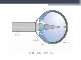
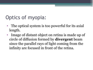
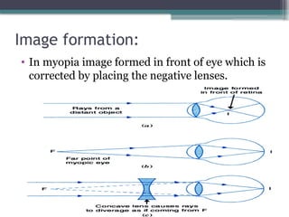
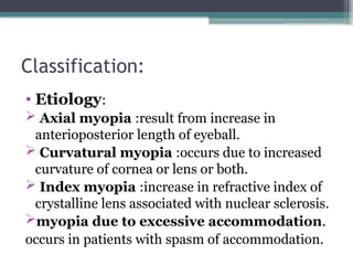
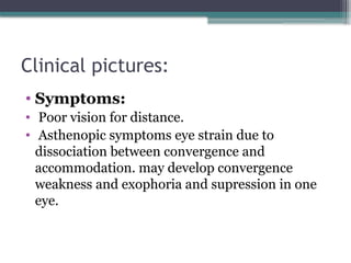
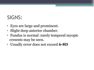
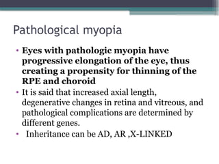
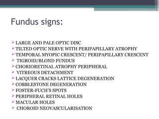
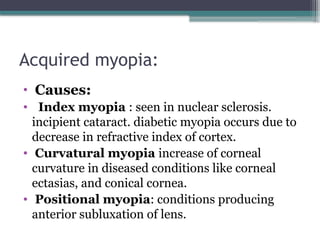
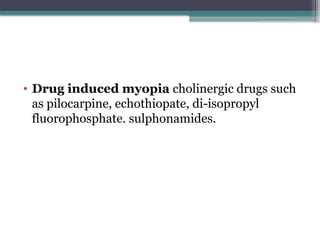
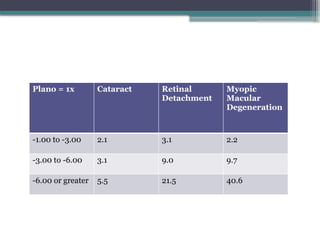
Myopia
By: Komal Yaseen
Contents:
• Introduction
• Optics of myopia
• Classification
• Management
Introduction:
• Derived from two Greek root words.
(Greek: μυωπία, muōpia, from myein "to shut" -
ops (gen. opos) "eye“)
• Myopia or shortsightedness is a type of
refractive error in which parallel rays of light
coming from infinity are focused in front of
retina with the accommodation is at rest.
Optics of myopia:
• The optical system is too powerful for its axial
length.
• Image of distant object on retina is made up of
circle of diffusion formed by divergent beam
since the parallel rays of light coming from the
infinity are focused in front of the retina.
Image formation:
• In myopia image formed in front of eye which is
corrected by placing the negative lenses.
RISK FACTOR
• Genetic
• Near work
• • More near work = higher risk
• • Smart phone/computers > tablet TV
• Time outdoors
• • Less time = higher risk
• Ethnicity
• >70% in East Asians, 43% Caucasians, 34%
African Americans
Classification:
• Etiology:
 Axial myopia :result from increase in
anterioposterior length of eyeball.
 Curvatural myopia :occurs due to increased
curvature of cornea or lens or both.
 Index myopia :increase in refractive index of
crystalline lens associated with nuclear sclerosis.
myopia due to excessive accommodation.
occurs in patients with spasm of accommodation.
Continue…
Clinically:
• Congenital myopia.
• Simple or developmental myopia.
• Pathological or degenerative myopia.
• Acquired myopia.
1.Congenital myopia
• Present since birth. Diagnosed: 2-3 year
• Seen more frequently in children who were born
prematurely or with various birth defects.
• Usually error is about 8-10D, which mostly
remains constant.
• May sometimes be associated with other
congenital anomalies such as cataract,
microphthalmos, aniridia, megalocornea.
2.Simple or developmental myopia:
• Physiological or school myopia.
• Physiological error not associated with any
disease of eye.
• Etiology: result from normal biological variation
in the development of eye.
• Axial – A.P diameter
• Curvatural – underdevelopment of eye ball
• Diet – poor nutrition
• Genetic influence – one (or) both parents
Clinical pictures:
• Symptoms:
• Poor vision for distance.
• Asthenopic symptoms eye strain due to
dissociation between convergence and
accommodation. may develop convergence
weakness and exophoria and supression in one
eye.
SIGNS:
• Eyes are large and prominent.
• Slight deep anterior chamber.
• Fundus is normal :rarely temporal myopic
cresents may be seen.
• Usually error does not exceed 6-8D
Pathological myopia
• Eyes with pathologic myopia have
progressive elongation of the eye, thus
creating a propensity for thinning of the
RPE and choroid
• It is said that increased axial length,
degenerative changes in retina and vitreous, and
pathological complications are determined by
different genes.
• Inheritance can be AD, AR ,X-LINKED
Symptoms:
• Image Minification
• Anisometric Amblyopia
• Subnormal VISUAL ACUITY
• Visual Field defects
• Impaired dark adaptation
• Abnormal color discrimination
• Suboptimal binocularity
• Muscae Volitantes
Signs:
• PROMINENT EYE BALLS(Exophthalmus)
• CORNEA IS LARGE
• ANTERIOR CHAMBER IS DEEP
• LARGE AND SLUGGISH PUPILS
• VISUAL FIELD CONTRACTION
• ERG MAY BE SUBNORMAL DUE TO
CHORIORETINAL ATTROPHY
• Refractive error:
• Increase by as much as 4.00D yearly stabilizes at
about the age of 20 but occasionally may
progress until mid 30s`` frequently result in
myopia of 10-20D
Fundus signs:
LARGE AND PALE OPTIC DISC
TILTED OPTIC NERVE WITH PERIPAPILLARY ATROPHY
TEMPORAL MYOPIC CRESCENT/ PERIPAPILLARY CRESCENT
 TIGROID/BLOND FUNDUS
CHORIORETINAL ATROPHY PERIPHERAL
 VITREOUS DETACHMENT
LACQUER CRACKS LATTICE DEGENERATION
COBBLESTONE DEGENERATION
FOSTER-FUCH’S SPOTS
PERIPHERAL RETINAL HOLES
MACULAR HOLES
 CHOROID NEOVASCULARISATION
Lattice degeneration
Comlication:
• Retinal detachment.
• Complicated cataract
• Vitreous and choridal haemorrages
• Myopic foveoschisis
• Posterior staphyloma
Acquired myopia:
• Causes:
• Index myopia : seen in nuclear sclerosis.
incipient cataract. diabetic myopia occurs due to
decrease in refractive index of cortex.
• Curvatural myopia increase of corneal
curvature in diseased conditions like corneal
ectasias, and conical cornea.
• Positional myopia: conditions producing
anterior subluxation of lens.
• Consecutive myopia surgical overcorrection
of hypermetropia pseudophakia with
overcorrecting IOL.
• Pseudomyopia:also called artificial myopia.
produced in a conditions such as excessive
accommodation and spasm of accommodation.
may develop after too full a hypermetropic
correction in children.
• Drug induced myopia cholinergic drugs such
as pilocarpine, echothiopate, di-isopropyl
fluorophosphate. sulphonamides.
Types on degree of myopia
• According to amount:
• Very low : upto – 1.00D
• Low : (-1.00-3.00)D
• Medium : (-3.00-6.00)D
• High : (-6.00-10.00)D 
• Very high : above –10.00D
Plano = 1x Cataract Retinal
Detachment
Myopic
Macular
Degeneration
-1.00 to -3.00 2.1 3.1 2.2
-3.00 to -6.00 3.1 9.0 9.7
-6.00 or greater 5.5 21.5 40.6

myopia C.pptx . . . . . . . . . . . .

  • 1.
  • 2.
    Contents: • Introduction • Opticsof myopia • Classification • Management
  • 3.
    Introduction: • Derived fromtwo Greek root words. (Greek: μυωπία, muōpia, from myein "to shut" - ops (gen. opos) "eye“) • Myopia or shortsightedness is a type of refractive error in which parallel rays of light coming from infinity are focused in front of retina with the accommodation is at rest.
  • 5.
    Optics of myopia: •The optical system is too powerful for its axial length. • Image of distant object on retina is made up of circle of diffusion formed by divergent beam since the parallel rays of light coming from the infinity are focused in front of the retina.
  • 6.
    Image formation: • Inmyopia image formed in front of eye which is corrected by placing the negative lenses.
  • 7.
    RISK FACTOR • Genetic •Near work • • More near work = higher risk • • Smart phone/computers > tablet TV • Time outdoors • • Less time = higher risk • Ethnicity • >70% in East Asians, 43% Caucasians, 34% African Americans
  • 8.
    Classification: • Etiology:  Axialmyopia :result from increase in anterioposterior length of eyeball.  Curvatural myopia :occurs due to increased curvature of cornea or lens or both.  Index myopia :increase in refractive index of crystalline lens associated with nuclear sclerosis. myopia due to excessive accommodation. occurs in patients with spasm of accommodation.
  • 9.
  • 11.
    Clinically: • Congenital myopia. •Simple or developmental myopia. • Pathological or degenerative myopia. • Acquired myopia.
  • 12.
    1.Congenital myopia • Presentsince birth. Diagnosed: 2-3 year • Seen more frequently in children who were born prematurely or with various birth defects. • Usually error is about 8-10D, which mostly remains constant. • May sometimes be associated with other congenital anomalies such as cataract, microphthalmos, aniridia, megalocornea.
  • 14.
    2.Simple or developmentalmyopia: • Physiological or school myopia. • Physiological error not associated with any disease of eye. • Etiology: result from normal biological variation in the development of eye. • Axial – A.P diameter • Curvatural – underdevelopment of eye ball • Diet – poor nutrition • Genetic influence – one (or) both parents
  • 15.
    Clinical pictures: • Symptoms: •Poor vision for distance. • Asthenopic symptoms eye strain due to dissociation between convergence and accommodation. may develop convergence weakness and exophoria and supression in one eye.
  • 16.
    SIGNS: • Eyes arelarge and prominent. • Slight deep anterior chamber. • Fundus is normal :rarely temporal myopic cresents may be seen. • Usually error does not exceed 6-8D
  • 17.
    Pathological myopia • Eyeswith pathologic myopia have progressive elongation of the eye, thus creating a propensity for thinning of the RPE and choroid • It is said that increased axial length, degenerative changes in retina and vitreous, and pathological complications are determined by different genes. • Inheritance can be AD, AR ,X-LINKED
  • 19.
    Symptoms: • Image Minification •Anisometric Amblyopia • Subnormal VISUAL ACUITY • Visual Field defects • Impaired dark adaptation • Abnormal color discrimination • Suboptimal binocularity • Muscae Volitantes
  • 20.
    Signs: • PROMINENT EYEBALLS(Exophthalmus) • CORNEA IS LARGE • ANTERIOR CHAMBER IS DEEP • LARGE AND SLUGGISH PUPILS • VISUAL FIELD CONTRACTION • ERG MAY BE SUBNORMAL DUE TO CHORIORETINAL ATTROPHY
  • 21.
    • Refractive error: •Increase by as much as 4.00D yearly stabilizes at about the age of 20 but occasionally may progress until mid 30s`` frequently result in myopia of 10-20D
  • 22.
    Fundus signs: LARGE ANDPALE OPTIC DISC TILTED OPTIC NERVE WITH PERIPAPILLARY ATROPHY TEMPORAL MYOPIC CRESCENT/ PERIPAPILLARY CRESCENT  TIGROID/BLOND FUNDUS CHORIORETINAL ATROPHY PERIPHERAL  VITREOUS DETACHMENT LACQUER CRACKS LATTICE DEGENERATION COBBLESTONE DEGENERATION FOSTER-FUCH’S SPOTS PERIPHERAL RETINAL HOLES MACULAR HOLES  CHOROID NEOVASCULARISATION
  • 24.
  • 25.
    Comlication: • Retinal detachment. •Complicated cataract • Vitreous and choridal haemorrages • Myopic foveoschisis • Posterior staphyloma
  • 26.
    Acquired myopia: • Causes: •Index myopia : seen in nuclear sclerosis. incipient cataract. diabetic myopia occurs due to decrease in refractive index of cortex. • Curvatural myopia increase of corneal curvature in diseased conditions like corneal ectasias, and conical cornea. • Positional myopia: conditions producing anterior subluxation of lens.
  • 27.
    • Consecutive myopiasurgical overcorrection of hypermetropia pseudophakia with overcorrecting IOL. • Pseudomyopia:also called artificial myopia. produced in a conditions such as excessive accommodation and spasm of accommodation. may develop after too full a hypermetropic correction in children.
  • 28.
    • Drug inducedmyopia cholinergic drugs such as pilocarpine, echothiopate, di-isopropyl fluorophosphate. sulphonamides.
  • 29.
    Types on degreeof myopia • According to amount: • Very low : upto – 1.00D • Low : (-1.00-3.00)D • Medium : (-3.00-6.00)D • High : (-6.00-10.00)D  • Very high : above –10.00D
  • 30.
    Plano = 1xCataract Retinal Detachment Myopic Macular Degeneration -1.00 to -3.00 2.1 3.1 2.2 -3.00 to -6.00 3.1 9.0 9.7 -6.00 or greater 5.5 21.5 40.6