OBJECTIVES
• Define normalanatomy & physiology of bone.
• Explain bone tumors & enlist the risk factors.
• Explain TNM classification of bone tumors
• Explain the types of bone tumors.
• Explain the clinical manifestations of bone tumors.
• Explain the diagnostic factors for bone tumors.
• Explain the management for bone tumors
3.
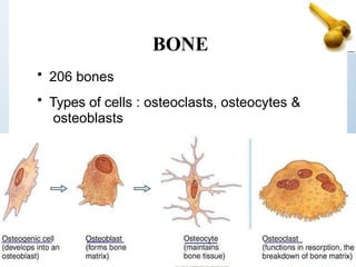
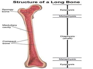
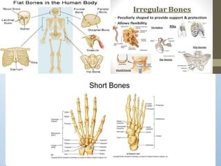
BONE
• 206 bones
•Types of cells : osteoclasts, osteocytes &
osteoblasts
6.
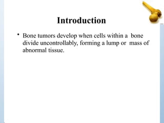
Introduction
• Bone tumorsdevelop when cells within a bone
divide uncontrollably, forming a lump or mass of
abnormal tissue.
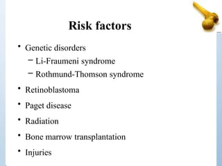
• The Li-Fraumenisyndrome makes people much
more likely to develop several types of
cancer,including breast cancer, brain cancer,
osteosarcoma, and other types of sarcoma.
• Most of those cases are caused by a mutation of the
p53 tumor suppressor gene, but some are caused by
mutations in the gene CHEK2.
10.
• Rothmund-Thomson syndrome:Children with
this syndrome are short, have skeletal problems,
and rashes. They also are more likely to develop
osteosarcoma. This syndrome is caused by
abnormal changes in the gene REQL4.
• Injuries: People have wondered whether
injury to a bone can cause cancer, but this has
never been proven.
11.
• Retinoblastoma isa rare eye cancer of children
that can be hereditary. The inherited form of
retinoblastoma is caused by a mutation of the
RB1 gene.
• Those with this mutation also have an
increased risk of developing bone or soft
tissue sarcomas.
• Also, if radiation therapy is used to treat the
retinoblastoma, the risk of osteosarcoma in the
bones around the eye is even higher.
12.
• Paget disease:seem to have a high risk of
chordomas during childhood.
• Radiation: Bones that have been exposed to
ionizing radiation may also have a higher risk of
developing bone cancer.
• Bone marrow transplantation: Osteosarcoma has
been reported in a few patients who have
undergone bone marrow (stem cell)
transplantation.
13.
Clinical manifestations
• cardinalsymptoms: Pain, swelling and general
discomfort
• limited mobility and spontaneous fracture may
also be important features.
• Other symptoms :fever and night sweats.
• painless mass or obvious bone growth
14.
• Varying degreeof disability, weight loss,
malaise.
• With spinal metastasis, spinal cord
compression may occur.
• Neurologic deficit e.g. progressive pain,
weakness, gait abnormality, paresthesia,
paraplegia, urinary retention, loss of bowel or
bladder control
15.
Pain
• first &most common symptom.
• may initially occur intermittently and only at rest
• become more intense, disturb sleep at night,
spread into the adjacent joint .
• A further intensification of pain is experienced as a
persistent and piercing pain.
• becomes excruciating and intolerable, requiring
opiate treatment.
• In case of pressure on nerve trunks or nerve
plexuses, the patient may experience radiating
pain.
16.
Swelling
• very longduration, especially in benign
neoplasms & cause no additional complaints.
• In malignant tumours, swelling develops more
rapidly.
• may also cause skin changes, including tensed
shining skin with prominent veins, livid
colouring, hyperthermia, as well as striation of
the skin and eventually, ulceration.
• The mobility of the skin & musculature above the
tumour should also be assessed. The less the
mobility, the more likely is this factor a criterion
of malignancy.
17.
Limitation of movement
•Mobility may be limited in cases of lesions
close to the joint
• In tumours such as osteoblastoma,
chrondroblastoma, giant cell tumours and all
types of sarcomas.
• Occasionally it is not the tumour but reactive
synovitis in the joint, especially in
chondroblastoma, that causes limitation of
movement and masks the true diagnosis.
18.
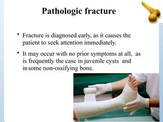
Pathologic fracture
• Fractureis diagnosed early, as it causes the
patient to seek attention immediately.
• It may occur with no prior symptoms at all, as
is frequently the case in juvenile cysts and
insome non-ossifying bone.
19.
Assessment & diagnosis
•The differential diagnosis is based on history,
physical examination & diagnostic studies.
• Age : it is useful information before age of 5, a
malignant tumour is often metastatic
neuroblastoma; between 5 and 15 years old,
osteosarcoma or Ewing sarcoma; and after 40
years, metastasis or myeloma.
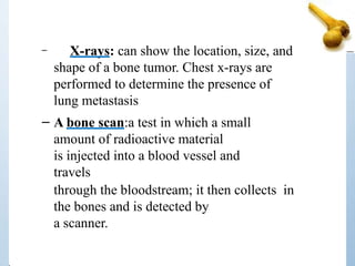
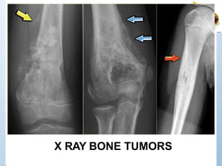
– X-rays: canshow the location, size, and
shape of a bone tumor. Chest x-rays are
performed to determine the presence of
lung metastasis
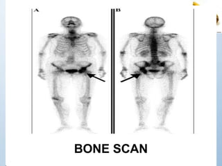
– A bone scan:a test in which a small
amount of radioactive material
is injected into a blood vessel and
travels
through the bloodstream; it then collects in
the bones and is detected by
a scanner.
– A computedtomography: a series of detailed
pictures of areas inside the body, taken from
different angles, that are created by a computer
linked to an x-ray machine.
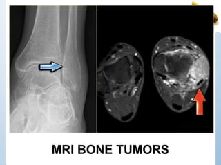
– A magnetic resonance imaging :which uses a
powerful magnet linked to a computer to create
detailed pictures of areas inside the body without
using x-rays.
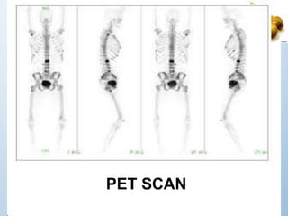
– A positron emission tomography: a small amount
of radioactive glucose (sugar) is injected into a vein,
and a scanner is used to make detailed, computerized
pictures of areas inside the body where the glucose is
used. Because cancer cells often use more glucose
than normal cells, the pictures can be used to find
cancer cells in the body.
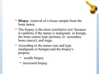
– Biopsy: removalof a tissue sample from the
bone tumor.
– The biopsy is the most conclusive test because
it confirms if the tumor is malignant or benign,
the bone cancer type (primary or secondary
bone cancer), and stage.
– According to the tumor size and type
(malignant or benign) and the biopsy's
purpose
– needle biopsy
– incisional biopsy
28.
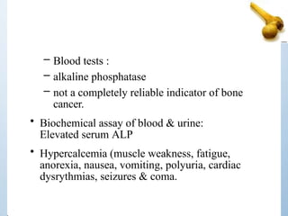
– Blood tests:
– alkaline phosphatase
– not a completely reliable indicator of bone
cancer.
• Biochemical assay of blood & urine:
Elevated serum ALP
• Hypercalcemia (muscle weakness, fatigue,
anorexia, nausea, vomiting, polyuria, cardiac
dysrythmias, seizures & coma.
29.
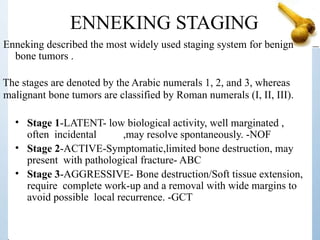
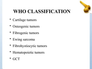
ENNEKING STAGING
Enneking describedthe most widely used staging system for benign
bone tumors .
The stages are denoted by the Arabic numerals 1, 2, and 3, whereas
malignant bone tumors are classified by Roman numerals (I, II, III).
• Stage 1-LATENT- low biological activity, well marginated ,
often incidental ,may resolve spontaneously. -NOF
• Stage 2-ACTIVE-Symptomatic,limited bone destruction, may
present with pathological fracture- ABC
• Stage 3-AGGRESSIVE- Bone destruction/Soft tissue extension,
require complete work-up and a removal with wide margins to
avoid possible local recurrence. -GCT
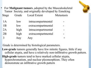
30.
• For Malignanttumors ,adopted by the Musculoskeletal
Tumor Society, and originally developed by Enneking
Stage Grade Local Extent Metastasis
1A low intracompartmental -
1B low extracompartmental -
2A high intracompartmental -
2B high extracompartmental -
3 Any Any +
Grade is determined by histological parameters.
Low-grade tumors generally have few mitotic figures, little if any
cellular atypia, and have a relatively non-infiltrative growth pattern.
High-grade tumors tend to have marked cellular atypia,
hyperchromatism, and nuclear pleomorphism. They often
demonstrate an infiltrative growth pattern
31.
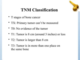
TNM Classification
• Tstages of bone cancer
• TX: Primary tumor can’t be measured
• T0: No evidence of the tumor
• T1: Tumor is 8 cm (around 3 inches) or less
• T2: Tumor is larger than 8 cm
• T3: Tumor is in more than one place on
the same bone
32.
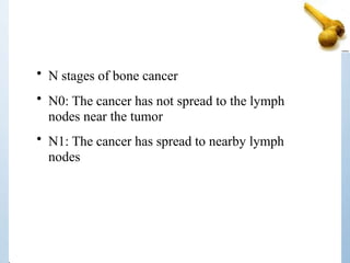
• N stagesof bone cancer
• N0: The cancer has not spread to the lymph
nodes near the tumor
• N1: The cancer has spread to nearby lymph
nodes
33.
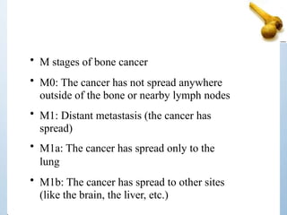
• M stagesof bone cancer
• M0: The cancer has not spread anywhere
outside of the bone or nearby lymph nodes
• M1: Distant metastasis (the cancer has
spread)
• M1a: The cancer has spread only to the
lung
• M1b: The cancer has spread to other sites
(like the brain, the liver, etc.)
Secondary/metastatic tumors
• Tumorsarising from elsewhere in the body
may invade the bone & produce localized
bone destruction or bone overgrowth.
41.
Types of cancerthat begin elsewhere
and commonly spread to bone
include:
• Breast
• Lung
• Thyroid
• Renal
• Prostate
• Metastatic tumor most commonly attack the
skull, spine, pelvis, femur, humerus & often
involve more than one bone (polyostotic).
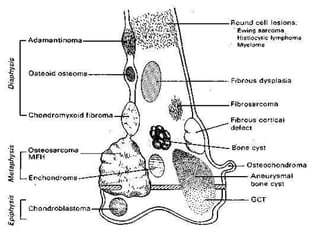
42.
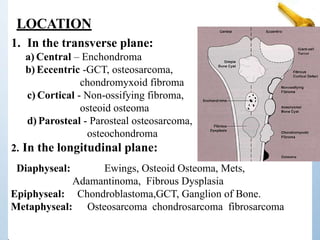
LOCATION
1. In thetransverse plane:
a) Central – Enchondroma
b) Eccentric -GCT, osteosarcoma,
chondromyxoid fibroma
c) Cortical - Non-ossifying fibroma,
osteoid osteoma
d) Parosteal - Parosteal osteosarcoma,
osteochondroma
2. In the longitudinal plane:
Diaphyseal: Ewings, Osteoid Osteoma, Mets,
Adamantinoma, Fibrous Dysplasia
Epiphyseal: Chondroblastoma,GCT, Ganglion of Bone.
Metaphyseal: Osteosarcoma chondrosarcoma fibrosarcoma
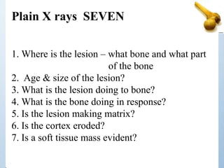
1. Where isthe lesion – what bone and what part
of the bone
2. Age & size of the lesion?
3. What is the lesion doing to bone?
4. What is the bone doing in response?
5. Is the lesion making matrix?
6. Is the cortex eroded?
7. Is a soft tissue mass evident?
Plain X rays SEVEN
45.
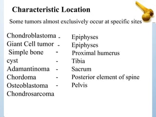
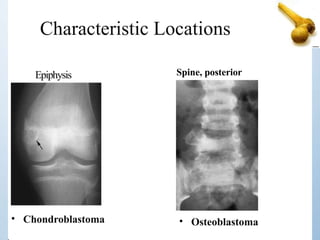
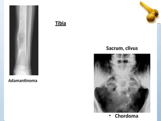
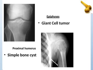
Chondroblastoma
Giant Cell tumor
Simplebone
cyst
Adamantinoma
Chordoma
Osteoblastoma
Chondrosarcoma
- Epiphyses
- Epiphyses
- Proximal humerus
- Tibia
- Sacrum
- Posterior element of spine
- Pelvis
Characteristic Location
Some tumors almost exclusively occur at specific sites
• Giant Celltumor
Epiphyses
Proximal humerus
• Simple bone cyst
49.
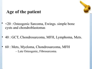
• <20 :Osteogenic Sarcoma, Ewings. simple bone
cysts and chondroblastomas
• 40 : GCT, Chondrosarcoma, MFH, Lymphoma, Mets.
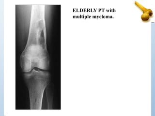
• 60 : Mets, Myeloma, Chondrosarcoma, MFH
– Late Osteogenic, Fibrosarcoma.
Age of the patient
50.
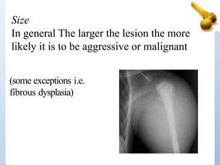
Size
In general Thelarger the lesion the more
likely it is to be aggressive or malignant
(some exceptions i.e.
fibrous dysplasia)
51.
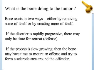
Bone reacts intwo ways -- either by removing
some of itself or by creating more of itself.
If the disorder is rapidly progressive, there may
only be time for retreat (defense).
If the process is slow growing, then the bone
may have time to mount an offense and try to
form a sclerotic area around the offender.
What is the bone doing to the tumor ?
52.
Aperiosteal reaction willoccur whenever the
periosteum is irritated.
This may occur due to a malignant
tumor, benign tumor, infection or trauma.
Two types Benign or Aggressive.
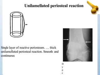
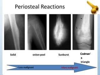
Periostitis
• Benign
– None
– Solid
Aggressive or malignant
– Lamellated or onion peel
– Sunburst
– Codman’s triangle
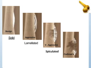
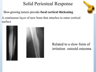
Solid Periosteal Response
Relatedto a slow form of
irritation osteoid osteoma
Slow-growing tumors provoke focal cortical thickening
A continuous layer of new bone that attaches to outer cortical
surface
Aggressive Periostitis
appearance ofaggressive
periostitis in Ewing’s
sarcoma
Layered, onion-skin, lamellated
• Alternating layers of opaque and
lucent densities
• Can be seen with slow growing
and aggressive tumors and
infections
growth
spurt.
57.
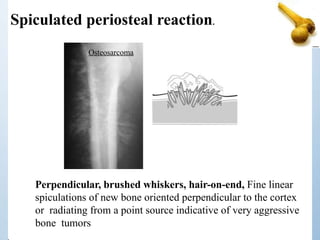
Spiculated periosteal reaction.
Perpendicular,brushed whiskers, hair-on-end, Fine linear
spiculations of new bone oriented perpendicular to the cortex
or radiating from a point source indicative of very aggressive
bone tumors
Osteosarcoma
58.
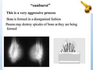
This is avery aggressive process
Bone is formed in a disorganized fashion
Process may destroy spicules of bone as they are being
formed
“sunburst”
59.
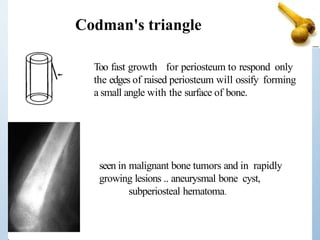
Too fast growthfor periosteum to respond only
the edges of raised periosteum will ossify forming
a small angle with the surface of bone.
Codman's triangle
seen in malignant bone tumors and in rapidly
growing lesions .. aneurysmal bone cyst,
subperiosteal hematoma.
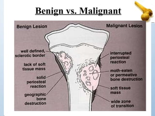
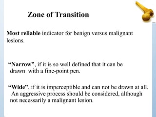
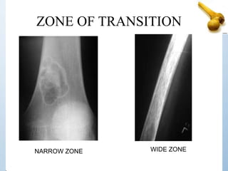
Zone of Transition
Mostreliable indicator for benign versus malignant
lesions.
“Narrow”, if it is so well defined that it can be
drawn with a fine-point pen.
“Wide”, if it is imperceptible and can not be drawn at all.
An aggressive process should be considered, although
not necessarily a malignant lesion.
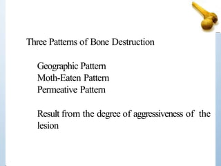
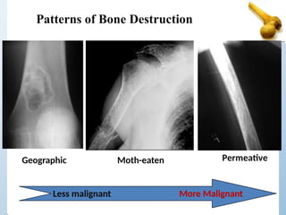
Three Patterns ofBone Destruction
Geographic Pattern
Moth-Eaten Pattern
Permeative Pattern
Result from the degree of aggressiveness of the
lesion
64.
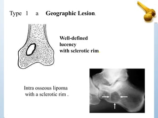
Type 1 aGeographic Lesion.
Intra osseous lipoma
with a sclerotic rim .
Well-defined
lucency
with sclerotic rim.
65.
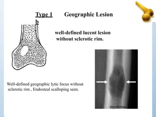
Type 1
b
Geographic Lesion
well-definedlucent lesion
without sclerotic rim.
Well-defined geographic lytic focus without
sclerotic rim , Endosteal scalloping seen.
myeloma
66.
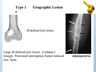
Large ill-defined lyticlesion , Codman’s
triangle Periosteal interruption,Tumor-induced
new bone .
.
Type 1
c
Geographic Lesion
ill-defined lytic lesion
osteosarcoma
67.
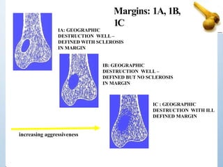
IA: GEOGRAPHIC
DESTRUCTION WELL–
DEFINED WITH SCLEROSIS
IN MARGIN
IB: GEOGRAPHIC
DESTRUCTION WELL –
DEFINED BUT NO SCLEROSIS
IN MARGIN
IC : GEOGRAPHIC
DESTRUCTION WITH ILL
DEFINED MARGIN
increasing aggressiveness
Margins: 1A, 1B,
1C
68.
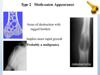
Type 2 Moth-eatenAppearance
Areas of destruction with
ragged borders
Implies more rapid growth
Probably a malignancy
osteosarcoma
69.
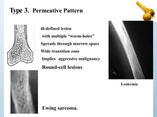
Type 3. PermeativePattern
Ewing sarcoma.
ill-defined lesion
with multiple “worm-holes”
Spreads through marrow space
Wide transition zone
Implies aggressive malignancy
Round-cell lesions
Leukemia
70.
Patterns of BoneDestruction
Geographic Moth-eaten Permeative
Less malignant More Malignant
71.
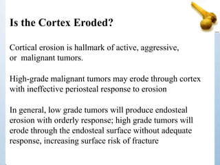
Is the CortexEroded?
Cortical erosion is hallmark of active, aggressive,
or malignant tumors.
High-grade malignant tumors may erode through cortex
with ineffective periosteal response to erosion
In general, low grade tumors will produce endosteal
erosion with orderly response; high grade tumors will
erode through the endosteal surface without adequate
response, increasing surface risk of fracture
72.
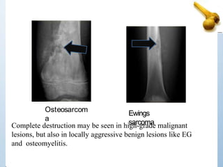
Ewings
sarcoma
Complete destruction maybe seen in high-grade malignant
lesions, but also in locally aggressive benign lesions like EG
and osteomyelitis.
Osteosarcom
a
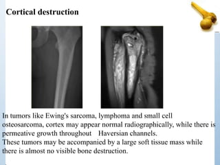
Cortical destruction
In tumorslike Ewing's sarcoma, lymphoma and small cell
osteosarcoma, cortex may appear normal radiographically, while there is
permeative growth throughout Haversian channels.
These tumors may be accompanied by a large soft tissue mass while
there is almost no visible bone destruction.
76.
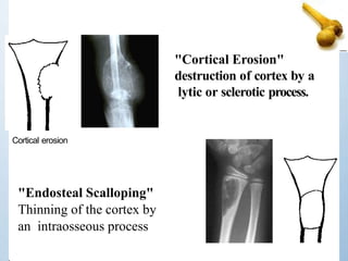
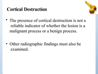
Cortical Destruction
• Thepresence of cortical destruction is not a
reliable indicator of whether the lesion is a
malignant process or a benign process.
• Other radiographic findings must also be
examined.
77.
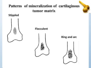
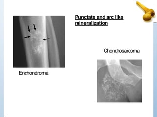
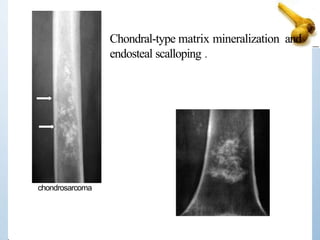
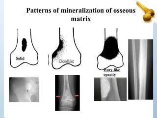
Is the lesionmaking matrix?
Matrix is the dominant internal extracellular substance
of a lesion.
Most tumor have soft tissue matrix-Radiolucent (lytic)
on X-ray.
Chondroid matrix -Calcified rings, arcs, dots.
Osteoid matrix- Bone forming
78.
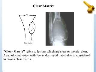
"Clear Matrix" refersto lesions which are clear or mostly clear.
A radiolucent lesion with few undestroyed trabeculae is considered
to have a clear matrix.
Clear Matrix
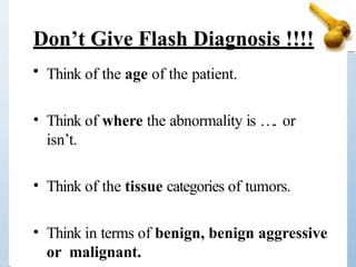
Don’t Give FlashDiagnosis !!!!
• Think of the age of the patient.
• Think of where the abnormality is …. or
isn’t.
• Think of the tissue categories of tumors.
• Think in terms of benign, benign aggressive
or malignant.
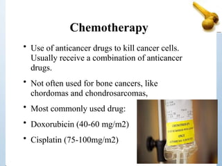
Chemotherapy
• Use ofanticancer drugs to kill cancer cells.
Usually receive a combination of anticancer
drugs.
• Not often used for bone cancers, like
chordomas and chondrosarcomas,
• Most commonly used drug:
• Doxorubicin (40-60 mg/m2)
• Cisplatin (75-100mg/m2)
95.
• Carboplatin
• Etoposide
•Ifosfamide (1.2g/m2)
• Cyclophosphamide 10-15mg/kg IV;1-5mg/kg oral)
• Methotrexate (oral/IV)
• Vincristine (1.4mg/m2)
• Usually, several drugs (2 or 3) are given together. For
example, a very common combination is cisplatin and
doxorubicin. Other combinations are ifosfamide and
etoposide or ifosfamide and doxorubicin
96.
Side effects ofchemotherapy
• Some common temporary side effects can
include nausea and vomiting, loss of appetite
,hair loss, mouth sores.
• Ifosfamide and cyclophosphamide can cause
hemorrhagic cystitis & can be prevented by
giving a drug called mesna along with the
chemo.
• Cisplatin may cause neuropathy leading to
problems with numbness, tingling, and even
pain in the hands and feet. Nephropathy can also
occur after treatment with cisplatin.
97.
Radiation therapy
• involvesthe use of high-energy x-rays to kill
cancer cells.
• may be used in combination with surgery, if
tumor is radiosensitive.
• often used to treat chondrosarcoma, which
cannot be treated with chemotherapy.
• may also be used for patients who refuse
surgery. Radiation can also reduce pain and
decrease the chance of bone fractures.
Intensity-modulated radiation
therapy (IMRT)
•an advanced form of external beam radiation therapy.
• With this technique, a computer matches the
radiation beams to the shape of the tumor and can adjust the
intensity (strength) of the beams.
• The radiation is delivered to the tumor from
several directions to reduce the amount of
radiation that goes through any one area of normal
tissue.
• Altogether, this makes it possible to reduce
radiation damage to normal tissues while increasing the
radiation dose to the cancer.
100.
Proton-beam radiation
• aspecial form of radiation that uses protons
instead of regular x-rays to kill cancer cells.
• cause little damage to the tissues they pass
through but are very good at killing cells at the
end of their path.
• This allows a high dose of radiation to be given to
the tumor without hurting the normal tissue around
it.
• very helpful in treating skull base
chondrosarcomas and chordomas.
101.
Side effects
• dependon what area of the body is being
treated and how much radiation is used.
• Common side effects include
Fatigue (tiredness),
Loss of appetite,
Skin changes, ranging from redness and
hair loss to blistering and peeling
102.
Surgical management
• usualtreatment for bone cancer.
• Removal of entire tumor with negative margins (no
cancer cells are found at the edge or border of the
tissue removed during surgery).
• special surgical techniques may be used to
minimize the amount of healthy tissue removed
with the tumor. It ranges from local excision to
amputation & disarticulation
• May include Amputation, limb salvage &
reconstructive surgery.
103.
Amputation
• surgery toremove part or all of a limb.
• amputation removes the limb part with the
tumor, some healthy tissue above it, and
everything below it.
• In the past, amputation was the main way to
treat bone cancers found in the arms or legs.
• For example, an amputation may be needed if
removing all of the cancer requires removing
essential nerves, arteries, or muscles that would
leave the limb without good function.
104.
Limb-salvage surgery
• goalis to remove all of the cancer and still leave a
working leg (or arm).
• Over 90% of patients with bone cancer in a limb are
able to have their limb spared.
• The challenge is to remove the entire tumor while still
saving the nearby tendons, nerves, and vessels.
• If a cancer has grown into these structures, they will
need to be removed along with the tumor. In that
case, amputation may be the best option.
• In this type of surgery, a wide-excision is done to
remove the tumor.
105.
• A bonegraft or an endoprosthesis is used to
replace the bone that is lost. May be used in
growing children, some can be made longer
without any extra surgery as the child grows.
• Further surgery could be needed if the bone
graft becomes infected, loose, or broken.
• May need more surgery during the following 5
years, and some may eventually need an
amputation.
• Rehabilitation is much more intense after limb-
salvage surgery than it is after amputation.
106.
Reconstructive surgery
• Ifthe leg must be amputated mid-thigh, the
lower leg and foot can be rotated and attached
to the thigh bone.
• The old ankle joint becomes the new knee
joint. This surgery is called rotationplasty.
• If the bone tumor is located in the upper
arm, the tumor may be removed and then the
lower arm attached again. This leaves the
patient with an arm that works but is much
shorter.
107.
Tumors in otherareas
• Bone cancer in the pelvis is treated with a wide-
excision when possible.
• If needed, bone grafts can be used to rebuild the
pelvic bones.
• For a tumor in the lower jaw bone, the entire lower
half of the jaw may be removed and later replaced
with bones from other parts of the body.
• For tumors in areas like the spine or the skull, it may
not be possible to safely do a wide-excision. Cancers in
these bones may require a combination of treatments
such as curettage, cryosurgery, and radiation.
108.
Surgical treatment ofmetastasis
• lungs are the most common site of distant
spread
• However, not all lung metastases can be removed.
Some tumors are too big or are too close to
important structures in the chest (such as large
blood vessels) to be removed safely.
• People whose general condition is not good (due
to poor nutritional status or problems with the
heart, liver, or kidneys) may not be able to
withstand the stress of anesthesia and surgery to
remove metastases
109.
• CONCLUSION:
• Toimprove quality of life,
Necessary nursing measures should be adopted to
intervene with postoperative functional rehabilitation
processes
110.
Targeted therapy
• Asresearchers have learned more about the
molecular and genetic changes in cells that cause
cancer, they have been able to develop newer
drugs that specifically target some of these
changes.
• These drugs, often called targeted therapy drugs,
work differently from standard chemotherapy
(chemo) drugs and have different side effects.
• Targeted drugs are especially important in
diseases such as chordomas and other bone
cancers, where chemo has not been very useful.
111.
• Imatinib: Somechordomas have gene mutations c- kit,
PDGFRA, and PDGFRB. The drug imatinib is a
targeted therapy drug that can block the signals from
these genes.
• This can make some tumors stop growing or even
shrink a little.
• used to treat chordomas that have spread or have
come back after treatment. This drug is given as a pill,
taken with food once a day.
• Common side effects are mild and can
include diarrhea, nausea, muscle pain, and fatigue.
These are generally mild. Some people taking the drug
have itchy skin rashes.
112.
• Denosumab: amonoclonal antibody that binds to
a protein called RANK ligand.
• RANK ligand normally tells osteoclasts to break
down bone,denosumab binds to it & action is
blocked.
• injected under the skin (sub-q or SQ), weekly for
4 weeks, and then every 4 weeks.
• can take months to see tumor shrinkage.
• side effects are mild and can include body
aches, headache, and nausea.
113.
• A rarebut very distressing side effect of
denosumab is damage to the jawbone
called osteonecrosis of the jaw (ONJ).
• Maintaining good oral hygiene by flossing,
brushing, making sure that dentures fit properly,
and having regular dental check-ups may help
prevent this.
• Most doctors recommend that patients have a
dental checkup and have any tooth or jaw
problems treated before they start taking this
drug.
114.
Other treatments
• Ifthe bone is weakened, structural support &
stabilization is needed to prevent pathologic
fractures.
• Bones are strengthened by prophylactic internal
fixation, arthroplasty or PMMA (bone cement)
reconstruction.
– Blood component therapy
– Pain management
115.
– Bisphosphonates tostabilize bones
– may be an acute reaction of a flu-like
syndrome with fever, chills, myalgia and
arthralgia, in approximately 50% of
patients.
– This typically occurs within 48 hours of the
infusion and resolves in 24 to 48 hours.
Acetaminophen or NSAIDs may be used to
relieve the symptoms and can be given
prophylactically prior to the infusion.
116.
Nursing management
• AcutePain r/t disease process
(compression/destruction of nerve tissue,
infiltration of nerves or their vascular supply,
obstruction of a nerve pathway, inflammation)
• Altered Nutrition: Less Than Body Requirements
r/t consequences of chemotherapy, radiation,
surgery, e.g., anorexia, gastric irritation, taste
distortions, nausea
117.
• Fatigue r/taltered body chemistry:side effects of pain &
other medications, chemotherapy
• Risk for Infection r/t immunosuppression
• Risk for Fluid Volume Deficit r/t excessive
losses(vomiting, diarrhea) or impaired intake.
• Risk for Altered Oral Mucous Membranes r/t Side
effect of some chemotherapeutic agents
• Risk for Impaired Skin Integrity r/t effects of radiation
and chemotherapy
• Situational Low Self-Esteem r/t feelings of lack of
control and doubt regarding acceptance by others
• Fear/Anxiety r/t threat of death
118.
Altered Nutrition: LessThan Body
Requirements
• Monitor daily food intake; have patient keep food diary as
indicated.
• Measure height, weight or other anthropometric
measurements as appropriate. Weigh daily or as indicated.
• Encourage patient to eat high-calorie, nutrient-rich diet, with
adequate fluid intake. Encourage use of supplements and
frequent or smaller meals spaced throughout the day.
• Create pleasant dining atmosphere; encourage patient to
share meals with family and friends.
119.
• Control environmentalfactors (strong or
noxious odors or noise). Avoid overly sweet,
fatty, or spicy foods.
• Encourage use of relaxation techniques,
visualization, guided imagery, moderate
exercise before meals.
• Administer antiemetic as appropriate.
• Insert and maintain NG or feeding tube for
enteric feedings, or central line for total
parenteral nutrition (TPN) if indicated.
120.
Risk for Infection
•Promote good handwashing procedures by
staff and visitors. Screen and limit visitors
who may have infections.
• Emphasize personal hygiene.
• Monitor temperature.
• Assess all systems (skin, respiratory,
genitourinary) for signs and symptoms of
infection on a continual basis.
121.
• Promote adequaterest and exercise periods.
• Stress importance of good oral hygiene.
• Avoid or limit invasive procedures. Adhere to
aseptic techniques.
• Monitor CBC with differential WBC and
granulocyte count, and platelets as indicated.
• Obtain cultures as indicated.
• Administer antibiotics as indicated.
122.
Risk for FluidVolume Deficit.
• Monitor I&O and specific gravity; include all
output sources, (emesis, diarrhea, draining
wounds. Calculate 24-hr balance).
• Weigh as indicated.
• Monitor vital signs. Evaluate peripheral
pulses, capillary refill.
• Assess skin turgor and moisture of mucous
membranes. Note reports of thirst.
123.
• Encourage increasedfluid intake to 3000 mL per
day as individually appropriate or tolerated.
• Observe for bleeding tendencies (oozing from
mucous membranes, puncture sites); presence of
ecchymosis or petechiae.
• Avoid trauma and apply pressure to puncture
sites.
• Provide IV fluids as indicated.
• Monitor laboratory studies (CBC, electrolytes,
serum albumin).
124.
Situational Low Self-Esteem
•Discuss with patient how the diagnosis and
treatment are affecting the patient’s personal life,
home and work activities.
• Review anticipated side effects associated with
a particular treatment, including possible effects
on sexual activity and sense of attractiveness
and desirability (alopecia, disfiguring surgery).
Tell patient that not all side effects occur, and
others may be minimized or controlled.
125.
• Encourage discussionof concerns about
effects of cancer and treatments on role as
homemaker, wage earner, parent, and so forth.
• Evaluate support structures available to and
used by patient.
• Provide emotional support for patient during
diagnostic tests and treatment phase.
126.
Fear/Anxiety
• Review patient’sprevious experience with
cancer.
• Encourage patient to share thoughts and
feelings.
• Provide open environment in which patient
feels safe to discuss feelings
• Maintain frequent contact with patient.
127.
• Be awareof effects of isolation on patient when
required by immunosuppression or radiation
implant.
• Assist patient in recognizing and clarifying fears to
begin developing coping strategies for dealing with
these fears.
• Provide accurate, consistent information regarding
diagnosis and prognosis. Avoid arguing about
patient’s perceptions of situation.
• Permit expressions of anger, fear, despair without
confrontation. Give information that feelings are
normal and are to be appropriately expressed.
128.
• Explain therecommended treatment, its purpose, and
potential side effects. Help patient prepare for
treatments.
• Explain procedures, providing opportunity for
questions and honest answers. Stay with patient
during anxiety-producing procedures and
consultations.
• Promote calm, quiet environment.
• Encourage and foster patient interaction with
support systems
129.
CONCLUSION
•Bone tumors developwhen cells within a bone
divide uncontrollably, forming a lump or mass of
abnormal tissue.
•Depending upon the type of tumor, treatment options are
wide-ranging—from simple observation to surgery to
remove the tumor. Some bone tumors are malignant.
Malignant bone tumors can metastasize—or cause cancer
cells to spread throughout the body.
•In almost all cases, treatment involves a combination of
chemotherapy, radiation, and surgery.