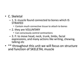
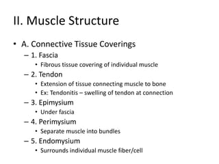
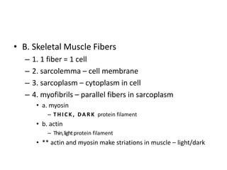
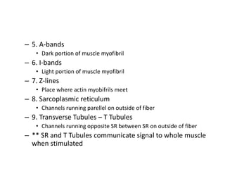
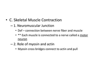
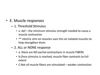
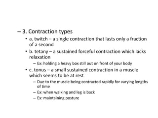
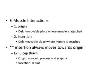
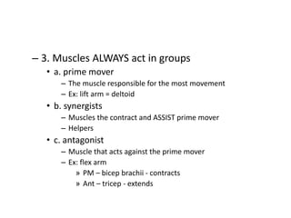
Muscular System
I. Muscle Types
• A. Smooth
– 1. S: muscle which LACKS STRIATIONS (stripes) and
is located in stomach, intestines, bladder, uterus,
and blood vessels
• Arranged in layers
– 2. It is INVOLUNTARY MUSCLE
• Unconsciously contracts
– 3. F: move food through digestive tract, constrict
blood vessels, contract uterus, empty bladder
• B. Cardiac
– 1. S: muscle which IS STRIATED located in the
heart and contains INTERCALATED DISKS
• Def of ID: junctions of cardiac muscle cells which
transmit impulses easily and allow the heart to beat as
one
• Cells look as though they are in figure 8 patterns
– 2. it is INVOLUNTARY muscle
– 3. F: to pump blood into heart chambers and
certain blood vessels
• C. Skeletal
– 1. S: muscle found connected to bones which IS
STRIATED
• Contain much connective tissue to attach to bones
– 2. they are VOLUNTARY
• Can consciously control contractions
– 3. F: to move head, neck, trunk, limbs, facial
expressions, and many actions like writing, chewing,
talking etc
• ** throughout this unit we will focus on structure
and function of SKELETAL muscle
II. Muscle Structure
• A. Connective Tissue Coverings
– 1. Fascia
• Fibrous tissue covering of individual muscle
– 2. Tendon
• Extension of tissue connecting muscle to bone
• Ex: Tendonitis – swelling of tendon at connection
– 3. Epimysium
• Under fascia
– 4. Perimysium
• Separate muscle into bundles
– 5. Endomysium
• Surrounds individual muscle fiber/cell
• B. Skeletal Muscle Fibers
– 1. 1 fiber = 1 cell
– 2. sarcolemma – cell membrane
– 3. sarcoplasm – cytoplasm in cell
– 4. myofibrils – parallel fibers in sarcoplasm
• a. myosin
– T H I C K , DA R K protein filament
• b. actin
– Thin,lightprotein filament
• ** actin and myosin make striations in muscle – light/dark
– 5. A-bands
• Dark portion of muscle myofibril
– 6. I-bands
• Light portion of muscle myofibril
– 7. Z-lines
• Place where actin myobifrils meet
– 8. Sarcoplasmic reticulum
• Channels running parellel on outside of fiber
– 9. Transverse Tubules – T Tubules
• Channels running opposite SR between SR on outside of fiber
– ** SR and T Tubules communicate signal to whole muscle
when stimulated
• C. Skeletal Muscle Contraction
– 1. Neuromuscular Junction
• Def – connection between nerve fiber and muscle
• ** Each muscle is connected to a nerve called a motor
neuron
– 2. Role of myosin and actin
• Myosin cross bridges connect to actin and pull
– 3. Process of Contraction
• a. motor neuron releases ACETYLCHOLINE
– Chemical needed for a muscle contraction
• b. actetylcholine stimulates impulses through the SR
and T Tubules
• c. impulses cause Ca+ to move through the muscle
• d. Myosin cross bridges connect to actin myofibrils
because of presence of Ca+
• e. bridges slide actin along myosin = CONTRACTION
• f. Ca+ leave muscle
• g. Myosin releases bridges = MUSCLE RELAXES
• D. O2 Debt, Fatigue, and Heat Production
– 1. O2 Debt
• a. if muscles are exercised strenuously, O2 cannot be
supplied fast enough
• b. Lactic Acid builds up
• c. ATP energy (body’s energy) decreases
• d. CO2 increases
• e.O2 must be replenished before more exercise can be done
– may take hours
– ** the O2 needed to replenish is O2 debt
• f. to replenish O2 you automatically breath deep and fast
– 2. Muscle fatigue
• a. def – MF occurs when a muscle loses its ability to
contract
• b. causes:
– Blood supply is cut off (no O2)
– Acetylcholine supply runs out in motor neuron
– Build up in lactic acid
• c. cramping results
– Muscle keeps contracting and can’t relax
– 3 . Heat Production
– Muscle contractions release heat
– Keeps body at 98.6
• E. Muscle responses
– 1. Threshold Stimulus
• a. def – the minimum stimulus strength needed to cause a
muscle contraction
• ** electric stim on muscles uses this on isolated muscles to
help strengthen them
– 2. ALL or NONE response
• a. there are NO partial contractions in muscle FIBERS
• b.Once stimulus is reached, muscle fiber contracts to full
extent
• C.Not all muscle fibers are stimulated – weaker contraction
– 3. Contraction types
• a. twitch – a single contraction that lasts only a fraction
of a second
• b. tetany – a sustained forceful contraction which lacks
relaxation
– Ex: holding a heavy box still out on front of your body
• c. tonus – a small sustained contraction in a muscle
which seems to be at rest
– Due to the muscle being contracted rapidly for varying lengths
of time
– Ex: when walking and leg is back
– Ex: maintaining posture
• F. Muscle Interactions
– 1. origin
• Def: immovable place where muscle is attached
– 2. insertion
• Def: moveable place where muscle is attached
• ** insertion always moves towards origin
– Ex: Bicep Brachii
• Origin: coracoid process and scapula
• Insertion: radius
– 3. Muscles ALWAYS act in groups
• a. prime mover
– The muscle responsible for the most movement
– Ex: lift arm = deltoid
• b. synergists
– Muscles the contract and ASSIST prime mover
– Helpers
• c. antagonist
– Muscle that acts against the prime mover
– Ex: flex arm
» PM – bicep brachii - contracts
» Ant – tricep - extends
– 4. muscle fiber types
• a. fast twitch
– Muscles which contract quickly and tire quickly
– Ex: sprinters
• b. slow twitch
– Muscles which contract slow and are more resistant to fatigue
– Ex: marathon runners, weight lifters
III. Major Skeletal Muscles
• A. Muscles of Facial Expression
– 1. frontalis
• Lies over frontal bone
– 2. occipitalis
• Lies over occipital bone
– 3. orbicularis oculi
• Around the eye orbit
– 4. orbicularis oris
• Around mouth orbit
– 5. buccinator
• Fish face
– 6. zygomaticus
• Smiling
– 7. platysma
• B. Muscles of chewing – mastication
– 1. masseter
• Main muscle of chewing
– 2. temporalis
• Lies over temporal bone
• C. Muscles that move the head
– 1. sternocleidomastoid
• Head flexion
– 2. splenis capitis
– 2. semispinalis capitis
• D. Muscles of the pectoral girdle
– 1. trapezius
• O:
– Occipital bone
– Spinous process of cervical and thoracic vertebrae
• I:
– Clavicle
– Scapula
• A:
– Move scapula
– Raise arm
– 2. Rhomboideus Major
– 3. serratus anterior
– 4. pectoralis minor
• O:
– Sternal ends of upper ribs
• I:
– Coracoid process
• A:
– Pull scapula down
– Pull scapula forward
– Raise ribs
• E. Muscles that move the Upper Arm
– 1. Pectoralis Major
• O:
– clavicle
– Sternum
– Costal cartilage
• I:
– Humerous
• A:
– Adduct arm
– Rotate humerus
– Pull arm forward
– 2. Teres Major
– 3. Latissimus Dorsi
• O:
– Spinous process of vertebrae
– Iliac crest
– Lower ribs
• I:
– Humerous
• A:
– Adducts arm
– Pulls shoulder down and back
– 4. supraspinatus
– 5. deltoid
• O:
– Acromion process
– Scapula spine
– Clavicle
• I:
– Humerous
• A:
– Abducts arm – main muscle to abduct
– 6. subscapularis
• On anterior side of scapula
– 7. infraspinatus
– 8. teres minor
• F. Muscles that move the forearm
– 1. biceps brachii
• O:
– Coracoid process
– Scapula
• I:
– Radius
• A:
– Flex arm at elbow
– Rotate hand laterally
– 2. Brachialis
– 3. brachioradialis
– 4. triceps brachii
• O:
– Lateral and medial surfaces of humerous
• I:
– Proximal ulna
• A:
– Extend arm at elbow
– 5. supinator
• In charge of supination
– 6. pronator teres
– 7. pronator quadratus
• G. Muscle that move the wrist, hand, and
fingers
– 1. flexor carpi radialis
– 2. flexor carpi ulnaris
– 3. extensor carpi radialis longus
– 4. extensor carpi radialis brevis
– 5. extensor carpi ulnaris
– 6. extensor digitorum
• H. Muscles of the abdomen
– 1. LINEA ALBA
• CONNECTIVE TISSUE which abdominal muscles connect
to
– 2. external oblique
• O:
– Outer surface of lower rib
• I:
– Linea alba, iliac crest
• A:
– Compress abdomen
– 3. internal oblique
• O:
– Iliac crest
• I:
– Rib cartilage, linea alba, pubis
• A:
– Compress abdomen
– 4. Transverse abdominus
– 5. Rectus abdominus
• O:
– Pubis
• I:
– Xyphoid process, costal cartilage
• A:
– Compress abdomen, flex vertebral column
• I. Muscles that move the Thigh
– 1. tensor fascia latae
– 2. gluteus maximus
• O:
– Sacrum, coccyx, ilium
• I:
– Posterior femur
• A:
– Extend leg at hip
– 3. gluteus medius
• Top of maximus over hip
– 4. adductor longus
– 5. adductor magnus
– 6. gracilis
– 7. fascia: sheet of connective tissue which muscle
may connect to
• J. Muscles that move the lower leg
– ** the next 3 muscles make up the HAMSTRING
– 1. biceps femoris
• O:
– Ischium and posterior surface of femur
• I:
– Fibula, tibia
• A:
– Flex/rotate leg, extend thigh
– 2. semitendinosous
– 3. semimembranous
– 4. sartorius
• Longest muscle in body
• Crosses 2 joints
– 5. quadraceps femoris
• Rectus femoris
• Vastus lateralis
• Vastus medialis
• Vastus intermedius
• K. Muscles that move ankle, foot, and toes
– 1. tibialis anterior
• O:
– Lateral surface of tibia
• I:
– Tarsals, 1st metatarsal
• A:
– Dorsal flexion and inversion of foot
– 2. extensor digitorum longus
• Extends toes -digits
– 3. gastrochnemius
• O:
– Lateral/medial condyles of femur
• I:
– Posterior surface of calcaneous
• A:
– Plantar flexion of foot and flexion of leg at knee
– 4. soleus
– 5. flexor digitorum longus
– 6. peroneous longus
P&A - Muscular system

P&A - Muscular system

  • 1.
  • 2.
    I. Muscle Types •A. Smooth – 1. S: muscle which LACKS STRIATIONS (stripes) and is located in stomach, intestines, bladder, uterus, and blood vessels • Arranged in layers – 2. It is INVOLUNTARY MUSCLE • Unconsciously contracts – 3. F: move food through digestive tract, constrict blood vessels, contract uterus, empty bladder
  • 3.
    • B. Cardiac –1. S: muscle which IS STRIATED located in the heart and contains INTERCALATED DISKS • Def of ID: junctions of cardiac muscle cells which transmit impulses easily and allow the heart to beat as one • Cells look as though they are in figure 8 patterns – 2. it is INVOLUNTARY muscle – 3. F: to pump blood into heart chambers and certain blood vessels
  • 4.
    • C. Skeletal –1. S: muscle found connected to bones which IS STRIATED • Contain much connective tissue to attach to bones – 2. they are VOLUNTARY • Can consciously control contractions – 3. F: to move head, neck, trunk, limbs, facial expressions, and many actions like writing, chewing, talking etc • ** throughout this unit we will focus on structure and function of SKELETAL muscle
  • 5.
    II. Muscle Structure •A. Connective Tissue Coverings – 1. Fascia • Fibrous tissue covering of individual muscle – 2. Tendon • Extension of tissue connecting muscle to bone • Ex: Tendonitis – swelling of tendon at connection – 3. Epimysium • Under fascia – 4. Perimysium • Separate muscle into bundles – 5. Endomysium • Surrounds individual muscle fiber/cell
  • 6.
    • B. SkeletalMuscle Fibers – 1. 1 fiber = 1 cell – 2. sarcolemma – cell membrane – 3. sarcoplasm – cytoplasm in cell – 4. myofibrils – parallel fibers in sarcoplasm • a. myosin – T H I C K , DA R K protein filament • b. actin – Thin,lightprotein filament • ** actin and myosin make striations in muscle – light/dark
  • 7.
    – 5. A-bands •Dark portion of muscle myofibril – 6. I-bands • Light portion of muscle myofibril – 7. Z-lines • Place where actin myobifrils meet – 8. Sarcoplasmic reticulum • Channels running parellel on outside of fiber – 9. Transverse Tubules – T Tubules • Channels running opposite SR between SR on outside of fiber – ** SR and T Tubules communicate signal to whole muscle when stimulated
  • 8.
    • C. SkeletalMuscle Contraction – 1. Neuromuscular Junction • Def – connection between nerve fiber and muscle • ** Each muscle is connected to a nerve called a motor neuron – 2. Role of myosin and actin • Myosin cross bridges connect to actin and pull
  • 9.
    – 3. Processof Contraction • a. motor neuron releases ACETYLCHOLINE – Chemical needed for a muscle contraction • b. actetylcholine stimulates impulses through the SR and T Tubules • c. impulses cause Ca+ to move through the muscle • d. Myosin cross bridges connect to actin myofibrils because of presence of Ca+ • e. bridges slide actin along myosin = CONTRACTION • f. Ca+ leave muscle • g. Myosin releases bridges = MUSCLE RELAXES
  • 10.
    • D. O2Debt, Fatigue, and Heat Production – 1. O2 Debt • a. if muscles are exercised strenuously, O2 cannot be supplied fast enough • b. Lactic Acid builds up • c. ATP energy (body’s energy) decreases • d. CO2 increases • e.O2 must be replenished before more exercise can be done – may take hours – ** the O2 needed to replenish is O2 debt • f. to replenish O2 you automatically breath deep and fast
  • 11.
    – 2. Musclefatigue • a. def – MF occurs when a muscle loses its ability to contract • b. causes: – Blood supply is cut off (no O2) – Acetylcholine supply runs out in motor neuron – Build up in lactic acid • c. cramping results – Muscle keeps contracting and can’t relax
  • 12.
    – 3 .Heat Production – Muscle contractions release heat – Keeps body at 98.6
  • 13.
    • E. Muscleresponses – 1. Threshold Stimulus • a. def – the minimum stimulus strength needed to cause a muscle contraction • ** electric stim on muscles uses this on isolated muscles to help strengthen them – 2. ALL or NONE response • a. there are NO partial contractions in muscle FIBERS • b.Once stimulus is reached, muscle fiber contracts to full extent • C.Not all muscle fibers are stimulated – weaker contraction
  • 14.
    – 3. Contractiontypes • a. twitch – a single contraction that lasts only a fraction of a second • b. tetany – a sustained forceful contraction which lacks relaxation – Ex: holding a heavy box still out on front of your body • c. tonus – a small sustained contraction in a muscle which seems to be at rest – Due to the muscle being contracted rapidly for varying lengths of time – Ex: when walking and leg is back – Ex: maintaining posture
  • 15.
    • F. MuscleInteractions – 1. origin • Def: immovable place where muscle is attached – 2. insertion • Def: moveable place where muscle is attached • ** insertion always moves towards origin – Ex: Bicep Brachii • Origin: coracoid process and scapula • Insertion: radius
  • 16.
    – 3. MusclesALWAYS act in groups • a. prime mover – The muscle responsible for the most movement – Ex: lift arm = deltoid • b. synergists – Muscles the contract and ASSIST prime mover – Helpers • c. antagonist – Muscle that acts against the prime mover – Ex: flex arm » PM – bicep brachii - contracts » Ant – tricep - extends
  • 17.
    – 4. musclefiber types • a. fast twitch – Muscles which contract quickly and tire quickly – Ex: sprinters • b. slow twitch – Muscles which contract slow and are more resistant to fatigue – Ex: marathon runners, weight lifters
  • 18.
    III. Major SkeletalMuscles • A. Muscles of Facial Expression – 1. frontalis • Lies over frontal bone – 2. occipitalis • Lies over occipital bone – 3. orbicularis oculi • Around the eye orbit – 4. orbicularis oris • Around mouth orbit
  • 19.
    – 5. buccinator •Fish face – 6. zygomaticus • Smiling – 7. platysma
  • 20.
    • B. Musclesof chewing – mastication – 1. masseter • Main muscle of chewing – 2. temporalis • Lies over temporal bone
  • 21.
    • C. Musclesthat move the head – 1. sternocleidomastoid • Head flexion – 2. splenis capitis – 2. semispinalis capitis
  • 22.
    • D. Musclesof the pectoral girdle – 1. trapezius • O: – Occipital bone – Spinous process of cervical and thoracic vertebrae • I: – Clavicle – Scapula • A: – Move scapula – Raise arm
  • 23.
    – 2. RhomboideusMajor – 3. serratus anterior – 4. pectoralis minor • O: – Sternal ends of upper ribs • I: – Coracoid process • A: – Pull scapula down – Pull scapula forward – Raise ribs
  • 24.
    • E. Musclesthat move the Upper Arm – 1. Pectoralis Major • O: – clavicle – Sternum – Costal cartilage • I: – Humerous • A: – Adduct arm – Rotate humerus – Pull arm forward
  • 25.
    – 2. TeresMajor – 3. Latissimus Dorsi • O: – Spinous process of vertebrae – Iliac crest – Lower ribs • I: – Humerous • A: – Adducts arm – Pulls shoulder down and back
  • 26.
    – 4. supraspinatus –5. deltoid • O: – Acromion process – Scapula spine – Clavicle • I: – Humerous • A: – Abducts arm – main muscle to abduct
  • 27.
    – 6. subscapularis •On anterior side of scapula – 7. infraspinatus – 8. teres minor
  • 28.
    • F. Musclesthat move the forearm – 1. biceps brachii • O: – Coracoid process – Scapula • I: – Radius • A: – Flex arm at elbow – Rotate hand laterally
  • 29.
    – 2. Brachialis –3. brachioradialis – 4. triceps brachii • O: – Lateral and medial surfaces of humerous • I: – Proximal ulna • A: – Extend arm at elbow
  • 30.
    – 5. supinator •In charge of supination – 6. pronator teres – 7. pronator quadratus
  • 31.
    • G. Musclethat move the wrist, hand, and fingers – 1. flexor carpi radialis – 2. flexor carpi ulnaris – 3. extensor carpi radialis longus – 4. extensor carpi radialis brevis – 5. extensor carpi ulnaris – 6. extensor digitorum
  • 32.
    • H. Musclesof the abdomen – 1. LINEA ALBA • CONNECTIVE TISSUE which abdominal muscles connect to – 2. external oblique • O: – Outer surface of lower rib • I: – Linea alba, iliac crest • A: – Compress abdomen
  • 33.
    – 3. internaloblique • O: – Iliac crest • I: – Rib cartilage, linea alba, pubis • A: – Compress abdomen – 4. Transverse abdominus
  • 34.
    – 5. Rectusabdominus • O: – Pubis • I: – Xyphoid process, costal cartilage • A: – Compress abdomen, flex vertebral column
  • 35.
    • I. Musclesthat move the Thigh – 1. tensor fascia latae – 2. gluteus maximus • O: – Sacrum, coccyx, ilium • I: – Posterior femur • A: – Extend leg at hip
  • 36.
    – 3. gluteusmedius • Top of maximus over hip – 4. adductor longus – 5. adductor magnus – 6. gracilis – 7. fascia: sheet of connective tissue which muscle may connect to
  • 37.
    • J. Musclesthat move the lower leg – ** the next 3 muscles make up the HAMSTRING – 1. biceps femoris • O: – Ischium and posterior surface of femur • I: – Fibula, tibia • A: – Flex/rotate leg, extend thigh
  • 38.
    – 2. semitendinosous –3. semimembranous
  • 39.
    – 4. sartorius •Longest muscle in body • Crosses 2 joints – 5. quadraceps femoris • Rectus femoris • Vastus lateralis • Vastus medialis • Vastus intermedius
  • 40.
    • K. Musclesthat move ankle, foot, and toes – 1. tibialis anterior • O: – Lateral surface of tibia • I: – Tarsals, 1st metatarsal • A: – Dorsal flexion and inversion of foot
  • 41.
    – 2. extensordigitorum longus • Extends toes -digits – 3. gastrochnemius • O: – Lateral/medial condyles of femur • I: – Posterior surface of calcaneous • A: – Plantar flexion of foot and flexion of leg at knee – 4. soleus – 5. flexor digitorum longus – 6. peroneous longus