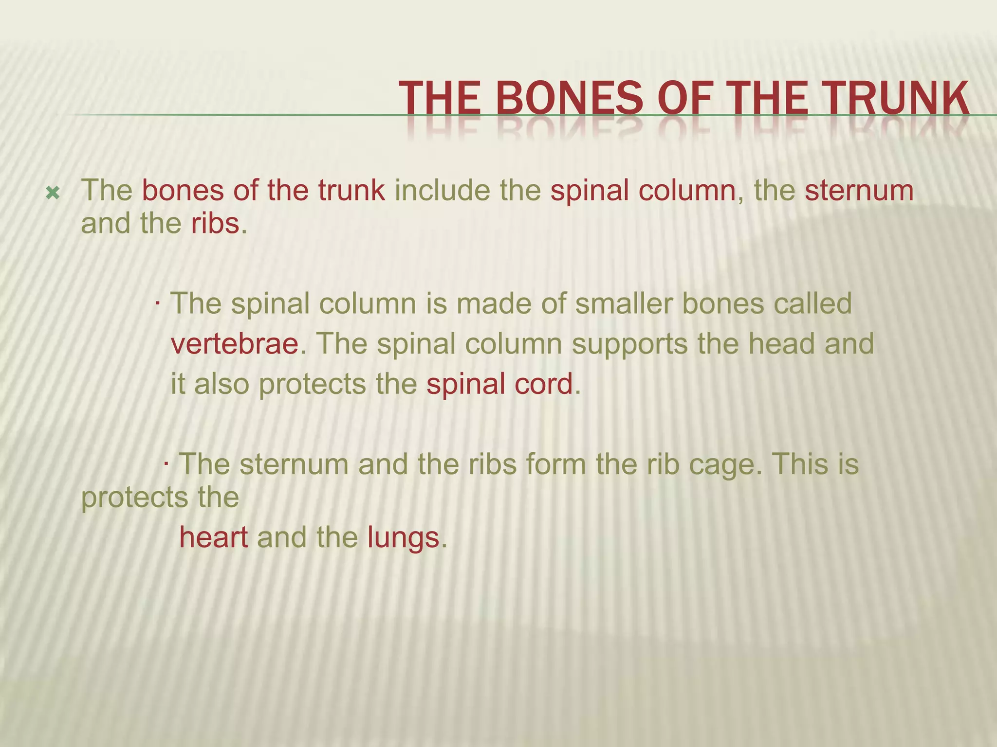
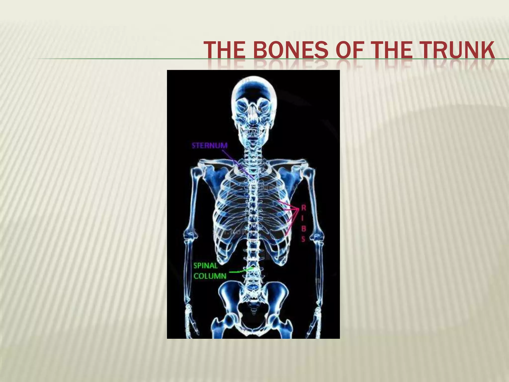
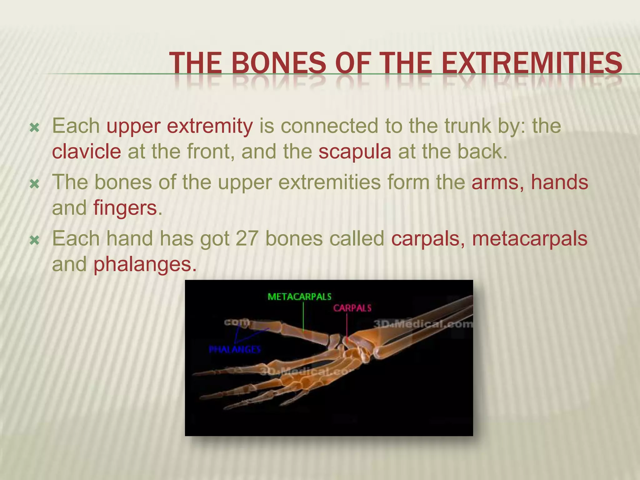
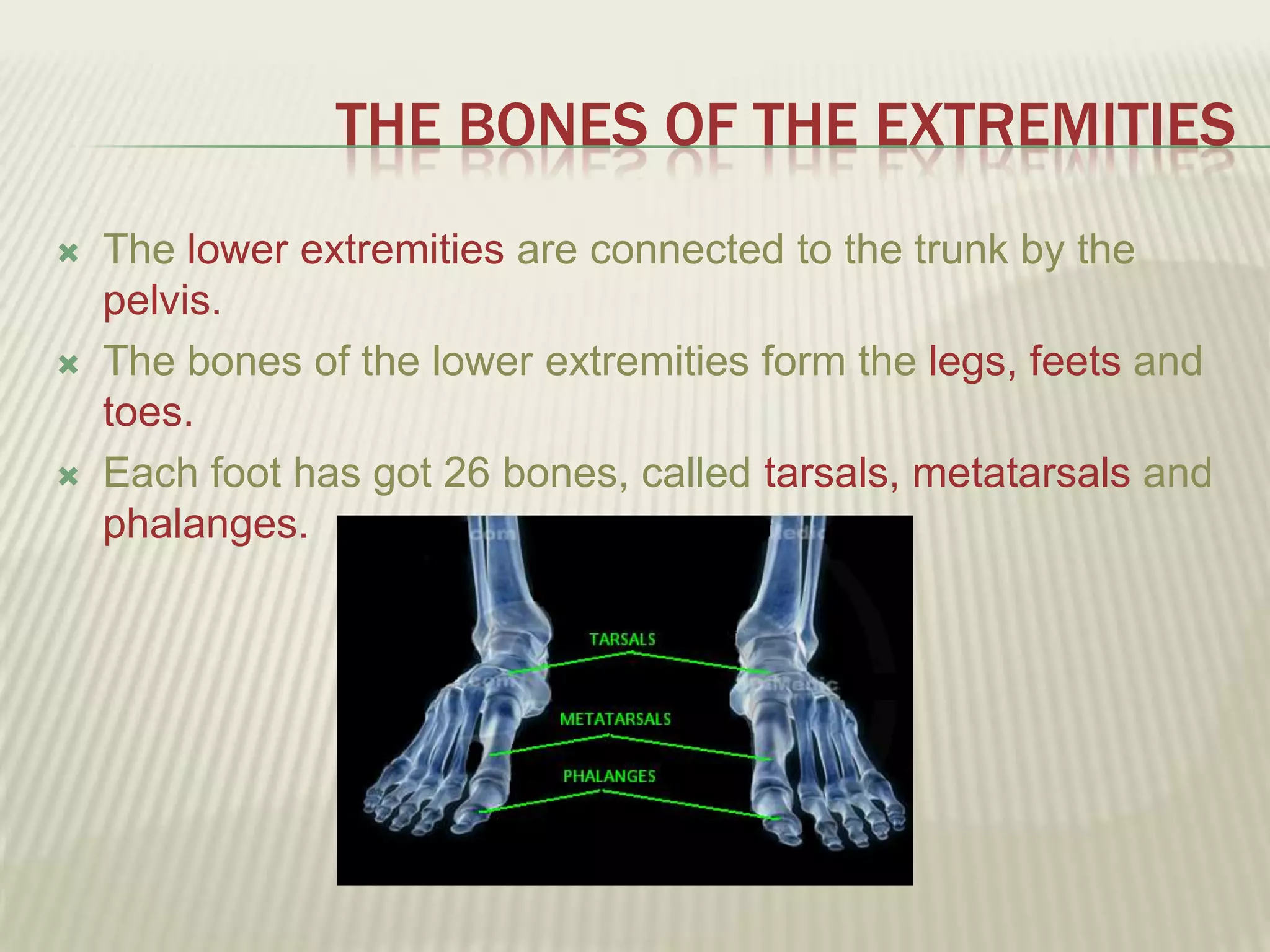
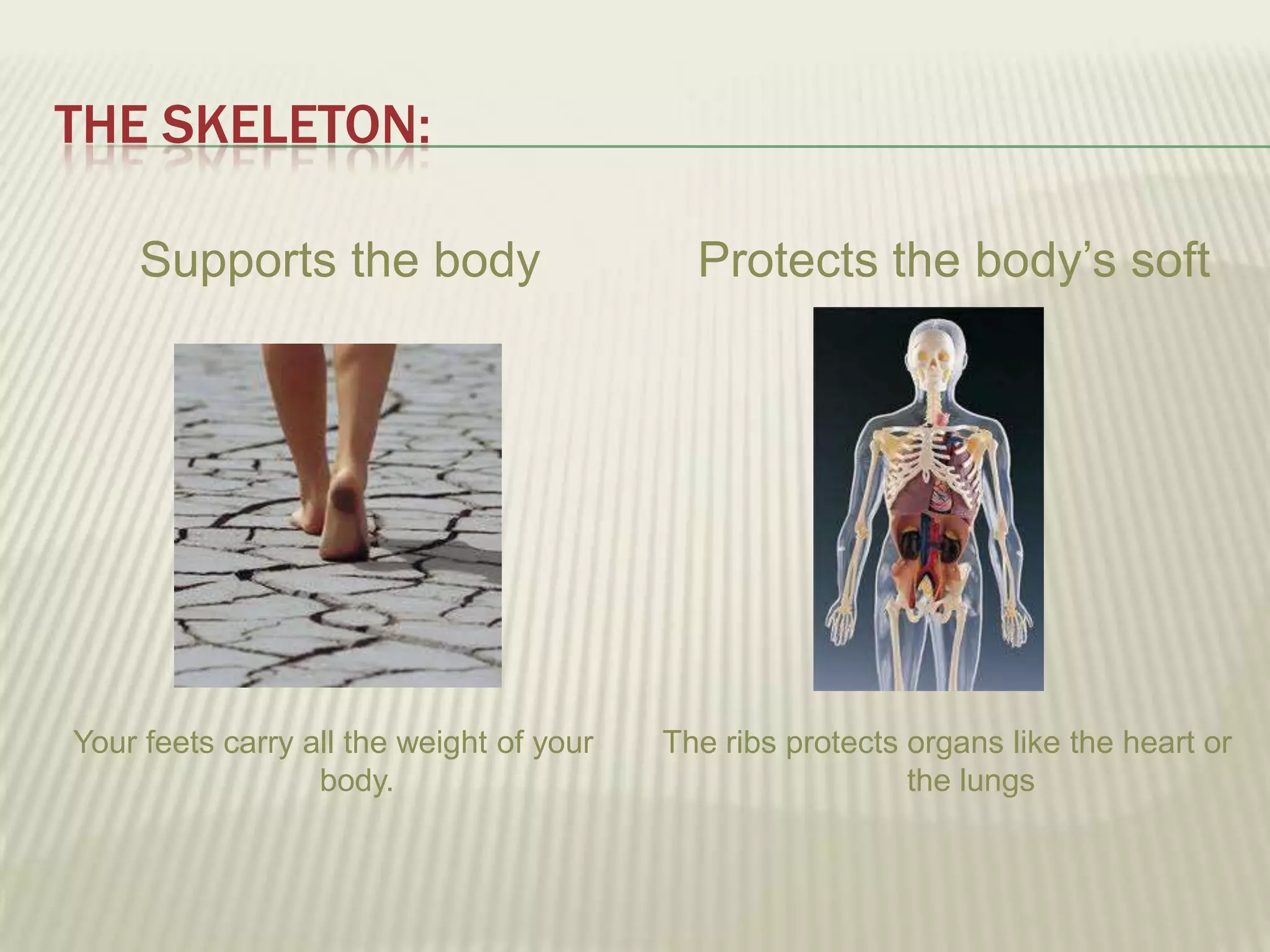
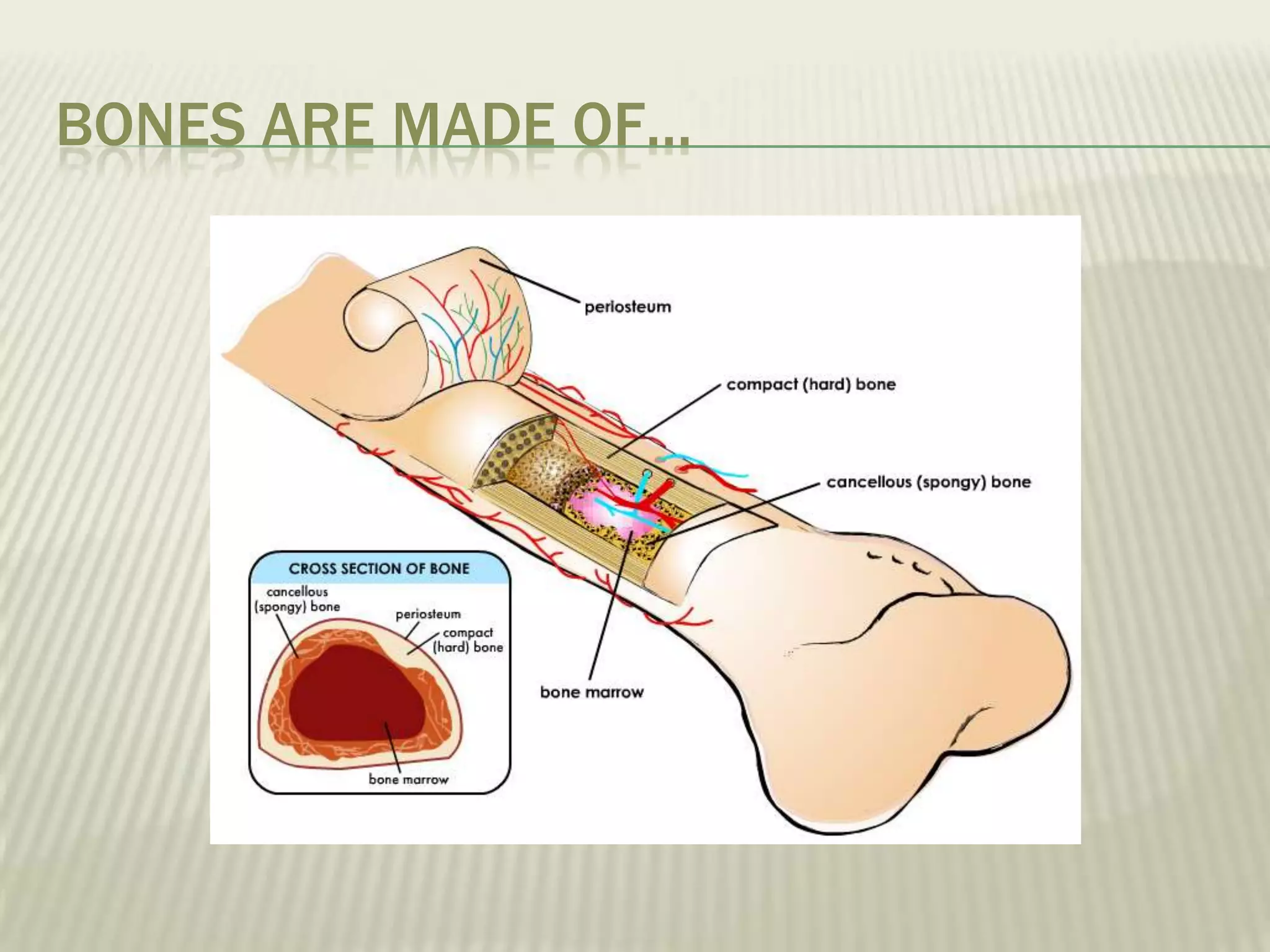
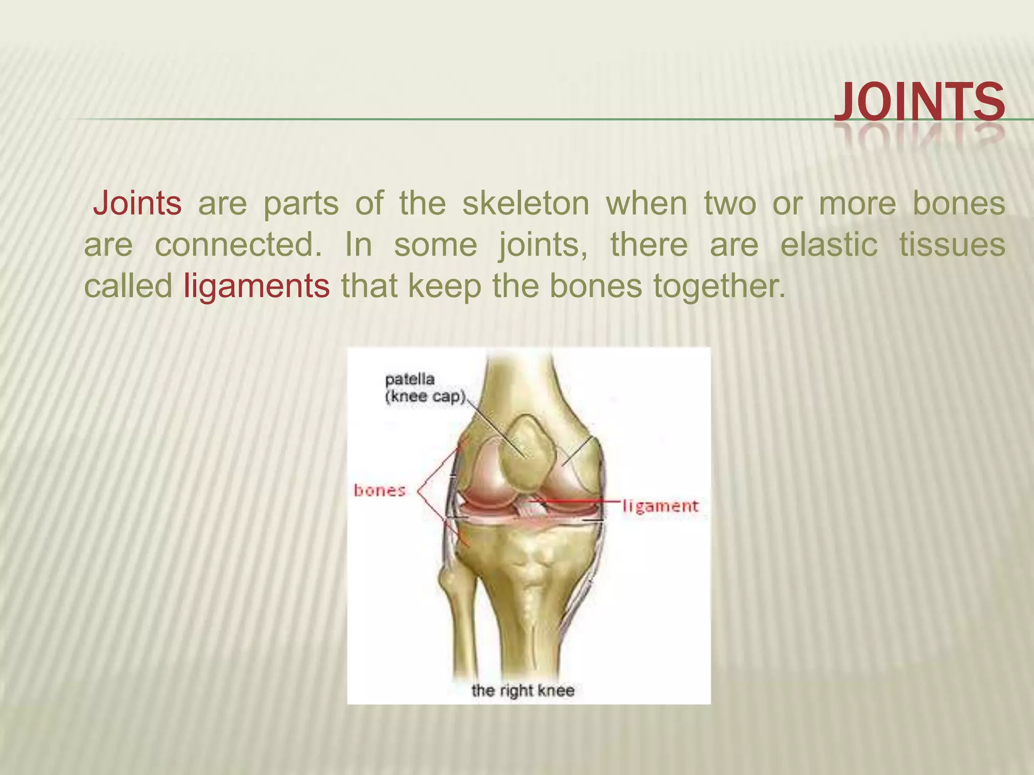
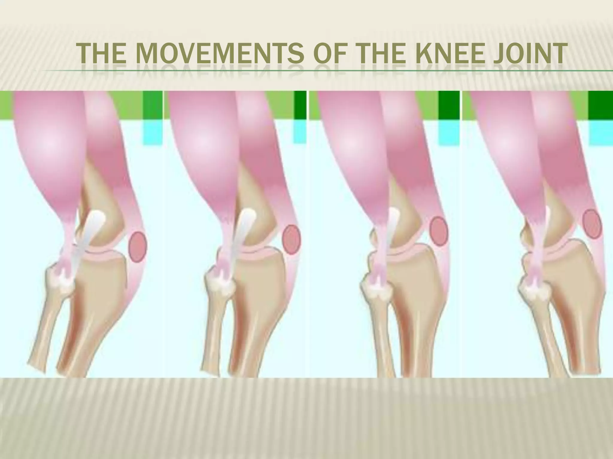
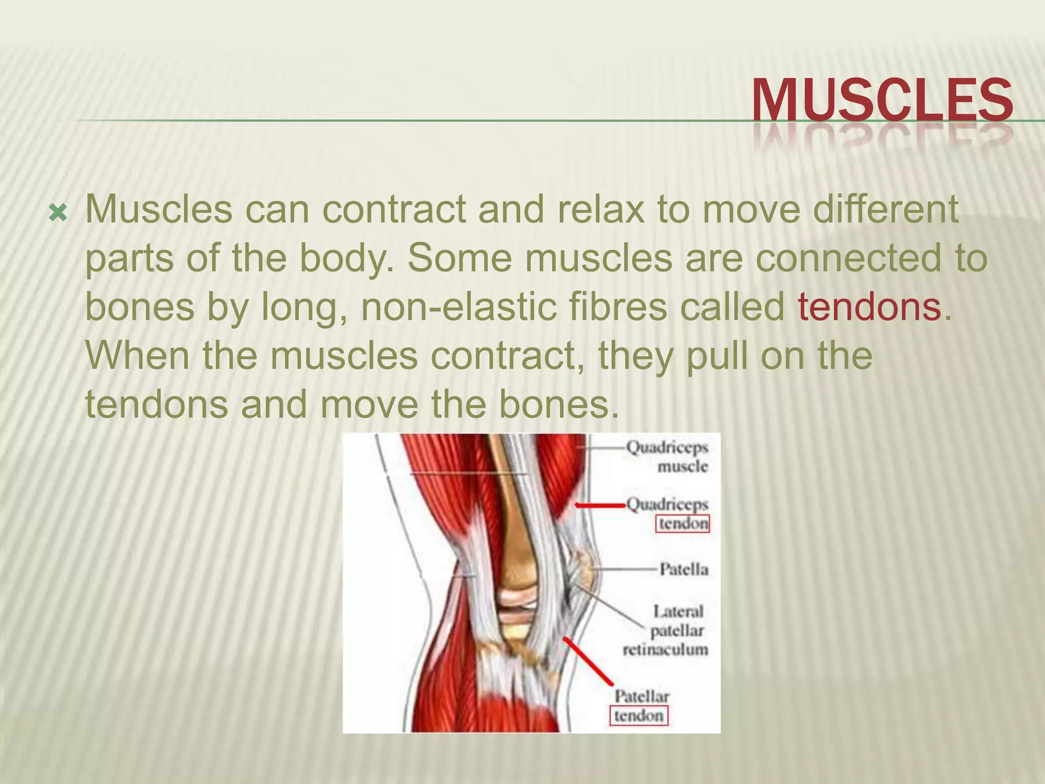
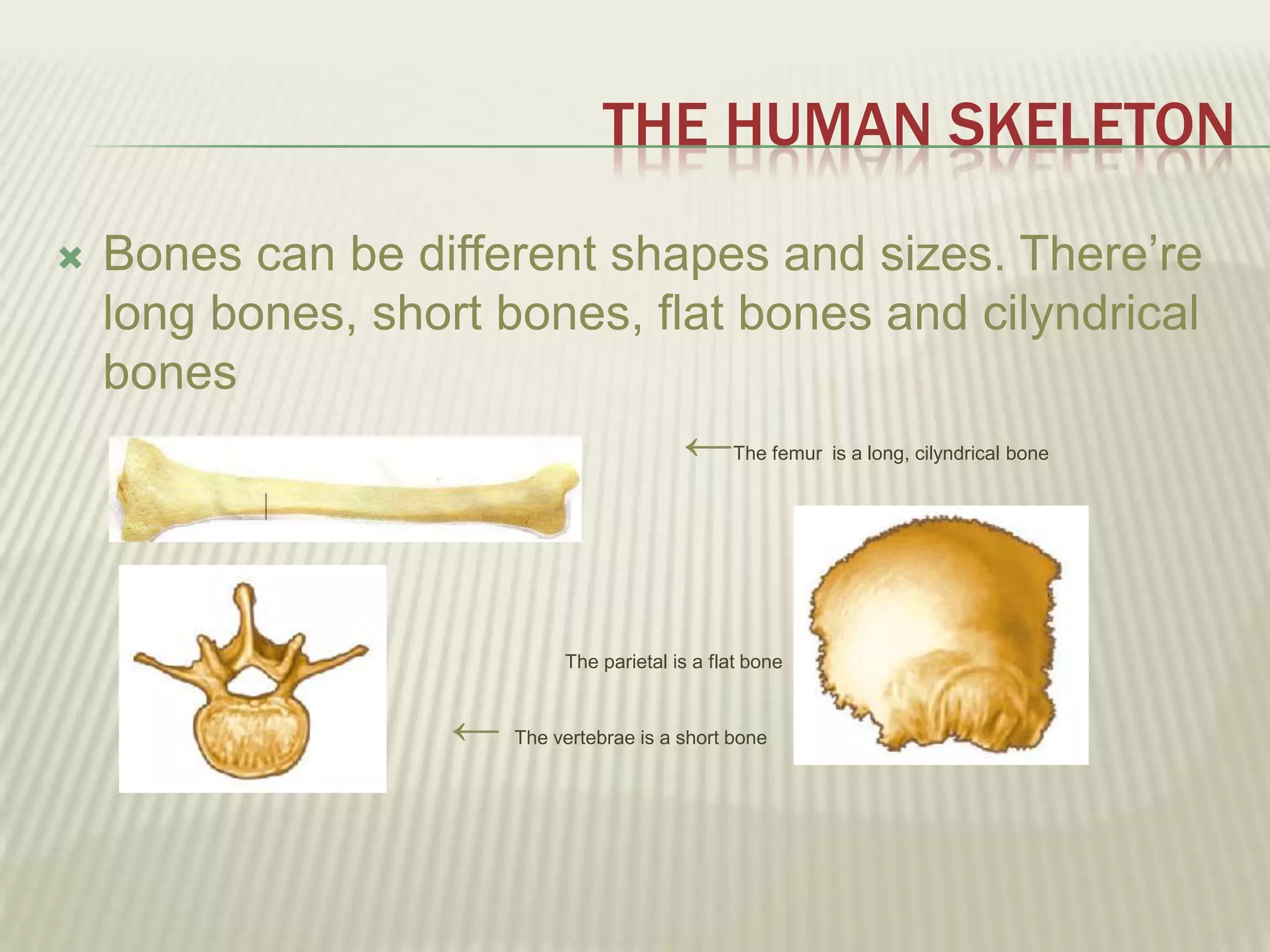
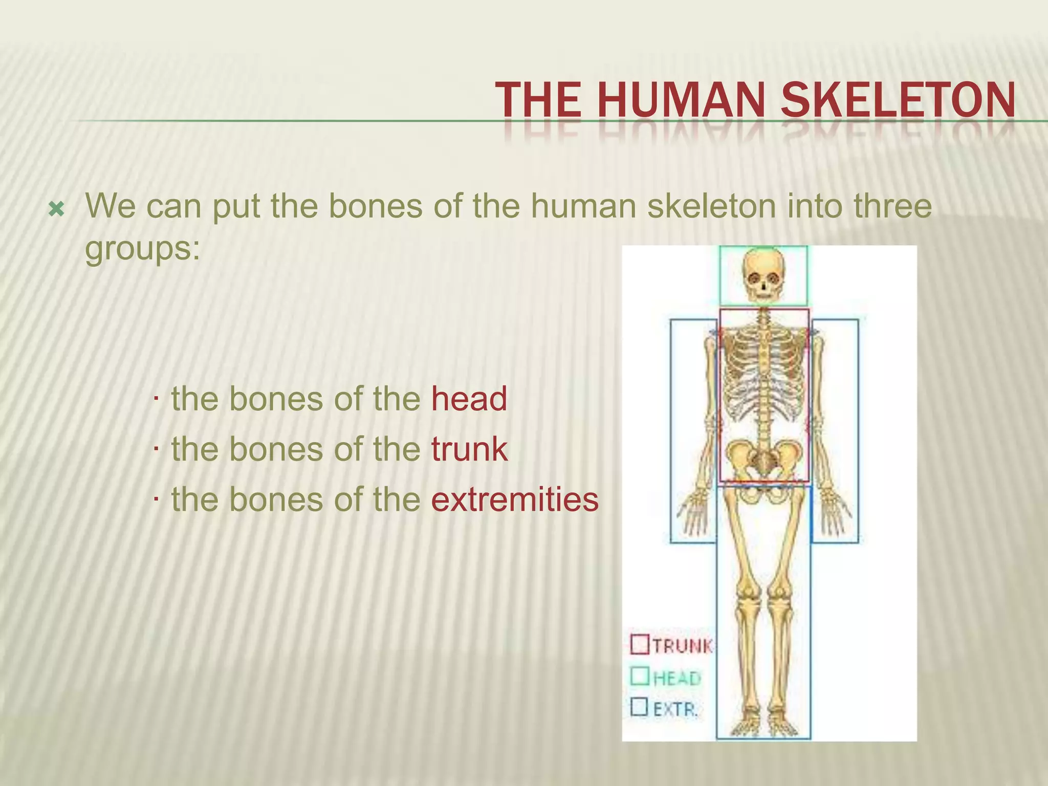
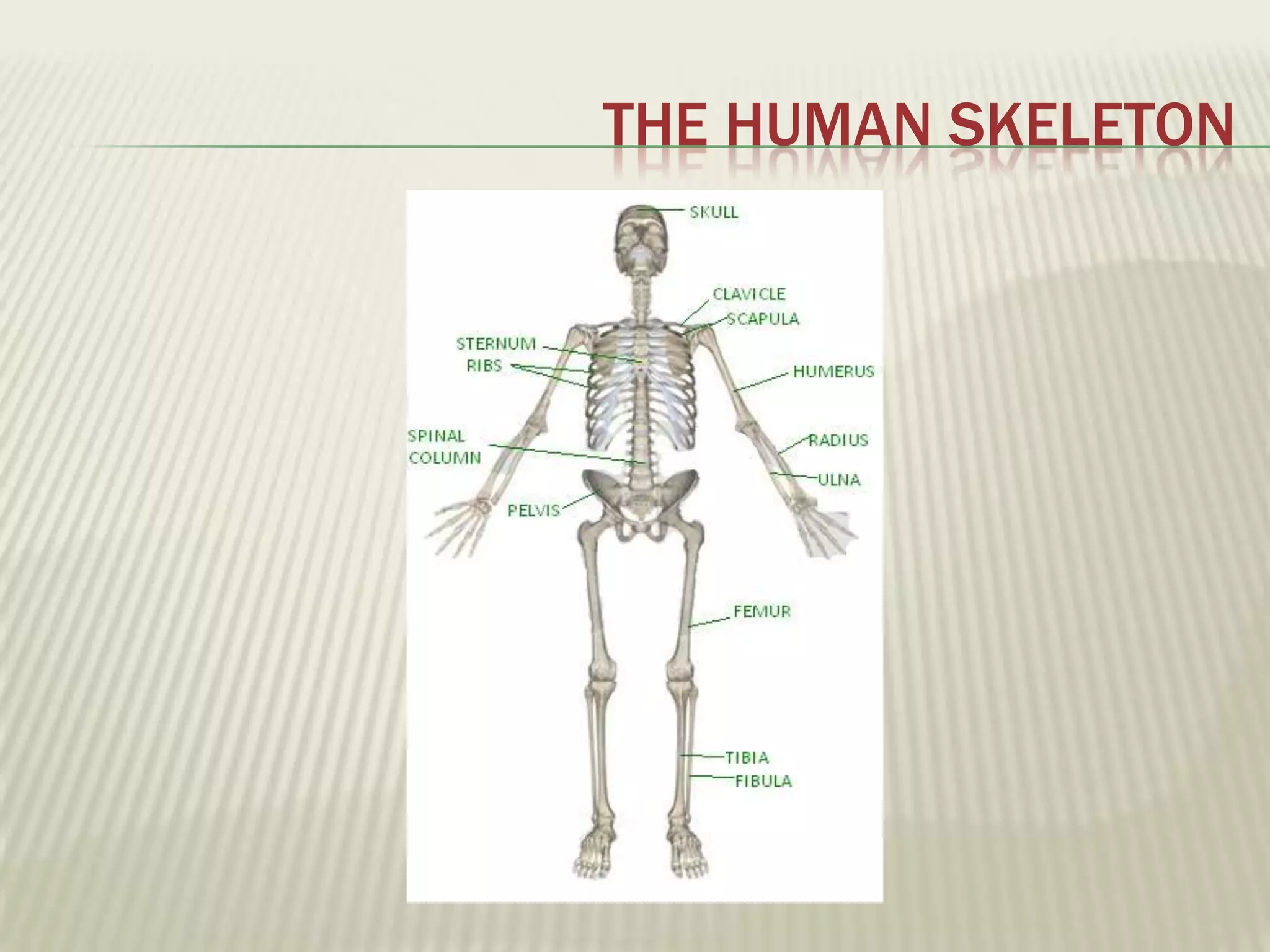
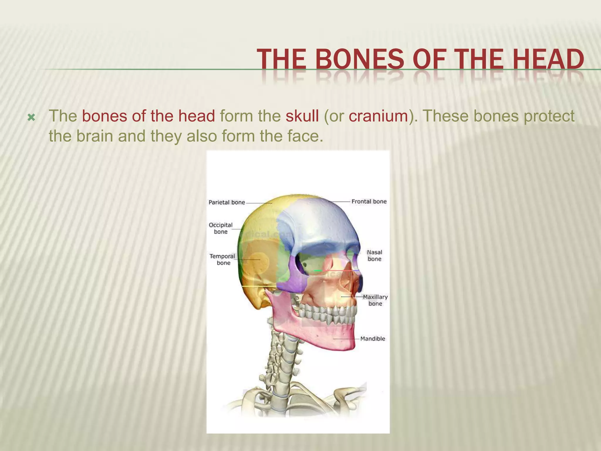
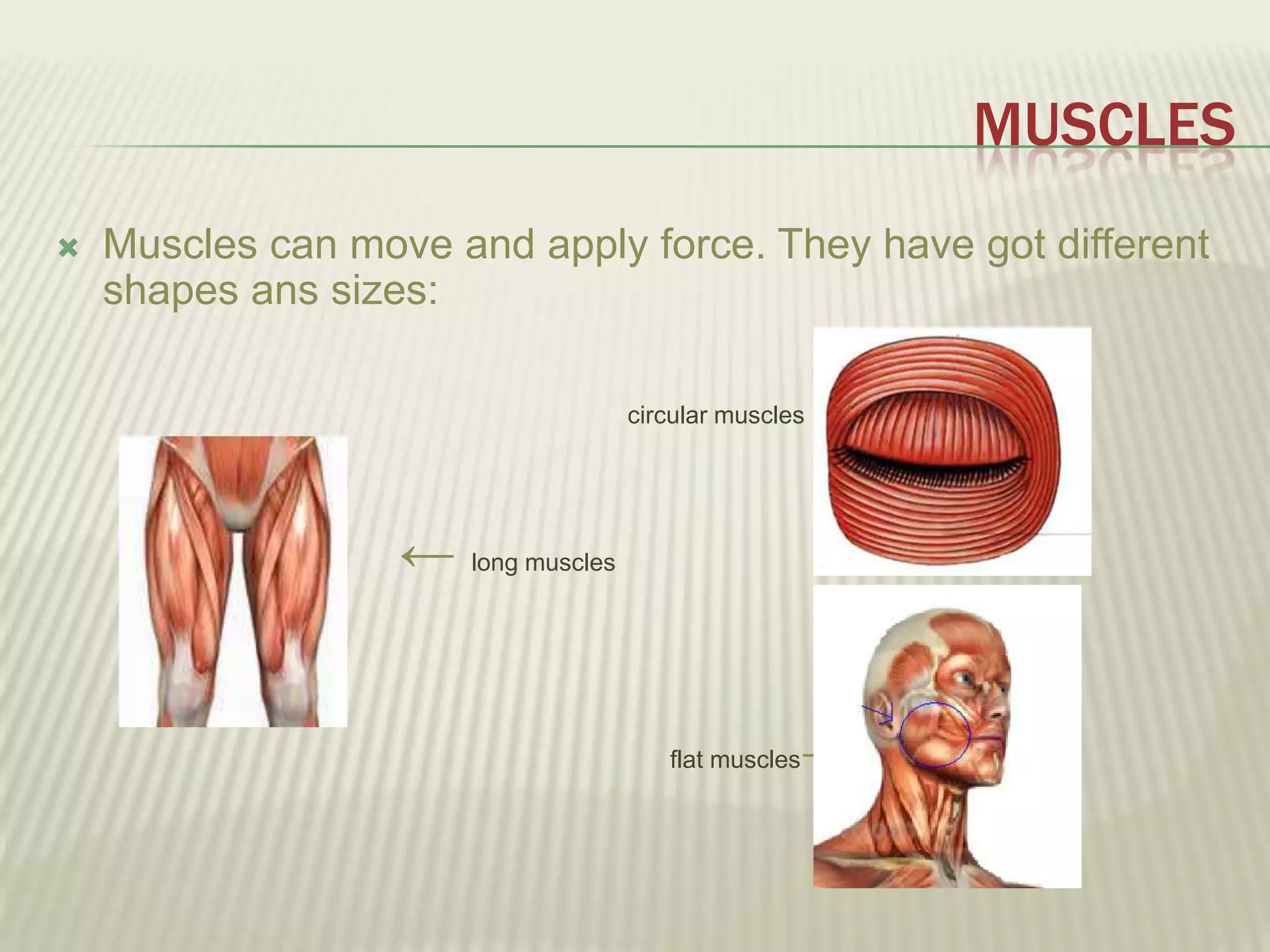
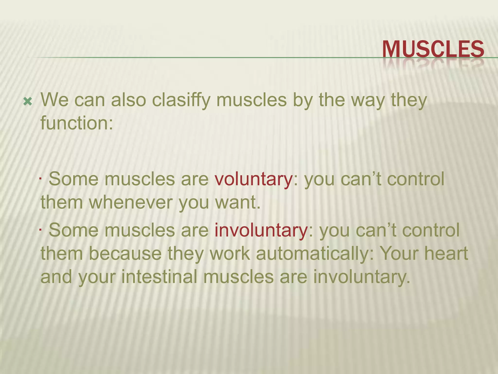
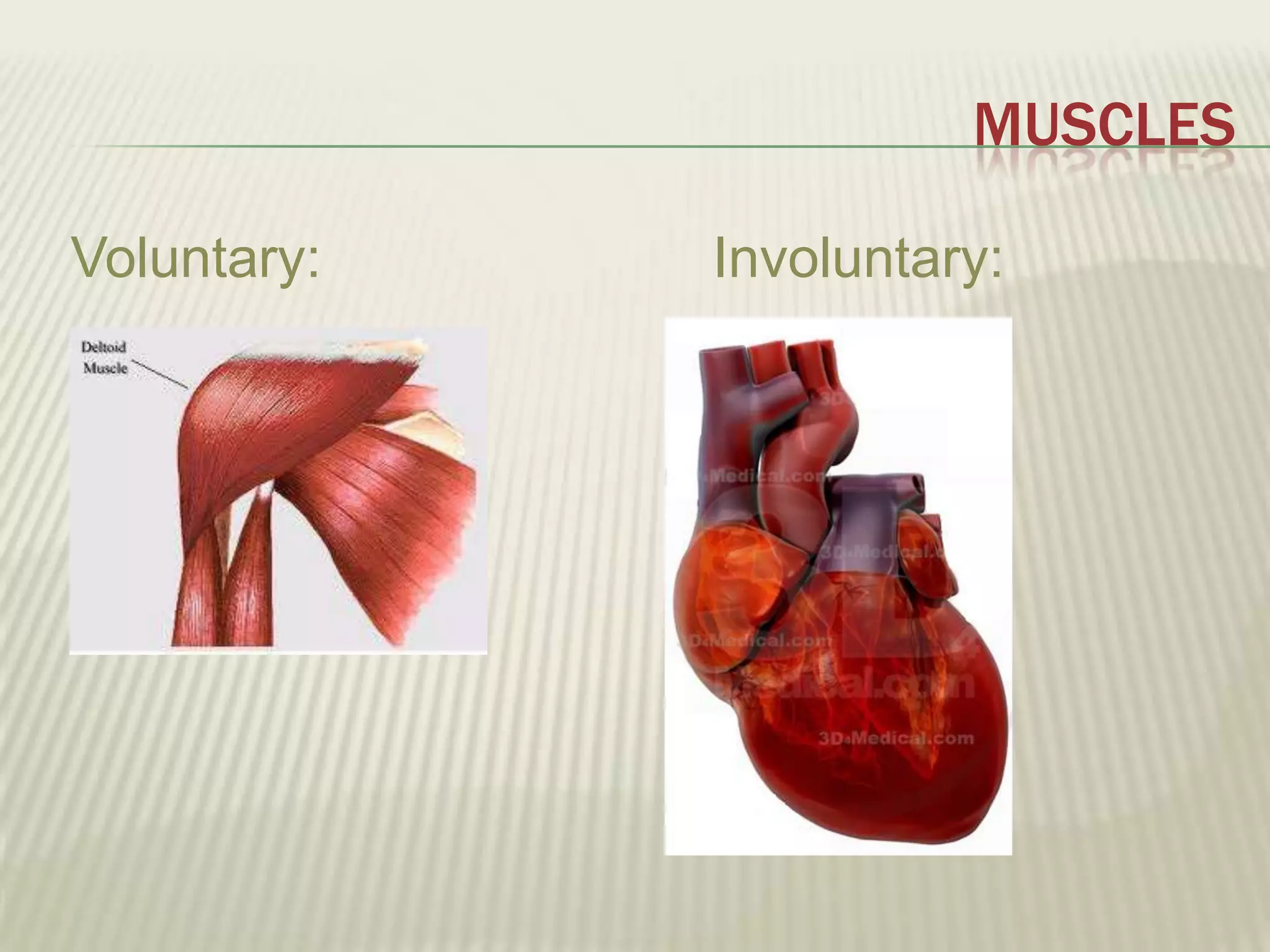
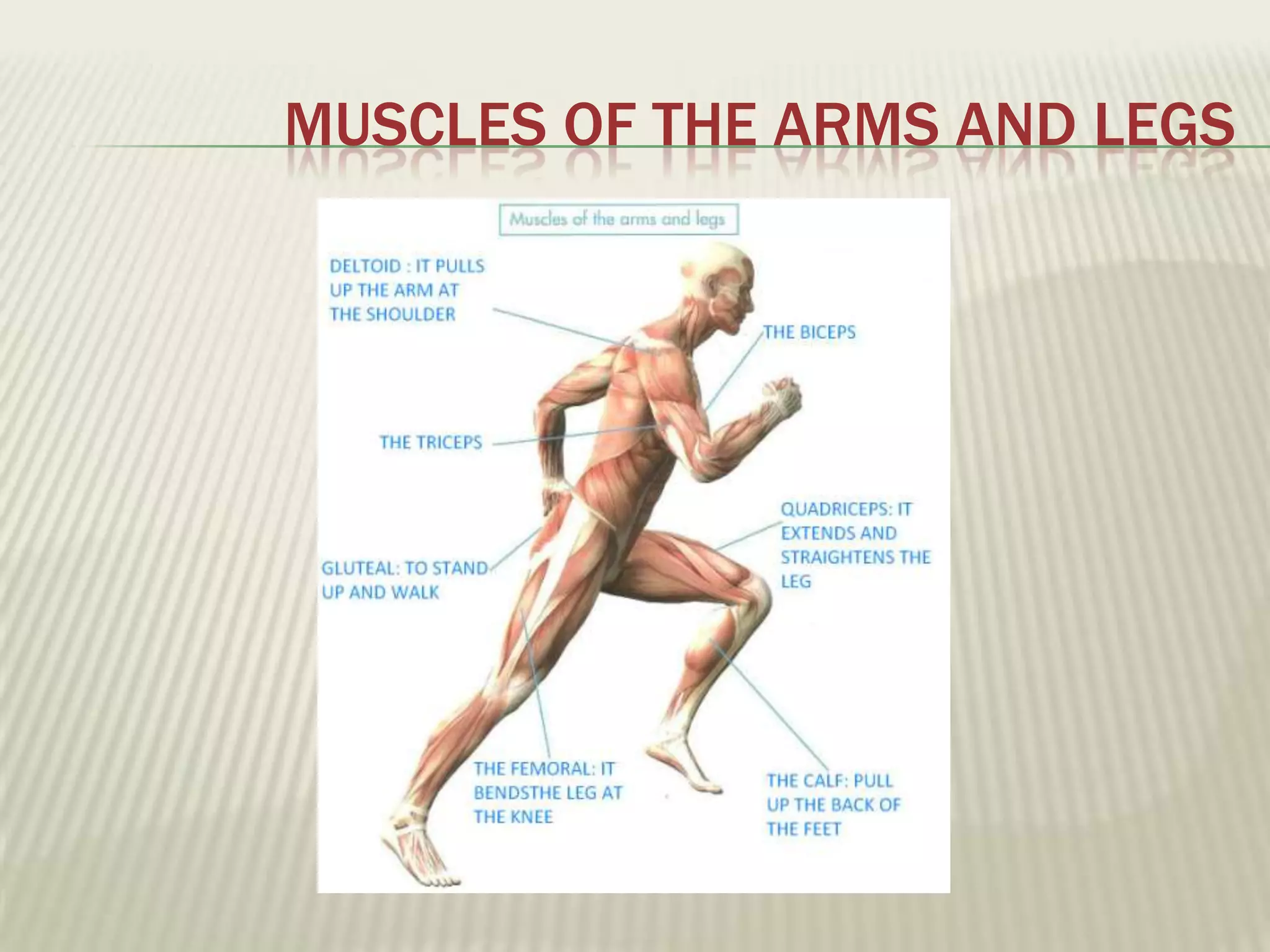
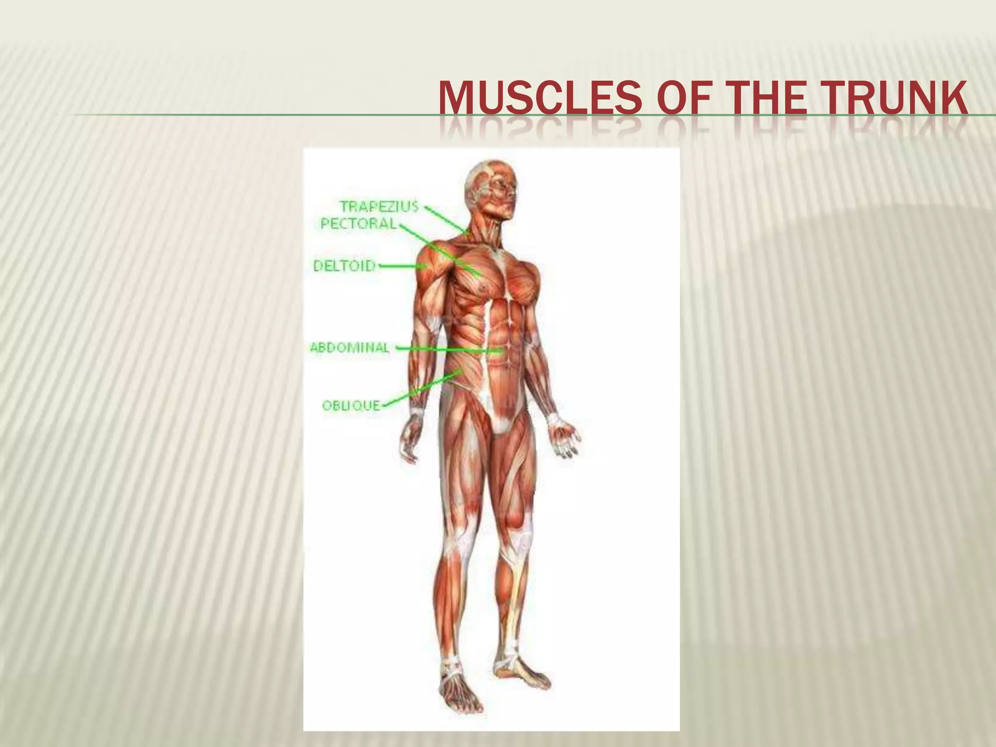
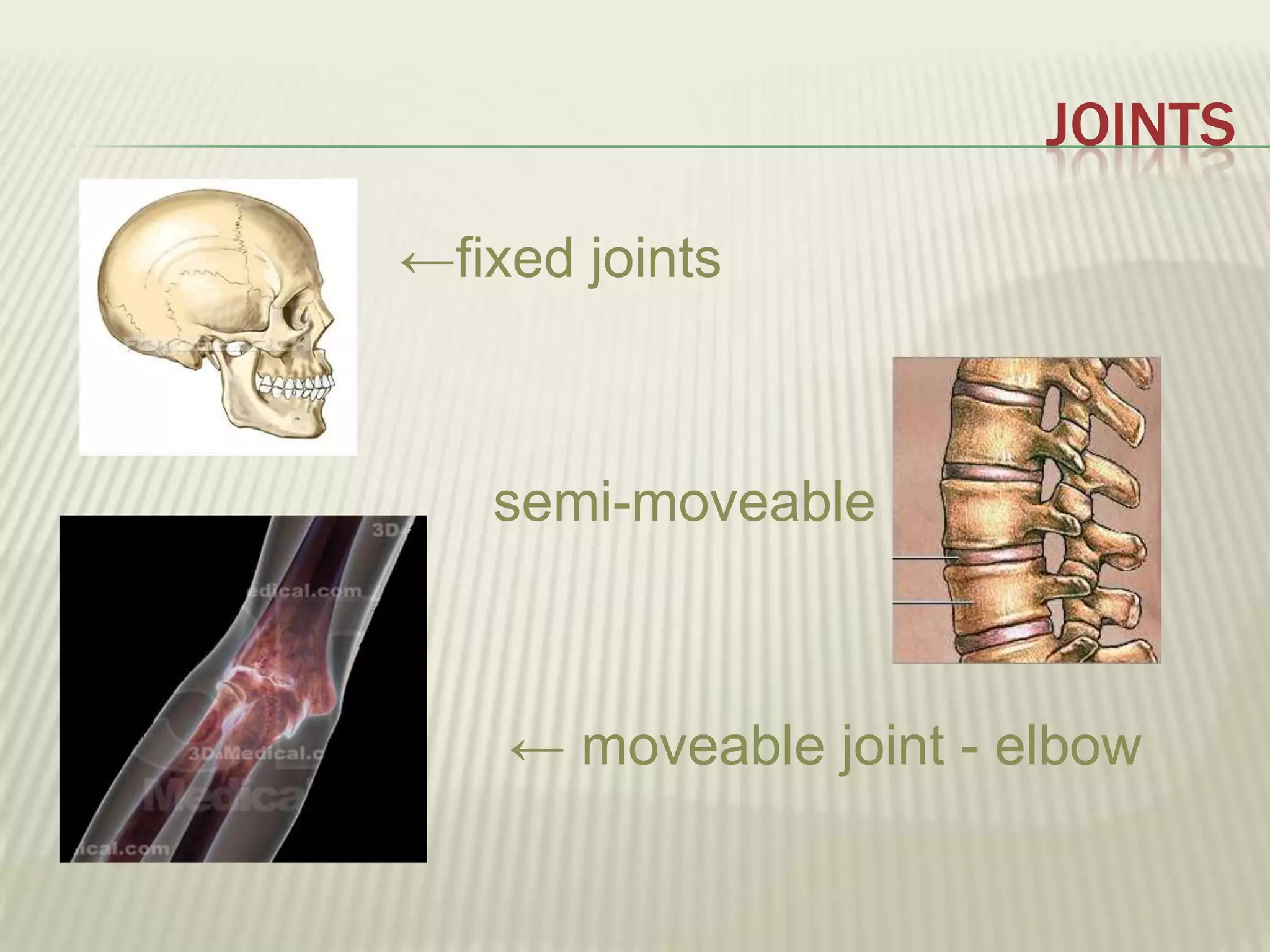
The musculoskeletal system allows humans to move using muscles and bones. It is composed of 206 bones that support the body, protect internal organs, and connect to muscles. Bones come in different shapes and sizes and can be grouped into those in the head, trunk, and extremities. Muscles contract and relax to move bones via tendons. Joints connect bones and vary in mobility. The skeleton and muscles work together to enable movement.
Review of Anatomy and Physiology of Musculoskeletal System / NursingAby Thankachan
Precise guide for DGNM, B.Sc Nursing & M.Sc Nursing Students .. regarding Review of Anatomy and Physiology of Musculoskeletal System . Highly recommended for II B.Sc Nursing Students.
Review of Anatomy and Physiology of Musculoskeletal System / NursingAby Thankachan
Precise guide for DGNM, B.Sc Nursing & M.Sc Nursing Students .. regarding Review of Anatomy and Physiology of Musculoskeletal System . Highly recommended for II B.Sc Nursing Students.
The Musculoskeletal System under the Unit HUMAN BODY
~now active with hyperlinks.
Please note that this presentation will be more appreciated if your computer is under Microsoft 2013. Kindly consider the compatibility for more convenient and pleasing slides.
The skeletal system works as a support structure for your body. It gives the body its shape, allows movement, makes blood cells, provides protection for organs and stores minerals.
Integrating Ayurveda into Parkinson’s Management: A Holistic ApproachAyurveda ForAll
Explore the benefits of combining Ayurveda with conventional Parkinson's treatments. Learn how a holistic approach can manage symptoms, enhance well-being, and balance body energies. Discover the steps to safely integrate Ayurvedic practices into your Parkinson’s care plan, including expert guidance on diet, herbal remedies, and lifestyle modifications.
Basavarajeeyam is a Sreshta Sangraha grantha (Compiled book ), written by Neelkanta kotturu Basavaraja Virachita. It contains 25 Prakaranas, First 24 Chapters related to Rogas& 25th to Rasadravyas.
Local Advanced Lung Cancer: Artificial Intelligence, Synergetics, Complex Sys...Oleg Kshivets
Overall life span (LS) was 1671.7±1721.6 days and cumulative 5YS reached 62.4%, 10 years – 50.4%, 20 years – 44.6%. 94 LCP lived more than 5 years without cancer (LS=2958.6±1723.6 days), 22 – more than 10 years (LS=5571±1841.8 days). 67 LCP died because of LC (LS=471.9±344 days). AT significantly improved 5YS (68% vs. 53.7%) (P=0.028 by log-rank test). Cox modeling displayed that 5YS of LCP significantly depended on: N0-N12, T3-4, blood cell circuit, cell ratio factors (ratio between cancer cells-CC and blood cells subpopulations), LC cell dynamics, recalcification time, heparin tolerance, prothrombin index, protein, AT, procedure type (P=0.000-0.031). Neural networks, genetic algorithm selection and bootstrap simulation revealed relationships between 5YS and N0-12 (rank=1), thrombocytes/CC (rank=2), segmented neutrophils/CC (3), eosinophils/CC (4), erythrocytes/CC (5), healthy cells/CC (6), lymphocytes/CC (7), stick neutrophils/CC (8), leucocytes/CC (9), monocytes/CC (10). Correct prediction of 5YS was 100% by neural networks computing (error=0.000; area under ROC curve=1.0).
Title: Sense of Taste
Presenter: Dr. Faiza, Assistant Professor of Physiology
Qualifications:
MBBS (Best Graduate, AIMC Lahore)
FCPS Physiology
ICMT, CHPE, DHPE (STMU)
MPH (GC University, Faisalabad)
MBA (Virtual University of Pakistan)
Learning Objectives:
Describe the structure and function of taste buds.
Describe the relationship between the taste threshold and taste index of common substances.
Explain the chemical basis and signal transduction of taste perception for each type of primary taste sensation.
Recognize different abnormalities of taste perception and their causes.
Key Topics:
Significance of Taste Sensation:
Differentiation between pleasant and harmful food
Influence on behavior
Selection of food based on metabolic needs
Receptors of Taste:
Taste buds on the tongue
Influence of sense of smell, texture of food, and pain stimulation (e.g., by pepper)
Primary and Secondary Taste Sensations:
Primary taste sensations: Sweet, Sour, Salty, Bitter, Umami
Chemical basis and signal transduction mechanisms for each taste
Taste Threshold and Index:
Taste threshold values for Sweet (sucrose), Salty (NaCl), Sour (HCl), and Bitter (Quinine)
Taste index relationship: Inversely proportional to taste threshold
Taste Blindness:
Inability to taste certain substances, particularly thiourea compounds
Example: Phenylthiocarbamide
Structure and Function of Taste Buds:
Composition: Epithelial cells, Sustentacular/Supporting cells, Taste cells, Basal cells
Features: Taste pores, Taste hairs/microvilli, and Taste nerve fibers
Location of Taste Buds:
Found in papillae of the tongue (Fungiform, Circumvallate, Foliate)
Also present on the palate, tonsillar pillars, epiglottis, and proximal esophagus
Mechanism of Taste Stimulation:
Interaction of taste substances with receptors on microvilli
Signal transduction pathways for Umami, Sweet, Bitter, Sour, and Salty tastes
Taste Sensitivity and Adaptation:
Decrease in sensitivity with age
Rapid adaptation of taste sensation
Role of Saliva in Taste:
Dissolution of tastants to reach receptors
Washing away the stimulus
Taste Preferences and Aversions:
Mechanisms behind taste preference and aversion
Influence of receptors and neural pathways
Impact of Sensory Nerve Damage:
Degeneration of taste buds if the sensory nerve fiber is cut
Abnormalities of Taste Detection:
Conditions: Ageusia, Hypogeusia, Dysgeusia (parageusia)
Causes: Nerve damage, neurological disorders, infections, poor oral hygiene, adverse drug effects, deficiencies, aging, tobacco use, altered neurotransmitter levels
Neurotransmitters and Taste Threshold:
Effects of serotonin (5-HT) and norepinephrine (NE) on taste sensitivity
Supertasters:
25% of the population with heightened sensitivity to taste, especially bitterness
Increased number of fungiform papillae
Rasamanikya is a excellent preparation in the field of Rasashastra, it is used in various Kushtha Roga, Shwasa, Vicharchika, Bhagandara, Vatarakta, and Phiranga Roga. In this article Preparation& Comparative analytical profile for both Formulationon i.e Rasamanikya prepared by Kushmanda swarasa & Churnodhaka Shodita Haratala. The study aims to provide insights into the comparative efficacy and analytical aspects of these formulations for enhanced therapeutic outcomes.
micro teaching on communication m.sc nursing.pdfAnurag Sharma
Microteaching is a unique model of practice teaching. It is a viable instrument for the. desired change in the teaching behavior or the behavior potential which, in specified types of real. classroom situations, tends to facilitate the achievement of specified types of objectives.
2. THE MUSCULOSKELETAL SYSTEM
The musculoskeletal system is composed of many muscles and bones.
Its lets you move different parts of your body and get you around from
one place to another.
Garfield can runs with his musculoskeletal system
3. THE FUNCTIONS OF THE SKELETON
Humans have got an internal skeleton. It is made of 206 different bones.
The skeleton perfoms three main functions:
· Its supports the body and helps to keep its shape.
· Its protects the body’s soft, internal organs.
· It is connected to muscles that move the different bones.
Bones are made of bone cells that form bone tissue. These cells can
grow and reproduce to repair broken bones.
4. THE SKELETON:
Supports the body Protects the body’s soft
Your feets carry all the weight of your The ribs protects organs like the heart or
body. the lungs
6. JOINTS
Joints are parts of the skeleton when two or more bones
are connected. In some joints, there are elastic tissues
called ligaments that keep the bones together.
8. MUSCLES
Muscles can contract and relax to move different
parts of the body. Some muscles are connected to
bones by long, non-elastic fibres called tendons.
When the muscles contract, they pull on the
tendons and move the bones.
9. THE HUMAN SKELETON
Bones can be different shapes and sizes. There’re
long bones, short bones, flat bones and cilyndrical
bones
← The femur is a long, cilyndrical bone
The parietal is a flat bone →
← The vertebrae is a short bone
10. THE HUMAN SKELETON
We can put the bones of the human skeleton into three
groups:
· the bones of the head
· the bones of the trunk
· the bones of the extremities
12. THE BONES OF THE HEAD
The bones of the head form the skull (or cranium). These bones protect
the brain and they also form the face.
13. THE BONES OF THE TRUNK
The bones of the trunk include the spinal column, the sternum
and the ribs.
· The spinal column is made of smaller bones called
vertebrae. The spinal column supports the head and
it also protects the spinal cord.
· The sternum and the ribs form the rib cage. This is
protects the
heart and the lungs.
15. THE BONES OF THE EXTREMITIES
Each upper extremity is connected to the trunk by: the
clavicle at the front, and the scapula at the back.
The bones of the upper extremities form the arms, hands
and fingers.
Each hand has got 27 bones called carpals, metacarpals
and phalanges.
16. THE BONES OF THE EXTREMITIES
The lower extremities are connected to the trunk by the
pelvis.
The bones of the lower extremities form the legs, feets and
toes.
Each foot has got 26 bones, called tarsals, metatarsals and
phalanges.
17. MUSCLES
Muscles can move and apply force. They have got different
shapes ans sizes:
circular muscles →
← long muscles
flat muscles →
18. MUSCLES
We can also clasiffy muscles by the way they
function:
· Some muscles are voluntary: you can’t control
them whenever you want.
· Some muscles are involuntary: you can’t control
them because they work automatically: Your heart
and your intestinal muscles are involuntary.
23. JOINTS
Joints are places when to or more bones are
connected.
We can clasiffy joints by how much they move:
· Fixed joints can’y move.
· Semi-moveable joints can’t move very much.
· Moveable joints can move a lot. Inside this
joints, there is a tough, flexible tissue called
cartilage.
25. JOINTS AND PAIRS OF MUSCLES
Muscles often works togheter in
pairs.They take turns contracting and
relaxing. When one of the muscles
contracts, it becomes shorter and pulls
on the other muscles. At the same time,
the other muscle relaxes and becomes
longer.