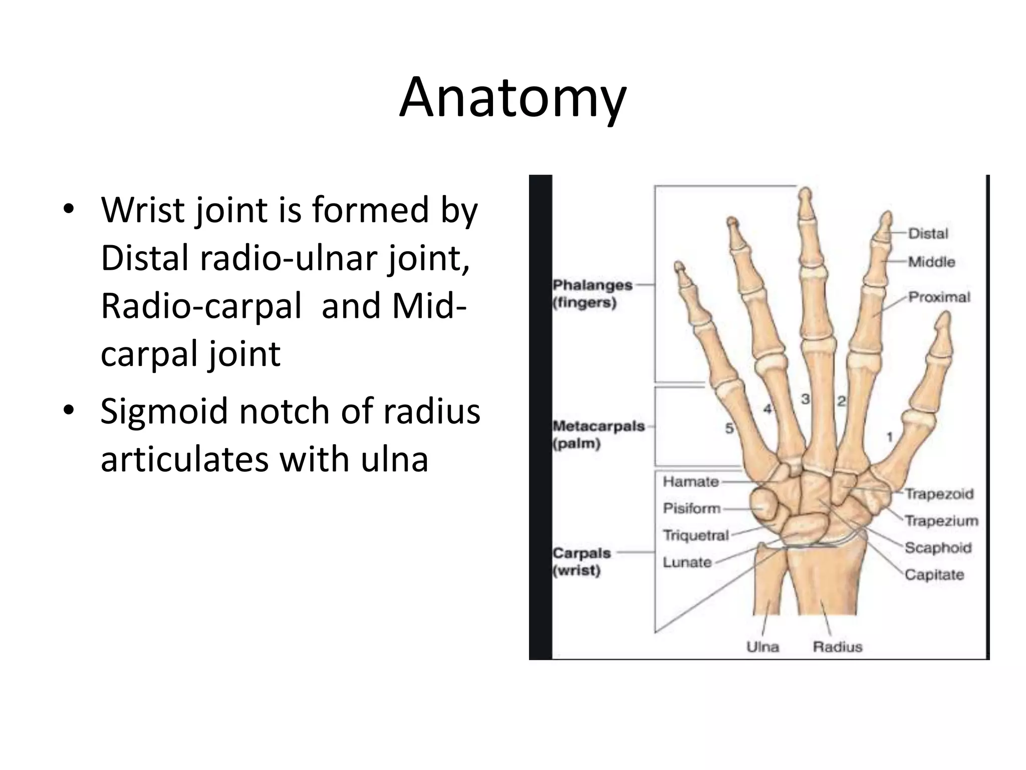
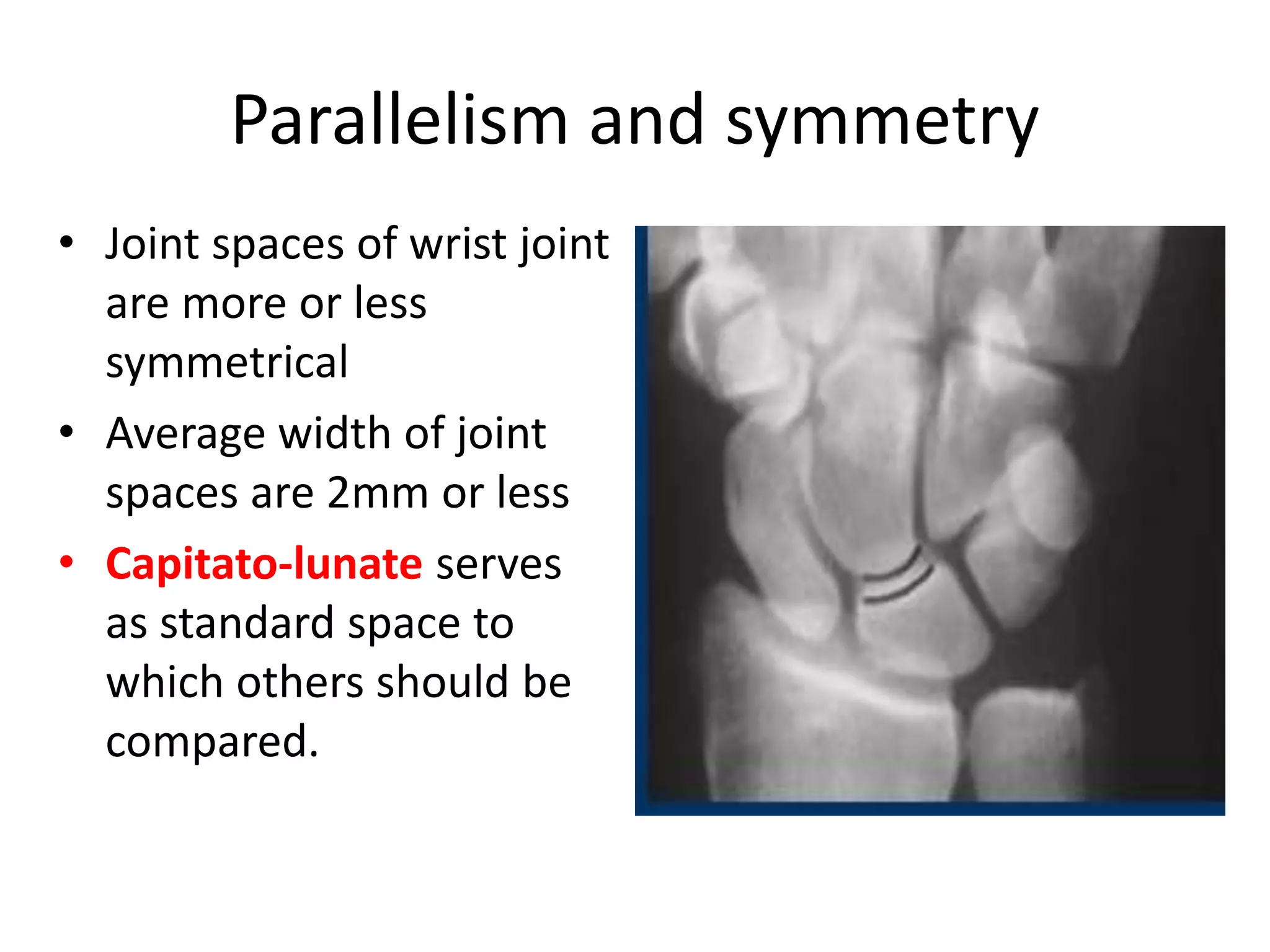
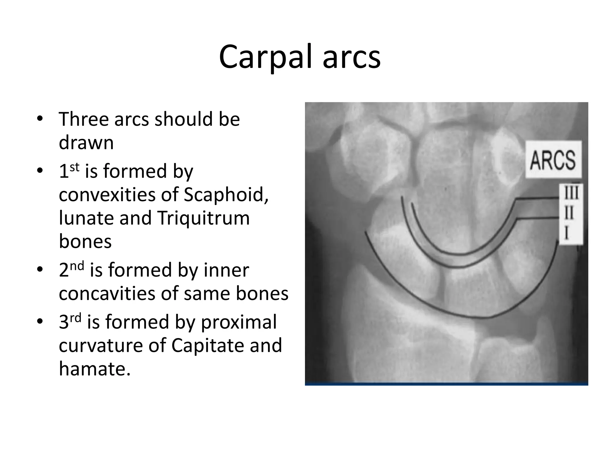
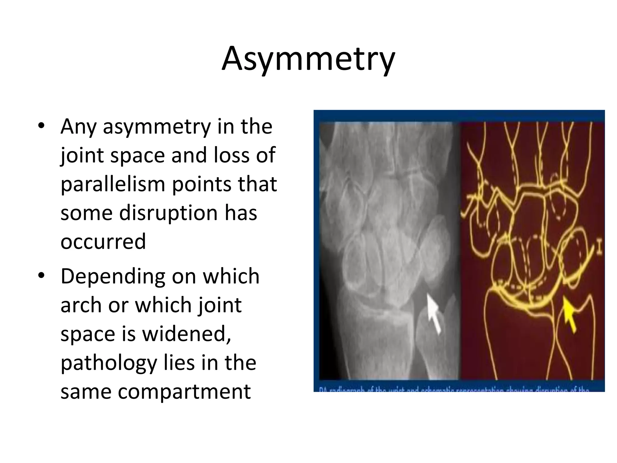
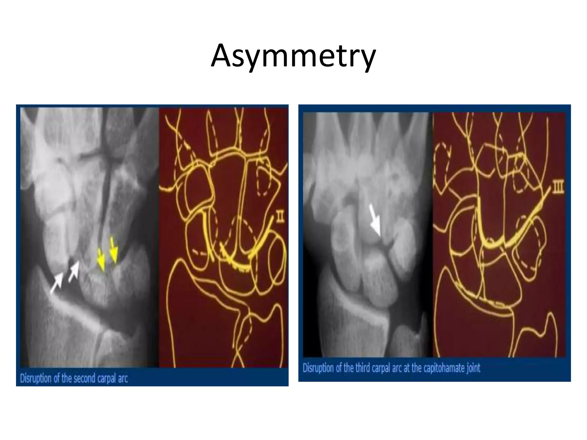
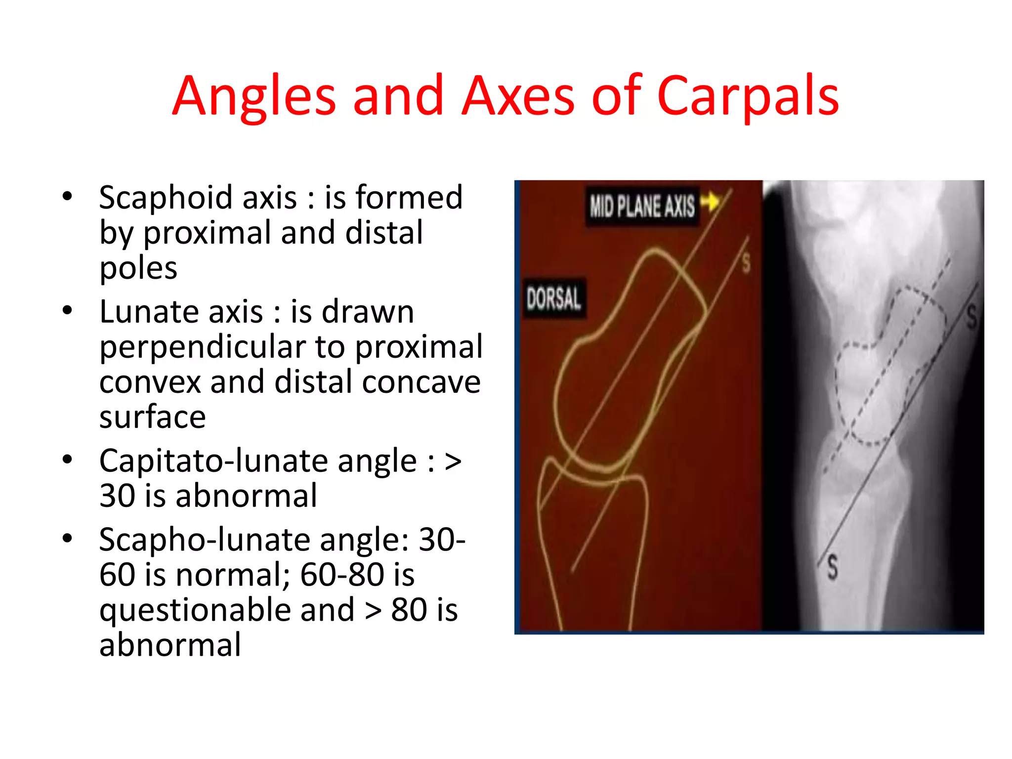
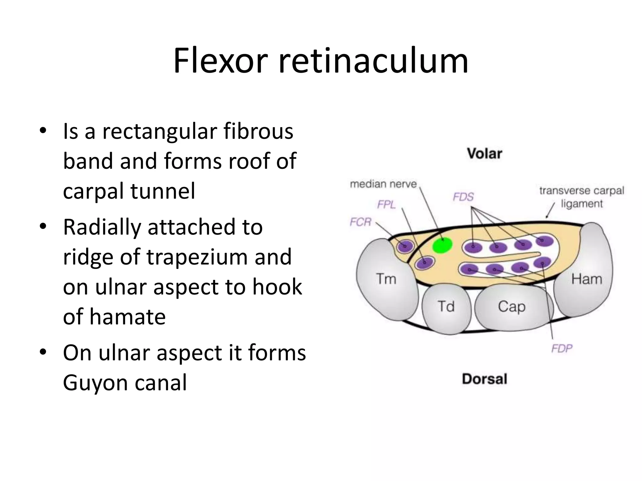
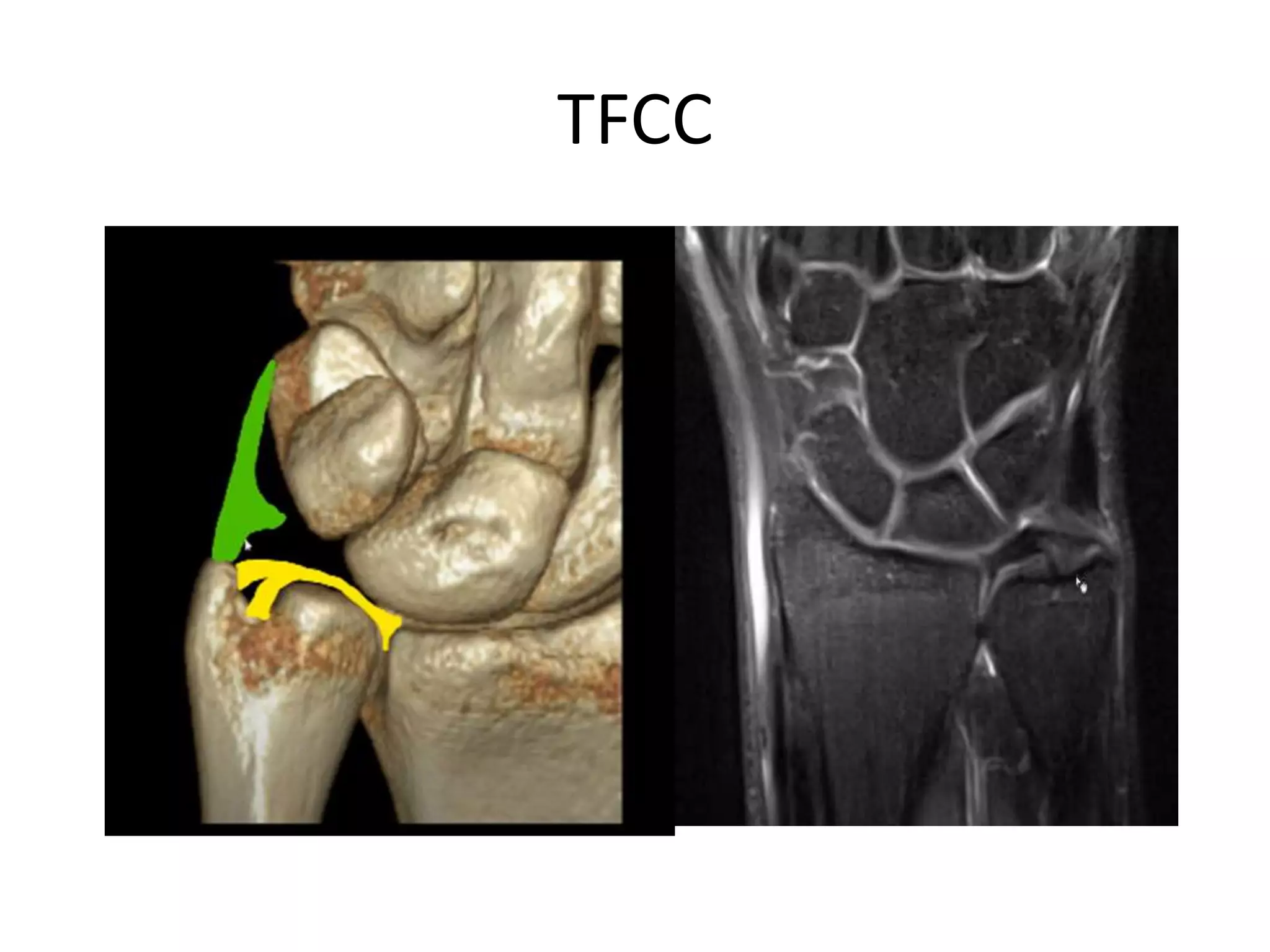
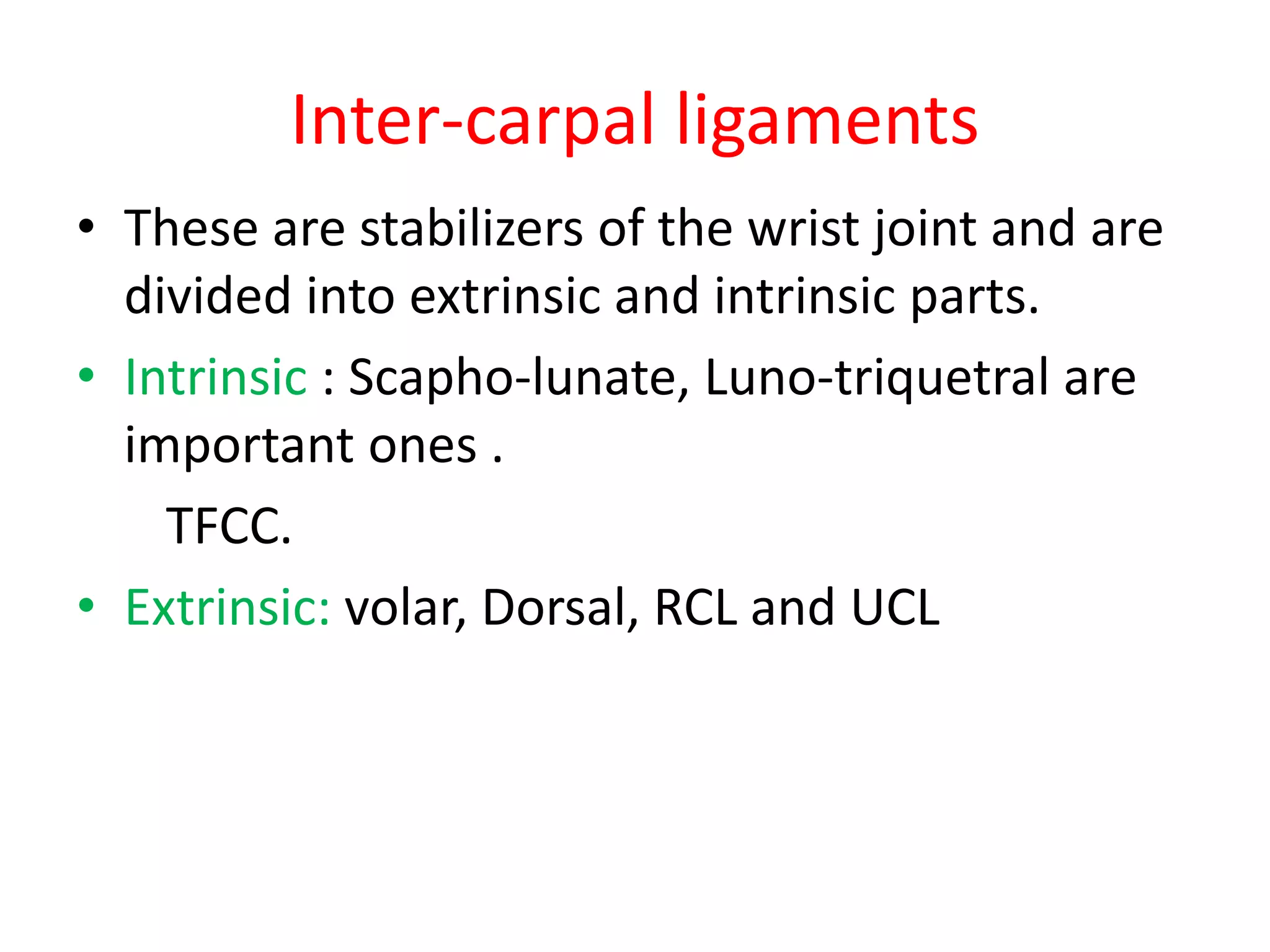
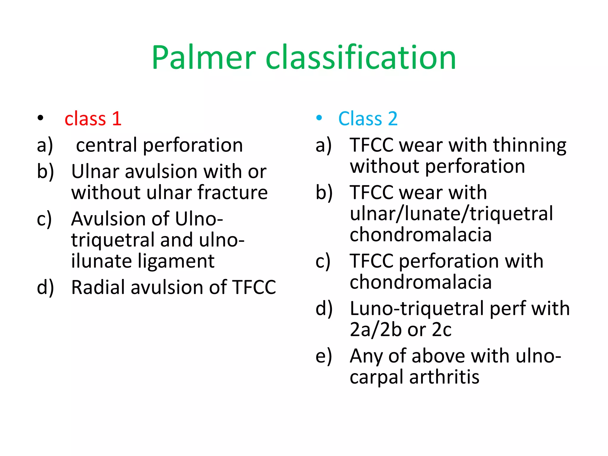
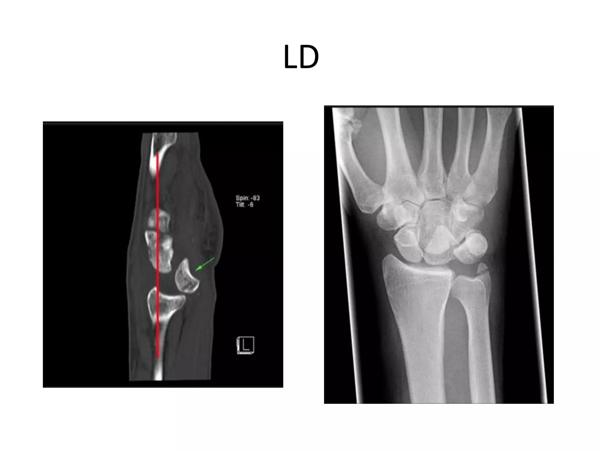
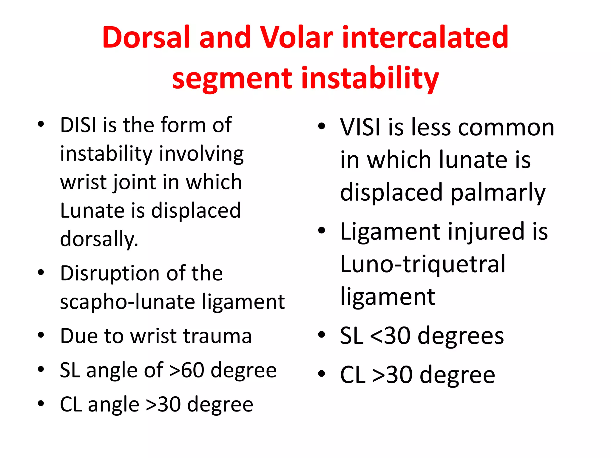
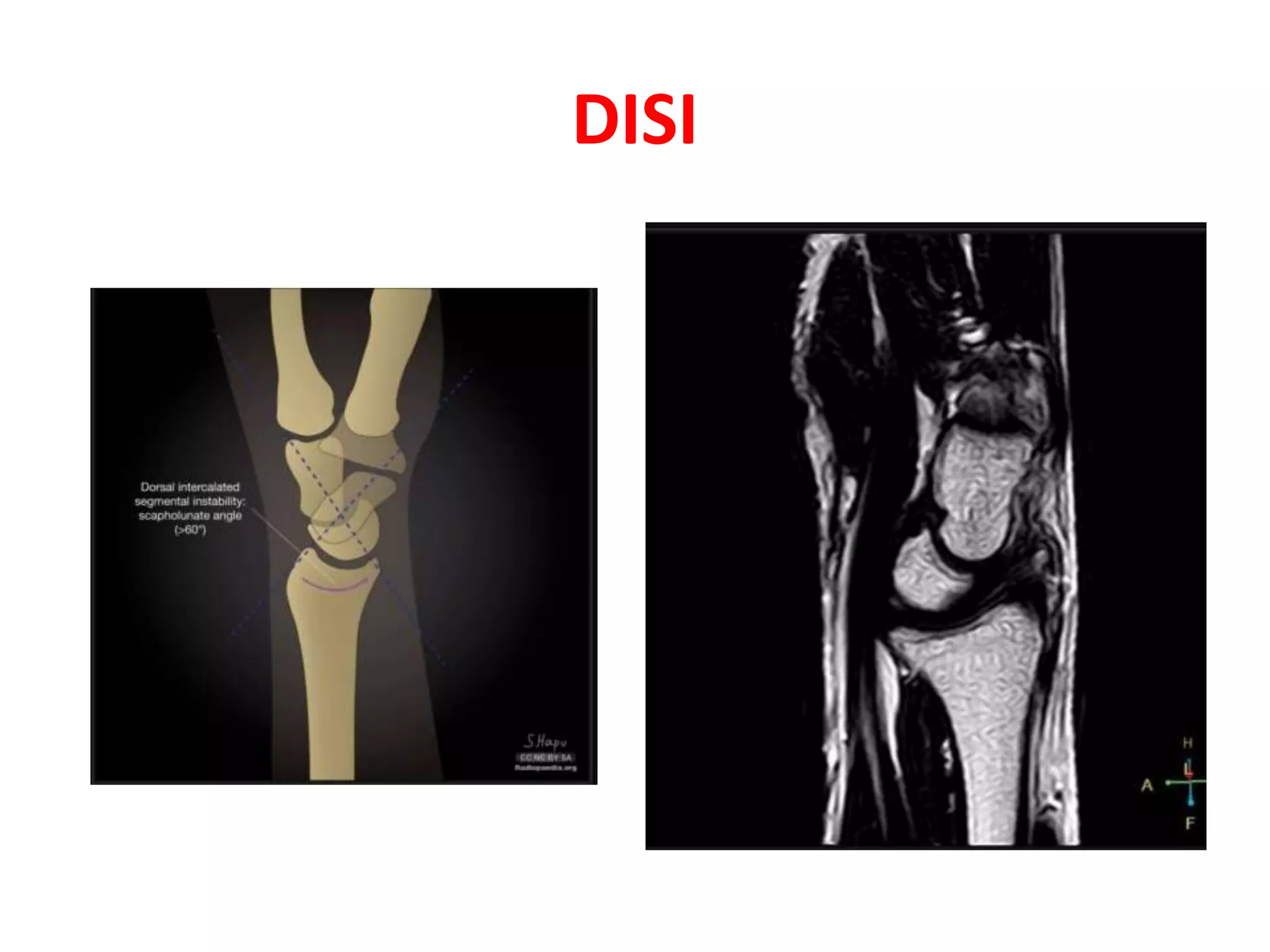
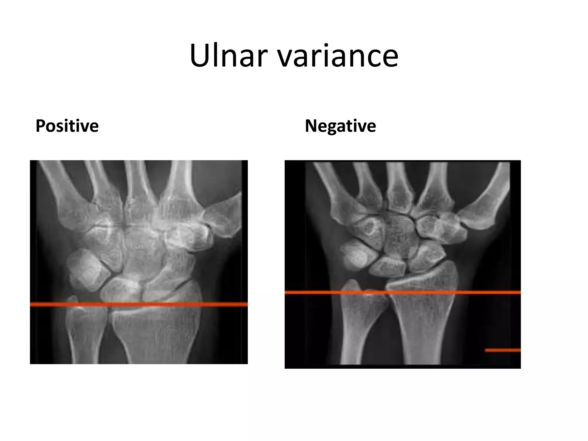
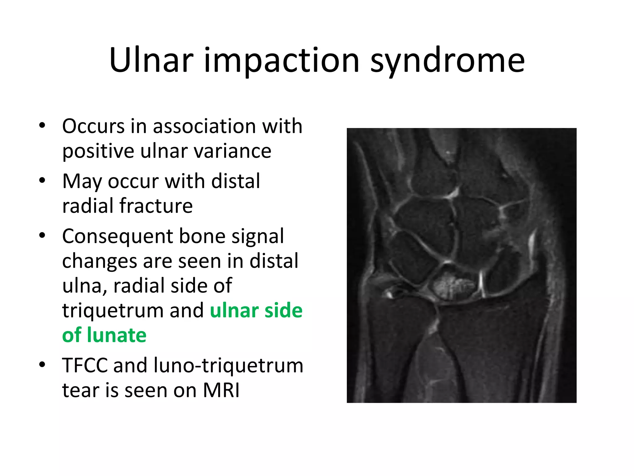
SlideShare a Scribd company logo
Imaging of Wrist joint
DR RAKIB AHMAD WANI
PG III YEAR
Introduction
• This joint is a complex joint in upper limb
• Consists of articulations of Radius, ulna,
carpus and carpo-metacarpal bones
• To evaluate and interpret wrist images one
should be fully aware of the anatomy
• X ray serves as basic tool while MRI serves to
answer the issues of soft tissues
Anatomy
• Wrist joint is formed by
Distal radio-ulnar joint,
Radio-carpal and Mid-
carpal joint
• Sigmoid notch of radius
articulates with ulna
X ray wrist
Parallelism and symmetry
• Joint spaces of wrist joint
are more or less
symmetrical
• Average width of joint
spaces are 2mm or less
• Capitato-lunate serves
as standard space to
which others should be
compared.
Carpal arcs
• Three arcs should be
drawn
• 1st is formed by
convexities of Scaphoid,
lunate and Triquitrum
bones
• 2nd is formed by inner
concavities of same bones
• 3rd is formed by proximal
curvature of Capitate and
hamate.
Asymmetry
• Any asymmetry in the
joint space and loss of
parallelism points that
some disruption has
occurred
• Depending on which
arch or which joint
space is widened,
pathology lies in the
same compartment
Asymmetry
Angles and Axes of Carpals
• Scaphoid axis : is formed
by proximal and distal
poles
• Lunate axis : is drawn
perpendicular to proximal
convex and distal concave
surface
• Capitato-lunate angle : >
30 is abnormal
• Scapho-lunate angle: 30-
60 is normal; 60-80 is
questionable and > 80 is
abnormal
Axes and Angles
Flexor retinaculum
• Is a rectangular fibrous
band and forms roof of
carpal tunnel
• Radially attached to
ridge of trapezium and
on ulnar aspect to hook
of hamate
• On ulnar aspect it forms
Guyon canal
Flexor retinaculum
Guyon’s canal
• Is a fibro-osseous
tunnel that contains
ulnar neuro-vascular
bundle
• Landmarks are deep to
FCU, Lateral to Pisiform
and at the hook of
hamate
Extensor Tendons
• On dorsal aspect,
tendons are divided
into 6 compartments
• Lister tubercle is
important landmark
that separates II and III
compartment.
Triangular Fibro-Cartilage complex
• It is a complex structure
has various parts :
Triangular fibro-
cartilage ; Radio-ulnar ;
ulno-carpal; Meniscal
homologue; ECU
tendon and ulnar
collateral ligament.
TFCC
TFCC
RU ligaments
Inter-carpal ligaments
• These are stabilizers of the wrist joint and are
divided into extrinsic and intrinsic parts.
• Intrinsic : Scapho-lunate, Luno-triquetral are
important ones .
TFCC.
• Extrinsic: volar, Dorsal, RCL and UCL
Inter-carpal ligaments
• The two important
ligaments are in the
proximal row : scapho-
lunate and luno-
triquitrum.
• The first one is
pyramidal and latter is
horse shoe in shape
Pathologies
TFCC Tear
• It can be traumatic in
young in radial end and
degenerative in elderly at
ulnar end
• Presents with ulnar side
wrist pain
• Seen as fluid intensity
signal along the ulna
• Discontinuity of complex
is seen on MR
arthrogram.
TFCC Tear
Palmer classification
• class 1
a) central perforation
b) Ulnar avulsion with or
without ulnar fracture
c) Avulsion of Ulno-
triquetral and ulno-
ilunate ligament
d) Radial avulsion of TFCC
• Class 2
a) TFCC wear with thinning
without perforation
b) TFCC wear with
ulnar/lunate/triquetral
chondromalacia
c) TFCC perforation with
chondromalacia
d) Luno-triquetral perf with
2a/2b or 2c
e) Any of above with ulno-
carpal arthritis
Scapho-lunate dissociation
• Is aka rotatory
subluxation of scaphoid.
• Refers to abnormal
orientation of scaphoid
and lunate and is due to
injury to the scapho-
lunate ligament
• Relative flexion of
scaphoid with relative
extension of lunate is
typical pattern
• On x ray widened
Scapho-lunate interval
of >4mm is seen
• Increase in Scapho-
lunate angle >60 degree
• On MRI examination, It
is seen as discontinuity
of the Scapho-lunate
ligament along with
joint effusion.
SL dissociation
Terry Thomas sign MRI imaging
SL Tear Grading
Lunate Dislocations
• These are uncommon traumatic wrist
injuries .
• Lunate is displaced and rotated volarly.
• Occurs due to fall on out-stretched hand.
• Lunate shows “Piece of Pie sign”
• Rest of the bones are well aligned with
maintained symmetry.
LD
Peri-lunate dislocation
• Are potentially closed wrist injuries and
involve the carpus relative to lunate
• involves scapho-lunate and Luno-
triquetral ligaments
• In this pathology, the Capitate (apple)
doesn’t sit in the concavity of Lunate
(Cup)
Dorsal and Volar intercalated
segment instability
• DISI is the form of
instability involving
wrist joint in which
Lunate is displaced
dorsally.
• Disruption of the
scapho-lunate ligament
• Due to wrist trauma
• SL angle of >60 degree
• CL angle >30 degree
• VISI is less common
in which lunate is
displaced palmarly
• Ligament injured is
Luno-triquetral
ligament
• SL <30 degrees
• CL >30 degree
DISI
VISI
Ulnar variance
• It is defined as the relative length of distal end of ulna
wrt radius
• If ulna is shorter than radius it is called Negative ulnar
variance
• If ulna is longer than radius it is called positive ulnar
variance
• If both are equal it is called neutral variance
• On radiographs Positive variance is when the level of
the ulna is >2.5 mm beyond the radius margin at the
distal radio-ulnar joint and negative variance is when
the ulna is ≤2.5 mm than the radius at the DRUJ
Ulnar variance
Positive Negative
Ulnar impaction syndrome
• Occurs in association with
positive ulnar variance
• May occur with distal
radial fracture
• Consequent bone signal
changes are seen in distal
ulna, radial side of
triquetrum and ulnar side
of lunate
• TFCC and luno-triquetrum
tear is seen on MRI
Ulnar impaction syndrome
Ulnar impingement syndrome
• Occurs in association with
Negative ulnar variance
• shortened distal ulna
results in contraction of
the extensor pollicis
brevis, abductor pollicis
longus, and pronator
quadratus muscles which
prevents normal
buttressing of the
radioulnar joint.
• Seen as edema where
ulna converges on radius
Ulnar impingement syndrome
AVN of Scaphoid bone
• Trauma of scaphoid
bone leads to AVN
• It has peculiar blood
supply where in
proximal pole receives
blood supply in
retrograde fashion
• It mnifests as sclerosis
of bone on wrist
radiographs
AVN of scaphoid
Kienbock disease
• Is associated with negative ulnar variance
together with a susceptible blood vessel
• Is defined as osteonecrosis of the lunate
where cortical blood supply is compromised
and results in infarction of the bone with
surrounding hyperemia
• Sclerosis with negative ulnar variance is seen
• Bone signal changes are seen along the central
and radial aspect of lunate
Kienbock disease
Stahl classification of Kienbock disease
• stage I: normal radiograph
• stage II: increased radio density of the lunate
with possible decrease of lunate height on the
radial side only
• stage III
– IIIa: lunate collapse, no scaphoid rotation
– IIIb: lunate collapse, fixed scaphoid rotation
• stage IV: degenerative changes around lunate
Kienbock disease
Stage II AP Stage II Lateral
Carpal coalition
• Refers to failure of separation of two or more
carpal bones
• Lunate and triquetrum are most common
• It may be osseous, cartilaginous or fibrous
• Associated with Holt oram, turner syndrome
etc
Carpal coalition
Carpal Tunnel Syndrome
• Results from the compression of Median Nerve in carpal tunnel
• Nocturnal ascending pain emanating from the wrist, is typical
• sensory symptoms affect the first three digits and, depending on
innervation patterns, the radial aspect of the fourth digit
• Positive Tinel test: paresthesias elicited by tapping the median
nerve at the wrist
• Occurs due to
mechanical overuse (considered the most common association)
osteoarthritis
trauma
acromegaly
• Ganglion cysts, primary nerve sheath tumors
synovial hypertrophy in rheumatoid arthritis
USG CTS
criteria
• Palmar bowing of Flexor
retinaculum of >2mm
• Flattening of nerve distally
with increased thickness
proximally
• Normal Cross section area is
9-11mm sq
• Difference of > 2mm sq at
CT and pronator quadratus
has high sensitivity
Image
Images
USG
MRI for CTS
• In carpal tunnel syndrome, MRI can demonstrate
palmar bowing of the flexor retinaculum,
enlargement of the median nerve at the level of
the pisiform, and flattening of the median nerve
at the level of the hook of hamate.
• Other signs are edema or loss of fat within the
carpal tunnel, and increased size or edema of the
nerve on fluid-sensitive sequences, and, in some
cases, contrast enhancement of the nerve
MRI
Fibrolipomatous hamartoma of MN
• It is a benign neoplasm
of median nerve
• Proliferation of fibro-
adipose tissue of sheath
• Imaging features are co-
axial cable like
appearance on axial and
spaghetti like on
coronal images
Guyon’s canal syndrome
• Results from compression
of ulnar nerve.
• Hook of hamate fracture,
Ganglion cyst are its
causes.
• On x ray hook of hamate
fracture is seen.
• On USG ganglion cyst,
lipoma or ulnar artery
thrombus is seen and on
MRI these findings are
seen with better accuracy
De Quervain TS
• Aka washerwoman’s
sprain
• Occurs due to
inflammation of EPB
and APL tendons
• On USG Thickening of
tendons with fluid in
extensor compartment
and increased flow on
CDI in peri-tendonous
region
MRI
• More sensitive and
specific
• Increased fluid in
tendon sheath
• Thickened edematous
retinaculum with peri
tendenous edema
• Sometimes features of
tendonosis are also
seen
Proximal intersection syndrome
• Is overuse tenosynovitis around the
intersection of 1st and 2nd compartment
extensor tendons
• Seen in weight lifters, skiers etc
• Fluid is seen within the involved tendons with
loss of hyper echoic plane between the
tendon compartments
• On MRI peri tendenous edema is noted 4cm
proximal to lister’s tubercle
Proximal intersection syndrome
USG MRI
Distal intersection syndrome
• Relates to teno-synovitis of EPL tendon where
it crosses the ECRB and ECRL tendon
• Crossover is located just distal to lister’s
tubercle
• Over us, trauma and RA are etiologic factors
• Edema is noted in peri-tendenous region at
intersection point
Images
Ganglion cyst
• is a non malignant
cystic mass and result
from myxoid
degenration of
connective tissue
assoiated with joint
capsule and tendon
sheath
• Seen as anechoic cyst
with PAE on USG and
cystic signal on MRI
Value of axial and coronal images
Axial
• Distal RU joint
• Carpal tunnel and contents
• Flexor and extensor tendons
• Ulnar nerve and median
nerve pathologies
Coronal
• TFCC
• Ulnar Variance
• Scapho-lunate and Luno-
triquetral ligaments
• Fractures
• AVN
• Arthropathies
Summary
History and clinical findings should be available
Assess the congruity of bones on bones, Look for
any asymmetry
Look for ulnar variance
Look for any bone marrow edema/effusion
Asses inter-carpal ligaments and TFCC
Look carefully for any pathology of
carpal/Guyon’s canal
Findings should be noted to reach to a specific
diagnosis
Thank
You

More Related Content

What's hot

MRI OF SHOULDER INJURY
MRI OF SHOULDER INJURYMRI OF SHOULDER INJURY
MRI OF SHOULDER INJURY
Krishna Kiran Karanth
 
Presentation1, radiological imaging of anterior knee pain.
Presentation1, radiological imaging of anterior knee pain.Presentation1, radiological imaging of anterior knee pain.
Presentation1, radiological imaging of anterior knee pain.
Abdellah Nazeer
 
MR ANATOMY OF WRIST AND ELBOW RV
MR ANATOMY OF WRIST AND ELBOW RVMR ANATOMY OF WRIST AND ELBOW RV
MR ANATOMY OF WRIST AND ELBOW RV
Roshan Valentine
 
Presentation1.pptx. shoulder joint.
Presentation1.pptx. shoulder joint.Presentation1.pptx. shoulder joint.
Presentation1.pptx. shoulder joint.
Abdellah Nazeer
 
75-Dr Ahmed Esawy imaging oral board mri ankle &amp;foot part II
75-Dr Ahmed Esawy imaging oral board mri ankle &amp;foot part II75-Dr Ahmed Esawy imaging oral board mri ankle &amp;foot part II
75-Dr Ahmed Esawy imaging oral board mri ankle &amp;foot part II
AHMED ESAWY
 
Transitional vertebrae radiology
Transitional vertebrae radiologyTransitional vertebrae radiology
Transitional vertebrae radiology
Dr. Mohit Goel
 
Patello femoral joint - MRI
Patello femoral joint - MRIPatello femoral joint - MRI
Patello femoral joint - MRI
Dr. Mohit Goel
 
MRI of the shoulder
MRI of the shoulderMRI of the shoulder
MRI of the shoulder
Thorsang Chayovan
 
Presentation1 radiological film reading of wrist joint.
Presentation1 radiological film reading of wrist joint.Presentation1 radiological film reading of wrist joint.
Presentation1 radiological film reading of wrist joint.
Abdellah Nazeer
 
71-Dr Ahmed Esawy imaging oral board of MRI hip joint part I
71-Dr Ahmed Esawy imaging oral board of  MRI hip joint part I71-Dr Ahmed Esawy imaging oral board of  MRI hip joint part I
71-Dr Ahmed Esawy imaging oral board of MRI hip joint part I
AHMED ESAWY
 
Ankle radiological anatomy
Ankle radiological anatomyAnkle radiological anatomy
Ankle radiological anatomy
Henock Negasi
 
72-Dr Ahmed Esawy imaging oral board of MRI hip joint part II
72-Dr Ahmed Esawy imaging oral board of  MRI hip joint part II72-Dr Ahmed Esawy imaging oral board of  MRI hip joint part II
72-Dr Ahmed Esawy imaging oral board of MRI hip joint part II
AHMED ESAWY
 
Diagnostic Imaging of Pediatric Hip Lesions
Diagnostic Imaging of Pediatric Hip LesionsDiagnostic Imaging of Pediatric Hip Lesions
Diagnostic Imaging of Pediatric Hip Lesions
Mohamed M.A. Zaitoun
 
Presentation1.pptx mri of elbow joint
Presentation1.pptx mri of elbow jointPresentation1.pptx mri of elbow joint
Presentation1.pptx mri of elbow jointAbdellah Nazeer
 
MRI KNEE JOINT ANATOMY
MRI KNEE JOINT ANATOMYMRI KNEE JOINT ANATOMY
MRI KNEE JOINT ANATOMY
Nikhil Bansal
 
Radiology of the Elbow Joint. Dr. Sumit Sharma
Radiology of the Elbow Joint. Dr. Sumit SharmaRadiology of the Elbow Joint. Dr. Sumit Sharma
Radiology of the Elbow Joint. Dr. Sumit Sharma
Sumit Sharma
 
Presentation1, radiological film reading of elbow joint.
Presentation1, radiological film reading of elbow joint.Presentation1, radiological film reading of elbow joint.
Presentation1, radiological film reading of elbow joint.
Abdellah Nazeer
 
Radiological anatomy of Knee joint.pptx
Radiological anatomy of Knee joint.pptxRadiological anatomy of Knee joint.pptx
Radiological anatomy of Knee joint.pptx
Alauddin Md
 
ELBOW JOINT X-RAY PROJECTIONS
ELBOW JOINT X-RAY PROJECTIONSELBOW JOINT X-RAY PROJECTIONS
ELBOW JOINT X-RAY PROJECTIONS
Jai Kumar
 
Presentation1.pptx. shoulder joint.
Presentation1.pptx. shoulder joint.Presentation1.pptx. shoulder joint.
Presentation1.pptx. shoulder joint.
Abdellah Nazeer
 

What's hot (20)

MRI OF SHOULDER INJURY
MRI OF SHOULDER INJURYMRI OF SHOULDER INJURY
MRI OF SHOULDER INJURY
 
Presentation1, radiological imaging of anterior knee pain.
Presentation1, radiological imaging of anterior knee pain.Presentation1, radiological imaging of anterior knee pain.
Presentation1, radiological imaging of anterior knee pain.
 
MR ANATOMY OF WRIST AND ELBOW RV
MR ANATOMY OF WRIST AND ELBOW RVMR ANATOMY OF WRIST AND ELBOW RV
MR ANATOMY OF WRIST AND ELBOW RV
 
Presentation1.pptx. shoulder joint.
Presentation1.pptx. shoulder joint.Presentation1.pptx. shoulder joint.
Presentation1.pptx. shoulder joint.
 
75-Dr Ahmed Esawy imaging oral board mri ankle &amp;foot part II
75-Dr Ahmed Esawy imaging oral board mri ankle &amp;foot part II75-Dr Ahmed Esawy imaging oral board mri ankle &amp;foot part II
75-Dr Ahmed Esawy imaging oral board mri ankle &amp;foot part II
 
Transitional vertebrae radiology
Transitional vertebrae radiologyTransitional vertebrae radiology
Transitional vertebrae radiology
 
Patello femoral joint - MRI
Patello femoral joint - MRIPatello femoral joint - MRI
Patello femoral joint - MRI
 
MRI of the shoulder
MRI of the shoulderMRI of the shoulder
MRI of the shoulder
 
Presentation1 radiological film reading of wrist joint.
Presentation1 radiological film reading of wrist joint.Presentation1 radiological film reading of wrist joint.
Presentation1 radiological film reading of wrist joint.
 
71-Dr Ahmed Esawy imaging oral board of MRI hip joint part I
71-Dr Ahmed Esawy imaging oral board of  MRI hip joint part I71-Dr Ahmed Esawy imaging oral board of  MRI hip joint part I
71-Dr Ahmed Esawy imaging oral board of MRI hip joint part I
 
Ankle radiological anatomy
Ankle radiological anatomyAnkle radiological anatomy
Ankle radiological anatomy
 
72-Dr Ahmed Esawy imaging oral board of MRI hip joint part II
72-Dr Ahmed Esawy imaging oral board of  MRI hip joint part II72-Dr Ahmed Esawy imaging oral board of  MRI hip joint part II
72-Dr Ahmed Esawy imaging oral board of MRI hip joint part II
 
Diagnostic Imaging of Pediatric Hip Lesions
Diagnostic Imaging of Pediatric Hip LesionsDiagnostic Imaging of Pediatric Hip Lesions
Diagnostic Imaging of Pediatric Hip Lesions
 
Presentation1.pptx mri of elbow joint
Presentation1.pptx mri of elbow jointPresentation1.pptx mri of elbow joint
Presentation1.pptx mri of elbow joint
 
MRI KNEE JOINT ANATOMY
MRI KNEE JOINT ANATOMYMRI KNEE JOINT ANATOMY
MRI KNEE JOINT ANATOMY
 
Radiology of the Elbow Joint. Dr. Sumit Sharma
Radiology of the Elbow Joint. Dr. Sumit SharmaRadiology of the Elbow Joint. Dr. Sumit Sharma
Radiology of the Elbow Joint. Dr. Sumit Sharma
 
Presentation1, radiological film reading of elbow joint.
Presentation1, radiological film reading of elbow joint.Presentation1, radiological film reading of elbow joint.
Presentation1, radiological film reading of elbow joint.
 
Radiological anatomy of Knee joint.pptx
Radiological anatomy of Knee joint.pptxRadiological anatomy of Knee joint.pptx
Radiological anatomy of Knee joint.pptx
 
ELBOW JOINT X-RAY PROJECTIONS
ELBOW JOINT X-RAY PROJECTIONSELBOW JOINT X-RAY PROJECTIONS
ELBOW JOINT X-RAY PROJECTIONS
 
Presentation1.pptx. shoulder joint.
Presentation1.pptx. shoulder joint.Presentation1.pptx. shoulder joint.
Presentation1.pptx. shoulder joint.
 

Similar to Imaging of wrist joint

wrist joint anatomy and biomechanics.pptx
wrist joint anatomy and biomechanics.pptxwrist joint anatomy and biomechanics.pptx
wrist joint anatomy and biomechanics.pptx
guneetkaur569647
 
Wrist disorders
Wrist disordersWrist disorders
Wrist disorders
yasim786
 
Perilunate dislocation, carpal injuries
Perilunate dislocation, carpal injuriesPerilunate dislocation, carpal injuries
Perilunate dislocation, carpal injuries
Kishore Vemula
 
anatomy of wrist and carpal bones
anatomy of wrist and carpal bones anatomy of wrist and carpal bones
anatomy of wrist and carpal bones
Rajesh Raj
 
Craniovertebral JUNCTION ANOMALIES
Craniovertebral JUNCTION ANOMALIESCraniovertebral JUNCTION ANOMALIES
Craniovertebral JUNCTION ANOMALIES
Uphar Gupta
 
carpal instability & management.pptx
carpal instability & management.pptxcarpal instability & management.pptx
carpal instability & management.pptx
Dr. Raghunath Reddy
 
carpal instability & management.pptx
carpal instability & management.pptxcarpal instability & management.pptx
carpal instability & management.pptx
Dr. Raghunath Reddy
 
DISTAL END OF RADIUS FRACTURE AND DISLOCATION MANAGEMENT.pptx
DISTAL END OF RADIUS FRACTURE AND DISLOCATION MANAGEMENT.pptxDISTAL END OF RADIUS FRACTURE AND DISLOCATION MANAGEMENT.pptx
DISTAL END OF RADIUS FRACTURE AND DISLOCATION MANAGEMENT.pptx
pradeepreddyseelam1
 
Kienbock's disease.ppt
Kienbock's disease.pptKienbock's disease.ppt
Kienbock's disease.ppt
ssuser0aca5c
 
carpal tunnel syndrome
carpal tunnel syndrome carpal tunnel syndrome
carpal tunnel syndrome
Anudeep Korada
 
Carpal instability
Carpal instabilityCarpal instability
Carpal instability
Raunak Milton
 
Wrist Joint
Wrist JointWrist Joint
Wrist Joint
Shweta Kotwani
 
Distal radius fractures
Distal radius fracturesDistal radius fractures
Distal radius fractures
Dr Padma C Anand
 
Lunate Fracture & Perilunate dislocation of carpals by Dr.@rpan Chaudhary (Re...
Lunate Fracture & Perilunate dislocation of carpals by Dr.@rpan Chaudhary (Re...Lunate Fracture & Perilunate dislocation of carpals by Dr.@rpan Chaudhary (Re...
Lunate Fracture & Perilunate dislocation of carpals by Dr.@rpan Chaudhary (Re...
CHAUDHARY ARPAN
 
Kienbock's disease
Kienbock's diseaseKienbock's disease
Kienbock's disease
Birimong Quinker
 
Craniovertebral junction
Craniovertebral junctionCraniovertebral junction
Craniovertebral junction
NeurologyKota
 
Radial club hand (Radial Dysplasia)
Radial club hand (Radial Dysplasia)Radial club hand (Radial Dysplasia)
Radial club hand (Radial Dysplasia)
Aiman Ali
 
Imaging of shoulder - Dr. Vishal Sankpal
Imaging of shoulder - Dr. Vishal SankpalImaging of shoulder - Dr. Vishal Sankpal
Imaging of shoulder - Dr. Vishal Sankpal
Vishal Sankpal
 
Elbow Injuries.pptx
Elbow Injuries.pptxElbow Injuries.pptx
Elbow Injuries.pptx
esicOrtho1
 
THORACIC%20OUTLET%20SYNDROME.pptx
THORACIC%20OUTLET%20SYNDROME.pptxTHORACIC%20OUTLET%20SYNDROME.pptx
THORACIC%20OUTLET%20SYNDROME.pptx
NEELESHCHOUDHARY4
 

Similar to Imaging of wrist joint (20)

wrist joint anatomy and biomechanics.pptx
wrist joint anatomy and biomechanics.pptxwrist joint anatomy and biomechanics.pptx
wrist joint anatomy and biomechanics.pptx
 
Wrist disorders
Wrist disordersWrist disorders
Wrist disorders
 
Perilunate dislocation, carpal injuries
Perilunate dislocation, carpal injuriesPerilunate dislocation, carpal injuries
Perilunate dislocation, carpal injuries
 
anatomy of wrist and carpal bones
anatomy of wrist and carpal bones anatomy of wrist and carpal bones
anatomy of wrist and carpal bones
 
Craniovertebral JUNCTION ANOMALIES
Craniovertebral JUNCTION ANOMALIESCraniovertebral JUNCTION ANOMALIES
Craniovertebral JUNCTION ANOMALIES
 
carpal instability & management.pptx
carpal instability & management.pptxcarpal instability & management.pptx
carpal instability & management.pptx
 
carpal instability & management.pptx
carpal instability & management.pptxcarpal instability & management.pptx
carpal instability & management.pptx
 
DISTAL END OF RADIUS FRACTURE AND DISLOCATION MANAGEMENT.pptx
DISTAL END OF RADIUS FRACTURE AND DISLOCATION MANAGEMENT.pptxDISTAL END OF RADIUS FRACTURE AND DISLOCATION MANAGEMENT.pptx
DISTAL END OF RADIUS FRACTURE AND DISLOCATION MANAGEMENT.pptx
 
Kienbock's disease.ppt
Kienbock's disease.pptKienbock's disease.ppt
Kienbock's disease.ppt
 
carpal tunnel syndrome
carpal tunnel syndrome carpal tunnel syndrome
carpal tunnel syndrome
 
Carpal instability
Carpal instabilityCarpal instability
Carpal instability
 
Wrist Joint
Wrist JointWrist Joint
Wrist Joint
 
Distal radius fractures
Distal radius fracturesDistal radius fractures
Distal radius fractures
 
Lunate Fracture & Perilunate dislocation of carpals by Dr.@rpan Chaudhary (Re...
Lunate Fracture & Perilunate dislocation of carpals by Dr.@rpan Chaudhary (Re...Lunate Fracture & Perilunate dislocation of carpals by Dr.@rpan Chaudhary (Re...
Lunate Fracture & Perilunate dislocation of carpals by Dr.@rpan Chaudhary (Re...
 
Kienbock's disease
Kienbock's diseaseKienbock's disease
Kienbock's disease
 
Craniovertebral junction
Craniovertebral junctionCraniovertebral junction
Craniovertebral junction
 
Radial club hand (Radial Dysplasia)
Radial club hand (Radial Dysplasia)Radial club hand (Radial Dysplasia)
Radial club hand (Radial Dysplasia)
 
Imaging of shoulder - Dr. Vishal Sankpal
Imaging of shoulder - Dr. Vishal SankpalImaging of shoulder - Dr. Vishal Sankpal
Imaging of shoulder - Dr. Vishal Sankpal
 
Elbow Injuries.pptx
Elbow Injuries.pptxElbow Injuries.pptx
Elbow Injuries.pptx
 
THORACIC%20OUTLET%20SYNDROME.pptx
THORACIC%20OUTLET%20SYNDROME.pptxTHORACIC%20OUTLET%20SYNDROME.pptx
THORACIC%20OUTLET%20SYNDROME.pptx
 

Recently uploaded

Flu Vaccine Alert in Bangalore Karnataka
Flu Vaccine Alert in Bangalore KarnatakaFlu Vaccine Alert in Bangalore Karnataka
Flu Vaccine Alert in Bangalore Karnataka
addon Scans
 
Non-respiratory Functions of the Lungs.pdf
Non-respiratory Functions of the Lungs.pdfNon-respiratory Functions of the Lungs.pdf
Non-respiratory Functions of the Lungs.pdf
MedicoseAcademics
 
Ocular injury ppt Upendra pal optometrist upums saifai etawah
Ocular injury  ppt  Upendra pal  optometrist upums saifai etawahOcular injury  ppt  Upendra pal  optometrist upums saifai etawah
Ocular injury ppt Upendra pal optometrist upums saifai etawah
pal078100
 
Light House Retreats: Plant Medicine Retreat Europe
Light House Retreats: Plant Medicine Retreat EuropeLight House Retreats: Plant Medicine Retreat Europe
Light House Retreats: Plant Medicine Retreat Europe
Lighthouse Retreat
 
Phone Us ❤85270-49040❤ #ℂall #gIRLS In Surat By Surat @ℂall @Girls Hotel With...
Phone Us ❤85270-49040❤ #ℂall #gIRLS In Surat By Surat @ℂall @Girls Hotel With...Phone Us ❤85270-49040❤ #ℂall #gIRLS In Surat By Surat @ℂall @Girls Hotel With...
Phone Us ❤85270-49040❤ #ℂall #gIRLS In Surat By Surat @ℂall @Girls Hotel With...
Savita Shen $i11
 
For Better Surat #ℂall #Girl Service ❤85270-49040❤ Surat #ℂall #Girls
For Better Surat #ℂall #Girl Service ❤85270-49040❤ Surat #ℂall #GirlsFor Better Surat #ℂall #Girl Service ❤85270-49040❤ Surat #ℂall #Girls
For Better Surat #ℂall #Girl Service ❤85270-49040❤ Surat #ℂall #Girls
Savita Shen $i11
 
HOT NEW PRODUCT! BIG SALES FAST SHIPPING NOW FROM CHINA!! EU KU DB BK substit...
HOT NEW PRODUCT! BIG SALES FAST SHIPPING NOW FROM CHINA!! EU KU DB BK substit...HOT NEW PRODUCT! BIG SALES FAST SHIPPING NOW FROM CHINA!! EU KU DB BK substit...
HOT NEW PRODUCT! BIG SALES FAST SHIPPING NOW FROM CHINA!! EU KU DB BK substit...
GL Anaacs
 
MANAGEMENT OF ATRIOVENTRICULAR CONDUCTION BLOCK.pdf
MANAGEMENT OF ATRIOVENTRICULAR CONDUCTION BLOCK.pdfMANAGEMENT OF ATRIOVENTRICULAR CONDUCTION BLOCK.pdf
MANAGEMENT OF ATRIOVENTRICULAR CONDUCTION BLOCK.pdf
Jim Jacob Roy
 
Are There Any Natural Remedies To Treat Syphilis.pdf
Are There Any Natural Remedies To Treat Syphilis.pdfAre There Any Natural Remedies To Treat Syphilis.pdf
Are There Any Natural Remedies To Treat Syphilis.pdf
Little Cross Family Clinic
 
planning for change nursing Management ppt
planning for change nursing Management pptplanning for change nursing Management ppt
planning for change nursing Management ppt
Thangamjayarani
 
Basavarajeeyam - Ayurvedic heritage book of Andhra pradesh
Basavarajeeyam - Ayurvedic heritage book of Andhra pradeshBasavarajeeyam - Ayurvedic heritage book of Andhra pradesh
Basavarajeeyam - Ayurvedic heritage book of Andhra pradesh
Dr. Madduru Muni Haritha
 
Physiology of Special Chemical Sensation of Taste
Physiology of Special Chemical Sensation of TastePhysiology of Special Chemical Sensation of Taste
Physiology of Special Chemical Sensation of Taste
MedicoseAcademics
 
Pictures of Superficial & Deep Fascia.ppt.pdf
Pictures of Superficial & Deep Fascia.ppt.pdfPictures of Superficial & Deep Fascia.ppt.pdf
Pictures of Superficial & Deep Fascia.ppt.pdf
Dr. Rabia Inam Gandapore
 
Ozempic: Preoperative Management of Patients on GLP-1 Receptor Agonists
Ozempic: Preoperative Management of Patients on GLP-1 Receptor Agonists  Ozempic: Preoperative Management of Patients on GLP-1 Receptor Agonists
Ozempic: Preoperative Management of Patients on GLP-1 Receptor Agonists
Saeid Safari
 
Maxilla, Mandible & Hyoid Bone & Clinical Correlations by Dr. RIG.pptx
Maxilla, Mandible & Hyoid Bone & Clinical Correlations by Dr. RIG.pptxMaxilla, Mandible & Hyoid Bone & Clinical Correlations by Dr. RIG.pptx
Maxilla, Mandible & Hyoid Bone & Clinical Correlations by Dr. RIG.pptx
Dr. Rabia Inam Gandapore
 
Pharma Pcd Franchise in Jharkhand - Yodley Lifesciences
Pharma Pcd Franchise in Jharkhand - Yodley LifesciencesPharma Pcd Franchise in Jharkhand - Yodley Lifesciences
Pharma Pcd Franchise in Jharkhand - Yodley Lifesciences
Yodley Lifesciences
 
New Directions in Targeted Therapeutic Approaches for Older Adults With Mantl...
New Directions in Targeted Therapeutic Approaches for Older Adults With Mantl...New Directions in Targeted Therapeutic Approaches for Older Adults With Mantl...
New Directions in Targeted Therapeutic Approaches for Older Adults With Mantl...
i3 Health
 
Cervical & Brachial Plexus By Dr. RIG.pptx
Cervical & Brachial Plexus By Dr. RIG.pptxCervical & Brachial Plexus By Dr. RIG.pptx
Cervical & Brachial Plexus By Dr. RIG.pptx
Dr. Rabia Inam Gandapore
 
ARTHROLOGY PPT NCISM SYLLABUS AYURVEDA STUDENTS
ARTHROLOGY PPT NCISM SYLLABUS AYURVEDA STUDENTSARTHROLOGY PPT NCISM SYLLABUS AYURVEDA STUDENTS
ARTHROLOGY PPT NCISM SYLLABUS AYURVEDA STUDENTS
Dr. Vinay Pareek
 
Knee anatomy and clinical tests 2024.pdf
Knee anatomy and clinical tests 2024.pdfKnee anatomy and clinical tests 2024.pdf
Knee anatomy and clinical tests 2024.pdf
vimalpl1234
 

Recently uploaded (20)

Flu Vaccine Alert in Bangalore Karnataka
Flu Vaccine Alert in Bangalore KarnatakaFlu Vaccine Alert in Bangalore Karnataka
Flu Vaccine Alert in Bangalore Karnataka
 
Non-respiratory Functions of the Lungs.pdf
Non-respiratory Functions of the Lungs.pdfNon-respiratory Functions of the Lungs.pdf
Non-respiratory Functions of the Lungs.pdf
 
Ocular injury ppt Upendra pal optometrist upums saifai etawah
Ocular injury  ppt  Upendra pal  optometrist upums saifai etawahOcular injury  ppt  Upendra pal  optometrist upums saifai etawah
Ocular injury ppt Upendra pal optometrist upums saifai etawah
 
Light House Retreats: Plant Medicine Retreat Europe
Light House Retreats: Plant Medicine Retreat EuropeLight House Retreats: Plant Medicine Retreat Europe
Light House Retreats: Plant Medicine Retreat Europe
 
Phone Us ❤85270-49040❤ #ℂall #gIRLS In Surat By Surat @ℂall @Girls Hotel With...
Phone Us ❤85270-49040❤ #ℂall #gIRLS In Surat By Surat @ℂall @Girls Hotel With...Phone Us ❤85270-49040❤ #ℂall #gIRLS In Surat By Surat @ℂall @Girls Hotel With...
Phone Us ❤85270-49040❤ #ℂall #gIRLS In Surat By Surat @ℂall @Girls Hotel With...
 
For Better Surat #ℂall #Girl Service ❤85270-49040❤ Surat #ℂall #Girls
For Better Surat #ℂall #Girl Service ❤85270-49040❤ Surat #ℂall #GirlsFor Better Surat #ℂall #Girl Service ❤85270-49040❤ Surat #ℂall #Girls
For Better Surat #ℂall #Girl Service ❤85270-49040❤ Surat #ℂall #Girls
 
HOT NEW PRODUCT! BIG SALES FAST SHIPPING NOW FROM CHINA!! EU KU DB BK substit...
HOT NEW PRODUCT! BIG SALES FAST SHIPPING NOW FROM CHINA!! EU KU DB BK substit...HOT NEW PRODUCT! BIG SALES FAST SHIPPING NOW FROM CHINA!! EU KU DB BK substit...
HOT NEW PRODUCT! BIG SALES FAST SHIPPING NOW FROM CHINA!! EU KU DB BK substit...
 
MANAGEMENT OF ATRIOVENTRICULAR CONDUCTION BLOCK.pdf
MANAGEMENT OF ATRIOVENTRICULAR CONDUCTION BLOCK.pdfMANAGEMENT OF ATRIOVENTRICULAR CONDUCTION BLOCK.pdf
MANAGEMENT OF ATRIOVENTRICULAR CONDUCTION BLOCK.pdf
 
Are There Any Natural Remedies To Treat Syphilis.pdf
Are There Any Natural Remedies To Treat Syphilis.pdfAre There Any Natural Remedies To Treat Syphilis.pdf
Are There Any Natural Remedies To Treat Syphilis.pdf
 
planning for change nursing Management ppt
planning for change nursing Management pptplanning for change nursing Management ppt
planning for change nursing Management ppt
 
Basavarajeeyam - Ayurvedic heritage book of Andhra pradesh
Basavarajeeyam - Ayurvedic heritage book of Andhra pradeshBasavarajeeyam - Ayurvedic heritage book of Andhra pradesh
Basavarajeeyam - Ayurvedic heritage book of Andhra pradesh
 
Physiology of Special Chemical Sensation of Taste
Physiology of Special Chemical Sensation of TastePhysiology of Special Chemical Sensation of Taste
Physiology of Special Chemical Sensation of Taste
 
Pictures of Superficial & Deep Fascia.ppt.pdf
Pictures of Superficial & Deep Fascia.ppt.pdfPictures of Superficial & Deep Fascia.ppt.pdf
Pictures of Superficial & Deep Fascia.ppt.pdf
 
Ozempic: Preoperative Management of Patients on GLP-1 Receptor Agonists
Ozempic: Preoperative Management of Patients on GLP-1 Receptor Agonists  Ozempic: Preoperative Management of Patients on GLP-1 Receptor Agonists
Ozempic: Preoperative Management of Patients on GLP-1 Receptor Agonists
 
Maxilla, Mandible & Hyoid Bone & Clinical Correlations by Dr. RIG.pptx
Maxilla, Mandible & Hyoid Bone & Clinical Correlations by Dr. RIG.pptxMaxilla, Mandible & Hyoid Bone & Clinical Correlations by Dr. RIG.pptx
Maxilla, Mandible & Hyoid Bone & Clinical Correlations by Dr. RIG.pptx
 
Pharma Pcd Franchise in Jharkhand - Yodley Lifesciences
Pharma Pcd Franchise in Jharkhand - Yodley LifesciencesPharma Pcd Franchise in Jharkhand - Yodley Lifesciences
Pharma Pcd Franchise in Jharkhand - Yodley Lifesciences
 
New Directions in Targeted Therapeutic Approaches for Older Adults With Mantl...
New Directions in Targeted Therapeutic Approaches for Older Adults With Mantl...New Directions in Targeted Therapeutic Approaches for Older Adults With Mantl...
New Directions in Targeted Therapeutic Approaches for Older Adults With Mantl...
 
Cervical & Brachial Plexus By Dr. RIG.pptx
Cervical & Brachial Plexus By Dr. RIG.pptxCervical & Brachial Plexus By Dr. RIG.pptx
Cervical & Brachial Plexus By Dr. RIG.pptx
 
ARTHROLOGY PPT NCISM SYLLABUS AYURVEDA STUDENTS
ARTHROLOGY PPT NCISM SYLLABUS AYURVEDA STUDENTSARTHROLOGY PPT NCISM SYLLABUS AYURVEDA STUDENTS
ARTHROLOGY PPT NCISM SYLLABUS AYURVEDA STUDENTS
 
Knee anatomy and clinical tests 2024.pdf
Knee anatomy and clinical tests 2024.pdfKnee anatomy and clinical tests 2024.pdf
Knee anatomy and clinical tests 2024.pdf
 

Imaging of wrist joint

  • 1. Imaging of Wrist joint DR RAKIB AHMAD WANI PG III YEAR
  • 2. Introduction • This joint is a complex joint in upper limb • Consists of articulations of Radius, ulna, carpus and carpo-metacarpal bones • To evaluate and interpret wrist images one should be fully aware of the anatomy • X ray serves as basic tool while MRI serves to answer the issues of soft tissues
  • 3. Anatomy • Wrist joint is formed by Distal radio-ulnar joint, Radio-carpal and Mid- carpal joint • Sigmoid notch of radius articulates with ulna
  • 5. Parallelism and symmetry • Joint spaces of wrist joint are more or less symmetrical • Average width of joint spaces are 2mm or less • Capitato-lunate serves as standard space to which others should be compared.
  • 6. Carpal arcs • Three arcs should be drawn • 1st is formed by convexities of Scaphoid, lunate and Triquitrum bones • 2nd is formed by inner concavities of same bones • 3rd is formed by proximal curvature of Capitate and hamate.
  • 7. Asymmetry • Any asymmetry in the joint space and loss of parallelism points that some disruption has occurred • Depending on which arch or which joint space is widened, pathology lies in the same compartment
  • 9. Angles and Axes of Carpals • Scaphoid axis : is formed by proximal and distal poles • Lunate axis : is drawn perpendicular to proximal convex and distal concave surface • Capitato-lunate angle : > 30 is abnormal • Scapho-lunate angle: 30- 60 is normal; 60-80 is questionable and > 80 is abnormal
  • 11. Flexor retinaculum • Is a rectangular fibrous band and forms roof of carpal tunnel • Radially attached to ridge of trapezium and on ulnar aspect to hook of hamate • On ulnar aspect it forms Guyon canal
  • 13. Guyon’s canal • Is a fibro-osseous tunnel that contains ulnar neuro-vascular bundle • Landmarks are deep to FCU, Lateral to Pisiform and at the hook of hamate
  • 14. Extensor Tendons • On dorsal aspect, tendons are divided into 6 compartments • Lister tubercle is important landmark that separates II and III compartment.
  • 15.
  • 16. Triangular Fibro-Cartilage complex • It is a complex structure has various parts : Triangular fibro- cartilage ; Radio-ulnar ; ulno-carpal; Meniscal homologue; ECU tendon and ulnar collateral ligament.
  • 17.
  • 18. TFCC
  • 19. TFCC
  • 21. Inter-carpal ligaments • These are stabilizers of the wrist joint and are divided into extrinsic and intrinsic parts. • Intrinsic : Scapho-lunate, Luno-triquetral are important ones . TFCC. • Extrinsic: volar, Dorsal, RCL and UCL
  • 22. Inter-carpal ligaments • The two important ligaments are in the proximal row : scapho- lunate and luno- triquitrum. • The first one is pyramidal and latter is horse shoe in shape
  • 24. TFCC Tear • It can be traumatic in young in radial end and degenerative in elderly at ulnar end • Presents with ulnar side wrist pain • Seen as fluid intensity signal along the ulna • Discontinuity of complex is seen on MR arthrogram.
  • 26.
  • 27. Palmer classification • class 1 a) central perforation b) Ulnar avulsion with or without ulnar fracture c) Avulsion of Ulno- triquetral and ulno- ilunate ligament d) Radial avulsion of TFCC • Class 2 a) TFCC wear with thinning without perforation b) TFCC wear with ulnar/lunate/triquetral chondromalacia c) TFCC perforation with chondromalacia d) Luno-triquetral perf with 2a/2b or 2c e) Any of above with ulno- carpal arthritis
  • 28. Scapho-lunate dissociation • Is aka rotatory subluxation of scaphoid. • Refers to abnormal orientation of scaphoid and lunate and is due to injury to the scapho- lunate ligament • Relative flexion of scaphoid with relative extension of lunate is typical pattern • On x ray widened Scapho-lunate interval of >4mm is seen • Increase in Scapho- lunate angle >60 degree • On MRI examination, It is seen as discontinuity of the Scapho-lunate ligament along with joint effusion.
  • 29. SL dissociation Terry Thomas sign MRI imaging
  • 31. Lunate Dislocations • These are uncommon traumatic wrist injuries . • Lunate is displaced and rotated volarly. • Occurs due to fall on out-stretched hand. • Lunate shows “Piece of Pie sign” • Rest of the bones are well aligned with maintained symmetry.
  • 32. LD
  • 33. Peri-lunate dislocation • Are potentially closed wrist injuries and involve the carpus relative to lunate • involves scapho-lunate and Luno- triquetral ligaments • In this pathology, the Capitate (apple) doesn’t sit in the concavity of Lunate (Cup)
  • 34.
  • 35. Dorsal and Volar intercalated segment instability • DISI is the form of instability involving wrist joint in which Lunate is displaced dorsally. • Disruption of the scapho-lunate ligament • Due to wrist trauma • SL angle of >60 degree • CL angle >30 degree • VISI is less common in which lunate is displaced palmarly • Ligament injured is Luno-triquetral ligament • SL <30 degrees • CL >30 degree
  • 36. DISI
  • 37. VISI
  • 38. Ulnar variance • It is defined as the relative length of distal end of ulna wrt radius • If ulna is shorter than radius it is called Negative ulnar variance • If ulna is longer than radius it is called positive ulnar variance • If both are equal it is called neutral variance • On radiographs Positive variance is when the level of the ulna is >2.5 mm beyond the radius margin at the distal radio-ulnar joint and negative variance is when the ulna is ≤2.5 mm than the radius at the DRUJ
  • 40. Ulnar impaction syndrome • Occurs in association with positive ulnar variance • May occur with distal radial fracture • Consequent bone signal changes are seen in distal ulna, radial side of triquetrum and ulnar side of lunate • TFCC and luno-triquetrum tear is seen on MRI
  • 42. Ulnar impingement syndrome • Occurs in association with Negative ulnar variance • shortened distal ulna results in contraction of the extensor pollicis brevis, abductor pollicis longus, and pronator quadratus muscles which prevents normal buttressing of the radioulnar joint. • Seen as edema where ulna converges on radius
  • 44. AVN of Scaphoid bone • Trauma of scaphoid bone leads to AVN • It has peculiar blood supply where in proximal pole receives blood supply in retrograde fashion • It mnifests as sclerosis of bone on wrist radiographs
  • 46. Kienbock disease • Is associated with negative ulnar variance together with a susceptible blood vessel • Is defined as osteonecrosis of the lunate where cortical blood supply is compromised and results in infarction of the bone with surrounding hyperemia • Sclerosis with negative ulnar variance is seen • Bone signal changes are seen along the central and radial aspect of lunate
  • 48. Stahl classification of Kienbock disease • stage I: normal radiograph • stage II: increased radio density of the lunate with possible decrease of lunate height on the radial side only • stage III – IIIa: lunate collapse, no scaphoid rotation – IIIb: lunate collapse, fixed scaphoid rotation • stage IV: degenerative changes around lunate
  • 49. Kienbock disease Stage II AP Stage II Lateral
  • 50. Carpal coalition • Refers to failure of separation of two or more carpal bones • Lunate and triquetrum are most common • It may be osseous, cartilaginous or fibrous • Associated with Holt oram, turner syndrome etc
  • 52. Carpal Tunnel Syndrome • Results from the compression of Median Nerve in carpal tunnel • Nocturnal ascending pain emanating from the wrist, is typical • sensory symptoms affect the first three digits and, depending on innervation patterns, the radial aspect of the fourth digit • Positive Tinel test: paresthesias elicited by tapping the median nerve at the wrist • Occurs due to mechanical overuse (considered the most common association) osteoarthritis trauma acromegaly • Ganglion cysts, primary nerve sheath tumors synovial hypertrophy in rheumatoid arthritis
  • 53. USG CTS criteria • Palmar bowing of Flexor retinaculum of >2mm • Flattening of nerve distally with increased thickness proximally • Normal Cross section area is 9-11mm sq • Difference of > 2mm sq at CT and pronator quadratus has high sensitivity Image
  • 55. USG
  • 56. MRI for CTS • In carpal tunnel syndrome, MRI can demonstrate palmar bowing of the flexor retinaculum, enlargement of the median nerve at the level of the pisiform, and flattening of the median nerve at the level of the hook of hamate. • Other signs are edema or loss of fat within the carpal tunnel, and increased size or edema of the nerve on fluid-sensitive sequences, and, in some cases, contrast enhancement of the nerve
  • 57. MRI
  • 58.
  • 59.
  • 60. Fibrolipomatous hamartoma of MN • It is a benign neoplasm of median nerve • Proliferation of fibro- adipose tissue of sheath • Imaging features are co- axial cable like appearance on axial and spaghetti like on coronal images
  • 61.
  • 62. Guyon’s canal syndrome • Results from compression of ulnar nerve. • Hook of hamate fracture, Ganglion cyst are its causes. • On x ray hook of hamate fracture is seen. • On USG ganglion cyst, lipoma or ulnar artery thrombus is seen and on MRI these findings are seen with better accuracy
  • 63. De Quervain TS • Aka washerwoman’s sprain • Occurs due to inflammation of EPB and APL tendons • On USG Thickening of tendons with fluid in extensor compartment and increased flow on CDI in peri-tendonous region
  • 64.
  • 65. MRI • More sensitive and specific • Increased fluid in tendon sheath • Thickened edematous retinaculum with peri tendenous edema • Sometimes features of tendonosis are also seen
  • 66. Proximal intersection syndrome • Is overuse tenosynovitis around the intersection of 1st and 2nd compartment extensor tendons • Seen in weight lifters, skiers etc • Fluid is seen within the involved tendons with loss of hyper echoic plane between the tendon compartments • On MRI peri tendenous edema is noted 4cm proximal to lister’s tubercle
  • 68. Distal intersection syndrome • Relates to teno-synovitis of EPL tendon where it crosses the ECRB and ECRL tendon • Crossover is located just distal to lister’s tubercle • Over us, trauma and RA are etiologic factors • Edema is noted in peri-tendenous region at intersection point
  • 70. Ganglion cyst • is a non malignant cystic mass and result from myxoid degenration of connective tissue assoiated with joint capsule and tendon sheath • Seen as anechoic cyst with PAE on USG and cystic signal on MRI
  • 71.
  • 72. Value of axial and coronal images Axial • Distal RU joint • Carpal tunnel and contents • Flexor and extensor tendons • Ulnar nerve and median nerve pathologies Coronal • TFCC • Ulnar Variance • Scapho-lunate and Luno- triquetral ligaments • Fractures • AVN • Arthropathies
  • 73. Summary History and clinical findings should be available Assess the congruity of bones on bones, Look for any asymmetry Look for ulnar variance Look for any bone marrow edema/effusion Asses inter-carpal ligaments and TFCC Look carefully for any pathology of carpal/Guyon’s canal Findings should be noted to reach to a specific diagnosis