SlideShare a Scribd company logo
Ultrasonido de tobillo
Dres. Héctor Domínguez Hernández y Victor Hugo Cruz
Residentes Imagenología, Diagnóstica y Terapéutica
Introducción
• La articulación del tobillo es la articulación que con
más frecuencia sufre lesión y el compartimiento
lateral es el que más se daña.
• US con un rango de frecuencia de 10 a 18MHz.
• Los huesos deben ser usados como punto de
referencia.
• El rayo debe de encontrarse lo mas perpendicular
posible a la estructura para analizar para evitar el
defecto de anisotropía.
longitudinal or transverse imaging, respectively, US pro
show the normal hyperechoic EDL tendon (white arrow) at the level of the
rows) appears as a thin curvilinear band of hypoechoic tissue superficial to
Fi
th
a
te
re
ar
fib
U
us
ce
SIN ANISOTROPÍA CON ANISOTROPÍA
Recordatorio antes de empezar.
• Paso 1
¿Que te paso? ¿Como te paso? ¿Hace cuanto
tiempo? ¿Tienes dificultad para caminar? ¿ El dolor
es con la marcha o cuando apoyas el pie? ¿Hay
algún lugar donde se acentúe el dolor?.
• Paso 2
Posición. El paciente puede
estar sentado con la rodilla
flexionada 45º.
and sweep it up
the tibialis ante
longus. These
from the myote
and the adjacen
• La articulación del tobillo “Hindfoot” esta estabilizada por
dos sistemas de ligamentos (el ligamento colateral
lateral (LCL) y el complejo ligamentario colateral
medial) y dos sistemas accesorios (ligamentos
anteriores y posteriores).
• La evaluación del tobillo se realiza por
compartimientos.
• ANTERIOR.
• LATERAL.
• MEDIAL.
• POSTERIOR.
• COMPARTIMIENTO ANTERIOR
Compartimiento Extensor ( Tibial Anterior, (TOM) Extensor
del Hallux (HARRY), Extensor de los Dígitos (DICK)).
Retinaculo extensor superior e inferior.
Sindesmosis anterior.
Ligamento Talo-Navicular.
Receso Anterior.
Arteria Pedia.
Nervios Peronéo Superficial.
2048 November-December 2013
Figure 1. Normal anatomy
of the ankle extensor com-
partment. (a) Frontal draw-
ing of the foot and ankle
shows the AT, EHL, and
EDL tendons.The deep pe-
roneal nerve ( ) and its
medial and lateral
branches are shown
descending deep to the trans-
versely oriented superior
extensor retinaculum
andY-shaped inferior
extensor retinaculum
. (b) Axial T1-weighted
MR image shows the normal
relationships of the AT, EHL,
and EDL tendons as well as
the superior extensor retinac-
ulum and the DPN
(*) within the neurovascular
• Origen: En el cóndilo lateral
de la tibia y el tercio superior
de la tibia lateral.
• Inserción: La superficie medial
e inferior del cuneiforme
medial (1er cuneiforme) y la
base del primer metatarsiano.
• Función: Dorsiflexión del pie y
el tobillo.
TIBIAL ANTERIOR
SUBASHKC/NMC-15TH/2014
• Origen: En el tercio medio de
la fíbula y la membrana
interósea.
• Inserción: Base dorsal del
primer dedo en la falánge
distal.
• Función: Extensión del dedo
gordo.
EXTENSOR DEL HALLUX
SUBASHKC/NMC-15TH/2014
• Origen: Condilo Lateral de la
tibia, Fíbula superior y anterior
y la membrana interósea.
• Inserción: En las falanges
medias y distales del 2-5o
dedo.
EXTENSOR LARGO DE LOS DEDOS
SUBASHKC/NMC-15TH/2014
it up and down over the dorsum of the ankle to examine
anterior, extensor hallucis longus and extensor digitorum
se tendons must be examined in their full length starting
yotendinous junction. Look at the tibialis anterior artery
acent deep peroneal nerve.
xamine the superior extensor retinaculum and the insertion of the ti
which lies distally and medially. Follow the tibialis anterior tendon u
onto the first cuneiform.
ta
edl
ehl
Talus
av
and sweep it u
the tibialis ante
longus. These
from the myot
and the adjace
Be sure to exa
rior tendon, wh
its insertion ont
TENDONES EXTENSORES
xamine
gitorum
starting
artery
ehl
av
En esta imagen se observa la arteria y vena tibial anterior,
con su íntima relación con el nervio peronéo profundo.
SUBASHKC/NMC-15TH/2014
ARTERIA TIBIAL ANTERIOR Y
NERVIO PERONÉO PROFUNDO
T. TIBIAL ANTERIOR
Leg
tery
long
sor
ta,
void
ant
tibia
sup
lum
per
o examine the superior extensor retinaculum and the insertion
n, which lies distally and medially. Follow the tibialis anterior
on onto the first cuneiform.
Cuneiform1
RECESO TIBIOTALAR ANTERIOR
sks, anterior fat pad; arrows, anterior recess of the tibiotalar
D, talar dome; TH, talar head
T
Talus
TH* *
TD
2
Place the transducer in the
mid longitudinal plane over
the dorsum of the ankle to
examine the anterior re-
cess of the tibiotalar joint.
Fluid may be shifted away
from this recess using ex-
cessive plantar flexion.
60%-70% of the talar dome
can be easily assessed by
moving the probe medially
and laterally.
Legend: aste
joint; T, tibia;
&,$"%-.%*%"/"++*.0*$1"*&,2'"*3.-,
A este nivel podremos identificar líquido más objetivamente
pidiendo al paciente que una flexión plantar máxima.
• IMPORTANTE:
• Las lesiones del compartimiento extensor del tobillo son
infravaloradas y pueden requerir un diagnóstico
temprano para preservar la función.
• El ligamento más frecuentemente lesionado es el tibial
anterior, seguido del extensor del Hallux.
LIGAMENTOS DEL
COMPARTIMIENTO ANTERIOR.
• Ligamento Tibiofibular Anterior Inferior.
• Origen: Se extiende oblicuamente hacia abajo y
lateral desde el margen anterior de la tubérculo
fíbular de la tibia.
• Inserción: Al borde anterior del eje distal del
maléolo lateral.
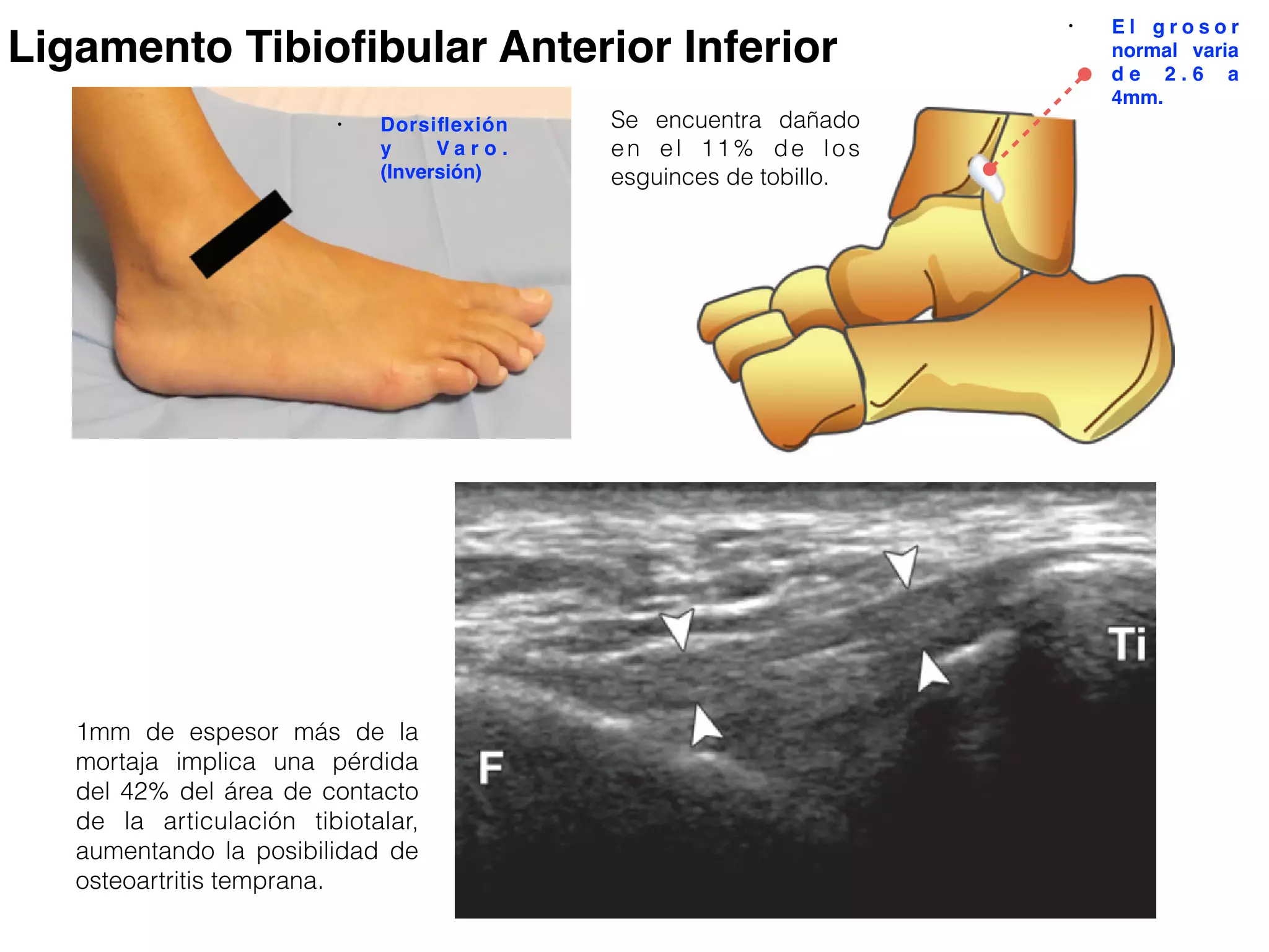
• El grosor normal varia de 2.6 a 4mm.
• Ligamento Tibiofibular Posterior Inferior. No
evaluado generalmente por ultrasonido.
LIGAMENTOS TIBIOFIBULARES
ary-February 2015 radiographics.rsna.org
Figure 1. Anterior inferior TFL. This ligament extends obliquely
downward and laterally from the anterior margin of the fibular
tubercle of the tibia to the anterior border of the distal fibular
shaft and lateral malleolus. (a) Schematic drawing of the anterior
inferior TFL anatomic structure. (b) Probe positioning on the lat-
eral ankle. (c) US scan of the anterior inferior TFL (arrowheads).
F = fibula, Ti = tibia.
probes that had lower performance parameters,
compared with more recent high-end equip-
ment.Thus, it is possible that the already high
detection values reported in the literature have
improved. Milz et al (41) reported visibility of
the anterior inferior TFL in 89.6% of cases in a
series of 48 ankle specimens.
The anterior inferiorTFL plays a crucial role
in increasing the stability of the distal tibiofibular
joint and is involved in up to 11% of ankle sprains
(10). In these cases, patients may complain about
instability resulting from the widening of the ankle
nostic accuracy of the detection of tears and
fferentiate a partial from a complete tear (36–
Pertinent dynamic maneuvers are discussed
e relevant sections of this article.
Ankle Ligaments
ofibular Ligaments
mechanics.—The tibia and fibula are articu-
d at their distal end at the inferior tibiofibular
.This joint is mechanically linked to the an-
eral ankle. (c) US scan of the anterior inferior TFL (arrowheads).
F = fibula, Ti = tibia.
rior inferior TFL. This ligament extends obliquely
laterally from the anterior margin of the fibular
tibia to the anterior border of the distal fibular
malleolus. (a) Schematic drawing of the anterior
omic structure. (b) Probe positioning on the lat-
S scan of the anterior inferior TFL (arrowheads).
bia.
Ligamento Tibiofibular Anterior Inferior
• E l g r o s o r
normal varia
d e 2 . 6 a
4mm.
Se encuentra dañado
en el 11% de los
esguinces de tobillo.
1mm de espesor más de la
mortaja implica una pérdida
del 42% del área de contacto
de la articulación tibiotalar,
aumentando la posibilidad de
osteoartritis temprana.
• Dorsiflexión
y V a r o .
(Inversión)
first sentence), keep the posterior edge of the
rotate its anterior edge upwards to image the
ucer will pass over a part of the talar cartilage,
ofibular ligament and the anterior tibiofibular
LM
Tibia
Legend: arrowheads, anterior tibiofibular ligament; LM, la
5
With the ankle lying on its medial aspect, p
oblique coronal plane with its superior edge
malleolus and its inferior margin slightly po
heel, while the foot is dorsiflexed to im
ligament.
pl
pb
Calcaneus
LM
/!,/!"$'*&)+,!%(,& !!$"#
Fig
of
The
mo
Luc
Mil
Ultrasonido que muestra una apariencia fibrótica de AITFL.
6
Look at the following midtarsal
ligaments: dorsal talonavicular,
dorsal calcaneocuboid and calca-
neo-cuboido-navicular ligament
(avulsion of the anterolateral tu-
bercle of the calcaneus).
Legend: arrowheads, dorsal talonavi-
cular ligament; NAV, navicular bone
$%&'!()!+$,!&'!()(+ !!./,'
NAVTalus
LIGAMENTO TALONAVICULAR
LIGAMENTOS DEL
COMPARTIMIENTO LATERAL.
• El LCL consiste en tres elementos separados:
• El ligamento talofibular anterior.
(peronéoastragalino)
• Ligamento calcáneofibular. (peronéocalcaneo)
• Peronéo largo y Peronéo corto.
• Ligamento Anterior Talofibular.
• Es el más débil y el más frecuentemente lastimado
de los tres componentes del LCL.
• Conecta el borde anterolateral del maléolo lateral y la
superficie lateral del cuello talar.
y-February 2015 radiographics.rsna.org
gure 4. Anterior talofibular ligament. This ligament originates
om the anterolateral border of the lateral malleolus and inserts
n the lateral surface of the talar neck. (a) Schematic drawing of
e anterior talofibular ligament anatomic structure. (b) Probe po-
ioning on the lateral ankle. (c) US scan of the anterior talofibular
ament (arrowheads). F = fibula, Ta = talus.
Figure 5. US appearances of injured anterior talofibular ligament. F = fibula, Ta = talus. (a) Acute full-thickness tear of
ligament (arrowheads). F = fibula, Ta = talus.
rior talofibular ligament. This ligament originates
ateral border of the lateral malleolus and inserts
rface of the talar neck. (a) Schematic drawing of
fibular ligament anatomic structure. (b) Probe po-
ateral ankle. (c) US scan of the anterior talofibular
heads). F = fibula, Ta = talus.
Sirve como un estabilizar del tálus,
previniendo el desplazamiento anterior
con respecto a la fíbula y tibia.
Puede haber una lesión aislada, o
asociada con CFL (Calcáneofibular),
cuando el pie es invertido.
• El grosor
normal es de 2
a 3.5mm.
Ligamento talofibular anterior
• Rotación Interna
!"#$%&'
(%&)"%*$"+$
* *
* *
1
2
2
1
!"#$%&'
(%&)"%*$"+$
* *
* *
1
2
2
1
erior talofibular ligament
Paciente con ruptura del ligamento Talofibular anterior
Evaluación del Cajón
Anterior
LM
When distinguishing a partial from a
complete tear is difficult, perform a so-
nographic anterior drawer test by pla-
cing the patient prone with the foot
hanging over the edge of the exami-
nation table while pulling the forefoot
anteriorly when in plantar flexion and
inversion. When the ligament is torn,
the anterior shift of the talus against
the tibia will open the gap in the sub-
stance of the ligament.
Legend: LM, l
anterior talofib
Figure 5. US appearances of injured anterior talofibular ligame
the anterior talofibular ligament in a 27-year-old basketball play
together with fluid effusion in the anterolateral recess (*). (b) F
after trauma. The ligament is remarkably thickened and inhom
over the proximal insertion (arrows). (Courtesy of Luca De Flavii
the LCL complex of the ankle joint. It connects
the anterolateral border of the lateral malleolus
and the lateral surface of the talar neck (Fig 4).
Along its course, it blends with some fibers of
the capsule of the tibiotalar joint.The anterior
talofibular ligament serves to stabilize the talus,
preventing anterior talar motion.The primary
function of the anterior talofibular ligament is
Becaus
plantar
freque
tion w
turned
diffuse
of the
linearr ligament. F = fibula, Ta = talus. (a) Acute full-thickness tear of
Lesión del ligamento
talofibular.
Lesión aguda.
Lesión crónica
Normal
• CFL. Es el más largo de los ligamentos en LCL. Es una
estructura fuerte que cruza oblicuamente debajo de los tendones
peronéo.
• Origen.Se extiende desde el borde inferior del maleo lateral.
• Inserción. Eminencia troclear en la superficie lateral del
calcáneo.
• Función. Estabilizador de la articulación subtalar. La función
principal es evitar la inversión del calcáneo con respecto a la
fíbula.
anuary-February 2015 radiographics.rsna.org
Figure 7. CFL. This ligament originates from the lateral mal-
leolar tip and inserts on the lateral surface of the calcaneus.
(a) Schematic drawing of the CFL anatomic structure. (b) Probe po-
sitioning on the lateral ankle. (c) US scan of the CFL (arrowheads).
C = calcaneus. F = fibula, PBT = peroneus brevis tendon, PLT =
peroneus longus tendon.
• Si no hay lesión del ligamento
talofibular anterior la lesión del CFL
es poco probable.
t parallel to the sole of the foot (Fig 4b).
nterior talofibular ligament can be seen as a
echoic fibrillar band in tension between the
l malleolus and the talus (Movie 2 [online]).
unctionality of the anterior talofibular liga-
can be tested by using the anterior drawer
This test consists of stressing the plantar
peroneus longus tendon.
Figure 8. US scan shows a partial tear of the CFL.
The ligament appears inhomogeneous and hy-
poechoic (arrowheads), with periligamentous effu-
sion (*). C = calcaneus, F = fibula, PBT = peroneus bre-
vis tendon, PLT = peroneus longus tendon, Ta = talus.
originates from the lateral mal-
teral surface of the calcaneus.
anatomic structure. (b) Probe po-
US scan of the CFL (arrowheads).
peroneus brevis tendon, PLT =
Ligamento Calcáneofibular.
Flexión dorsal
edial aspect, place the transducer in an
s superior edge over the tip of the lateral
argin slightly posterior to it, towards the
rsiflexed to image the calcaneofibular
Calcaneus
LM
pl
pb
pb LM
5
With the ankle lying on its medial aspect, place the transducer in an
oblique coronal plane with its superior edge over the tip of the lateral
malleolus and its inferior margin slightly posterior to it, towards the
heel, while the foot is dorsiflexed to image the calcaneofibular
igament.
Calcaneus
LM
pl
pbpl
pb
Calcaneus
LM
/!,/!"$'*&)+,!%(,& !!$"#
5
With the ankle lying on its medial aspect, place the transducer in an
oblique coronal plane with its superior edge over the tip of the lateral
malleolus and its inferior margin slightly posterior to it, towards the
heel, while the foot is dorsiflexed to image the calcaneofibular
igament.
Calcaneus
LM
pl
pbpl
pb
Calcaneus
LM
/!,/!"$'*&)+,!%(,& !!$"#5
With the ankle lying on its medial aspect, place the transducer in an
oblique coronal plane with its superior edge over the tip of the lateral
malleolus and its inferior margin slightly posterior to it, towards the
heel, while the foot is dorsiflexed to image the calcaneofibular
ligament.
Calcaneus
LM
pl
pbpl
pb
Calcaneus
LM
/!,/!"$'*&)+,!%(,& !!$"#
5
With the ankle lying on its medial aspect, place the transducer in an
oblique coronal plane with its superior edge over the tip of the lateral
malleolus and its inferior margin slightly posterior to it, towards the
heel, while the foot is dorsiflexed to image the calcaneofibular
gament.
Calcaneus
LM
pl
pbpl
pb
Calcaneus
LM
/!,/!"$'*&)+,!%(,& !!$"#
Ligamento Calcáneofibular
Dorsiflexión
Lesión parcial del CFL.
as a
the
e]).
a-
er
may
s
ent
of the probe should be held over the tip of the
lateral malleolus and the probe should be ro-
tated caudad to reach a coronal plane, with the
distal edge of the probe slightly posterior to
the CFL.
and hy-
us effu-
eus bre-
a = talus.
• Origen: Cabeza y los dos
tercios superiores de la fíbula
lateral.
• Inserción: Base del primer
metatarsiano y cuneiforme
medial.
• Función: Eversión y Flexión
plantar del pie.
PERONEO LARGOSUBASHKC/NMC-15TH/2014
• Origen: Dos tercios inferiores
de la fíbula lateral.
• Inserción: Base Dorsolateral
del 5o metatarsiano.
• Función: Eversión del pie.
PERONEO CORTOSUBASHKC/NMC-15TH/2014
or approximately 5 cm and downwards through the inframalleola
LM
!
" pbm
Check them at the level of the peroneal tubercle of calcaneus,
own to the area where the os peroneum can be found. Follow
he base of the 5th metatarsal. Look at the superior and inferior p
Legend: arr
curved arro
LM, lateral m
muscle; void
arrow, peron
!
#
peroneal tendons to examine
). Because these tendons arc
beam perpendicular to them
ollow these tendons upwards
eolar region.
LM
pbm
"
TENDONES PERONÉOS
7
Behind the lateral mall
them in their short-axis
around the malleolus, t
and avoid anisotropy a
for approximately 5 cm
!
"
0123410)156
"
Check them at the le
down to the area wh
the base of the 5th m
#
When intermittent
suspected clinically,
LM
evel of the peroneal tubercle of calcaneus, and the
ere the os peroneum can be found. Follow the per
etatarsal. Look at the superior and inferior peroneal
Legend: arrowheads, p
curved arrows, superio
LM, lateral malleolus; p
muscle; void arrow, per
arrow, peroneus longus
subluxation of the peroneals is
! "
#
• COMPARTIMIENTO MEDIAL
• Ligamento Deltoideo.
• Arteria y Nervio Tibial Posterior.
• Tibial posterior (TOM), Flexor del Hallux (HARRY) y
Flexor de los dedos (DICK) (orden supramaleolar).
TENDONES DEL
COMPARTIMIENTO MEDIAL.
• Origen: En la membrana interósea y
la superficie posterior de la tibia y el
peroné.
• Inserción: Navicular, Cuneiforme
medial, base del 2, 3 y 4o
metatarsianos.
• Función: Inversión y Flexión plantar.
TIBIAL POSTERIOR
SUBASHKC/NMC-15TH/2014
• Origen: Tibia medial y
posterior y sobre la
aponeurosis de la fíbula.
• Inserción: Base de las
falanges distales del 2o al 5o
dedo.
• Función: Flexión de los 4to
dedos.
FLEXOR LARGO DE LOS DEDOS
SUBASHKC/NMC-15TH/2014
• Origen: Fibula posterior e
inferior y la membrana
interósea.
• Inserción: Base de la falange
distal del Hallux.
FLEXOR LARGO DEL HALLUX
SUBASHKC/NMC-15TH/2014
skin over the medial ankle. The examination of tendons
formed first.
Legend: a, tibialis posterior artery; MM, medial malleolus; v, posterior tibial
digitorum longus tendon; white arrowheads, flexor retinaculum; white arrow
Behin
the tra
the ti
digitor
tibialis
ous ju
short-
ce of
on lon
of the
!#
##
$$
TENDONES DEL COMPARTIMIENTO FLEXOR
n of the medial ankle, the patient is seated with
ace of the foot rolled internally or in a “frog-leg”
atively, the patient may lie supine with the foot
aterally. A small pillow under the lateral malleo-
o improve the contact between transducer and
edial ankle. The examination of tendons is per-
Ank
)230-4156*6!(671"975:;69;1!<+1&(:=9;1+6 659;?!1(9< ?715:<+9<7
ARTERIA TIBIAL POSTERIOR Y NERVIO TIBIAL
5
at the flexor retinaculum, the posterior tibial vessels and the tibial nerve with its divisional
branches (medial and lateral plantar nerves). Compression may help to assess whether
the veins are patent.
Legend: AbdH, abductor hallucis muscle; curved arrow,
tibial nerve; fhl, flexor hallucis longus tendon; ST,
sustentaculum tali; straight arrows, flexor digitorum
longus tendon; void arrowhead, posterior tibial artery;
white arrowheads, posteiror tibial veins
%% %%&'( &'(
)*+&
nd: a, tibialis posterior artery; MM, medial malleolus; v, posterior tibial veins; void arrowheads, flexor
orum longus tendon; white arrowheads, flexor retinaculum; white arrows, tibialis posterior tendon
on long-axis scans over the insertion
of the tibialis posterior.
mine the flexor digitorum longus tendon down to reach the sustentaculum tali. Look
e flexor retinaculum, the posterior tibial vessels and the tibial nerve with its divisional
ches (medial and lateral plantar nerves). Compression may help to assess whether
veins are patent.
%% %%&'( &'(
)*+&
!;7!(15?<<:(1!<+156*6!(1<:;#:
Legend: a, tibialis posterior artery; MM, medial malleolus; v, posterior tibial ve
digitorum longus tendon; white arrowheads, flexor retinaculum; white arrows,
on long-
of the tib
9
Examine the flexor digitorum longus tendon down to reach
at the flexor retinaculum, the posterior tibial vessels and th
branches (medial and lateral plantar nerves). Compression
the veins are patent.
%% &'(
5!;7!(15?<<:(1!<+156*6!(1<:;#:
5
own to reach the sustentaculum tali. Look
ssels and the tibial nerve with its divisional
Compression may help to assess whether
%%
&'(
)*+&
entaculum tali. Look
rve with its divisional
p to assess whether
&'(
)*+&
Con Compresión
• TIBIONAVICULAR.
• TIBIOCALCÁNEO.
• TIBIOTALAR
(ANTERIOR Y
POSTERIOR).
LIGAMENTO DELTOIDEO
profundo11,39,41,42
. Los ligamentos que componen el plano
superficial cruzan dos articulaciones, la del tobillo y la sub-
Golanó P, et al. Anatomía
Figura 8. Representación esquemática de los componentes constantes
del ligamento colateral medial descritos por Milner y Soames41
. 1. Li-
gamento tibiospring. 2. Ligamento tibionavicular. 3. Ligamento tibiota-
lar posterior profundo. 4. Ligamento calcaneonavicular superomedial.
• Capa superficial.
• El ligamento tibionavicular se origina del borde
anterior del colículo del maléolo medial y se
inserta en el aspecto dorsomedial del hueso
navicular.
of the medial malleolus and inserts on the dorsomedial aspect of the navicular. (a) Schematic drawing of the
tibionavicular ligament anatomic structure. (b) Probe positioning on the medial ankle. (c) US scan of the tibi-
onavicular ligament (arrowheads). N = navicular, Ta = talus, Ti = tibia.
tendons superficially.This is an indirect sign of
continuity of the CFL. If displacement is absent
of the deltoid ligament act as a unit prov
support to the ankle. Insufficiency of th
collateral ligament may lead to osteoarth
the ankle joint (51).
Lesions of the deltoid ligament occur
severe eversion injuries and are usually a
ated with fractures of the lateral malleol
with lateral displacement of the talus. L
monly, deltoid ligament injuries are obs
in association with avulsion fractures of
medial malleolus at the site of attachme
superficial portion. Rupture of the delto
of
sup
col
the
sev
ate
LIGAMENTO TIBIONAVICULAR
• El ligamento tibiocalcáneo se origina del
aspecto medial del colículo anterior del
maléolo medial, desciende verticalmente y se
inserta en el borde medial del sustentaculum
tali.
l ligament.
om the me-
colliculus of
cends verti-
edial border
a) Schematic
eal ligament
be position-
(c) US scan
ment (arrow-
= posterior
ament.The tibionavicular
rted to be present in ap-
general population (52).
ment connects the ante-
us to the superior border
ortion of the spring liga-
gament originates from
e anterior colliculus of the
ular
ap-
52).
nte-
rder
iga-
om
of the
LIGAMENTO TIBIOCALCÁNEO
RG • Volume 35 Number 1
Figure
the tib
portion
echotex
ligamen
with so
The dis
homog
• Capa profunda.
• El ligamento anterior tibiotalar. Es muy delgado.
Se origina de la punta del colículo anterior y la
parte anterior del surco intercolicular del maléolo
medial y se inserta en la superficie medial del
tálus.
Figure 13. Anterior tibiotalar ligament.
This ligament originates from the tip of the
anterior colliculus and the anterior part of
the intercollicular groove of the medial mal-
leolus and inserts on the medial surface of
the talus just distal to the anterior part of the
medial talar articular surface. (a) Schematic
drawing of the anterior tibiotalar ligament
anatomic structure. (b) Probe positioning
on the medial ankle. (c) US scan of the an-
terior tibiotalar ligament (arrowheads). Ta =
talus, Ti = tibia.
The distal insertion of the ligament is also in-
homogeneous and hypoechoic (**).
Figure 13. Anterior tibiotalar ligament.
This ligament originates from the tip of the
anterior colliculus and the anterior part of
the intercollicular groove of the medial mal-
leolus and inserts on the medial surface of
the talus just distal to the anterior part of the
medial talar articular surface. (a) Schematic
drawing of the anterior tibiotalar ligament
anatomic structure. (b) Probe positioning
on the medial ankle. (c) US scan of the an-
terior tibiotalar ligament (arrowheads). Ta =
talus, Ti = tibia.
Figure 13. Ante
This ligament orig
anterior colliculus
the intercollicular g
leolus and inserts
the talus just distal
medial talar articul
drawing of the an
anatomic structure
on the medial ank
terior tibiotalar liga
talus, Ti = tibia.
LIGAMENTO TIBIOTALAR ANTERIOR
• El ligamento tibiotalar posterior es el más grueso
de los ligamentos de la región medial. Este
ligamento se origina en el segmento superior de la
superficie posterior del colículo anterior, y se
inserta en la superficie medial del tálus por debajo
de la faceta articular.
ligament (arrowheads). PTT = posterior tibial tendon, Ta = ta
Figure 15. Dorsal calcaneocuboid liga-
t. This ligament originates from the upper seg-
rior colliculus, the intercollicular groove, and the
of the medial malleolus. The fibers insert on the
l of the articular facet, up to the posteromedial
the posterior tibiotalar ligament anatomic struc-
ial ankle. (c) US scan of the posterior tibiotalar
ibial tendon, Ta = talus, Ti = tibia.
radiographics.rsna.org
per seg-
and the
rt on the
romedial
mic struc-
tibiotalar
• REGIÓN POSTERIOR
• Tendón de Aquiles.
• Grasa de Kagger.
• Bursa PA Y RA.
• Fascia Plantar.
TENDÓN DE AQUÍLESOn a prone position, let the foot hanging out of the examination
table. Look clinically to the position of the foot, comparing both sid-
es to see any differences that can lead to the diagnosis of Achilles
tendon full-thickness tear. Then, examine the Achilles tendon from
its myotendinous junction to its calcanear insertion by means of
transverse and longitudinal planes. While scanning the Achilles
tendon on short-axis planes, tilt the probe on each side of the tend-
on to assess the peritendinous envelope. Measure the size of the
Achilles tendon only on transverse planes. The Achilles tendon has
to be followed down to its calcanear insertion. Check the retroachil-
les and the retrocalcanear bursae.
Legend: arrowheads, Achilles tendon; asterisk, anisotropy; fhl, flexor
hallucis longus muscle
soleus
fhl
Kager Calcaneus
*
!
"
#
$
Check the plantaris tendon. In cases of complete Achilles tendon tear, the plantaris may
mimic residual intact fibers of the Achilles. Dynamic scanning during passive dorsal and
plantar flexion help to distinguish partial from complete Achilles tendon tears.
On a prone position, let the foot hanging out of the examination
table. Look clinically to the position of the foot, comparing both sid-
es to see any differences that can lead to the diagnosis of Achilles
tendon full-thickness tear. Then, examine the Achilles tendon from
its myotendinous junction to its calcanear insertion by means of
transverse and longitudinal planes. While scanning the Achilles
tendon on short-axis planes, tilt the probe on each side of the tend-
on to assess the peritendinous envelope. Measure the size of the
Achilles tendon only on transverse planes. The Achilles tendon has
to be followed down to its calcanear insertion. Check the retroachil-
les and the retrocalcanear bursae.
Legend: arrowheads, Achilles tendon; asterisk, anisotropy; fhl, flexor
hallucis longus muscle
soleus
fhl
Kager Calcaneus
*
!
"
#
$
Check the plantaris tendon. In cases of complete Achilles tendon tear, the plantaris may
mimic residual intact fibers of the Achilles. Dynamic scanning during passive dorsal and
plantar flexion help to distinguish partial from complete Achilles tendon tears.
BURSA PREAQUÍLEA
e este paciente se apreciaba en el examen lon-
tudinal (Figura 1) un tendón moderadamente
El estudio dinámico es una de las ventajas
aporta la ecografía y precisamente a travé
GURA 1. Realizando un corte longitudinal del tendón de Aquiles
aprecia en el interior una imagen anecoica que corresponde a
na rotura parcial (r). Además en la parte profunda del tendón se
precia la ocupación de la bursa preaquílea (B)
FIGURA 2. El examen longitudinal realizado sobre el tendó
aquileo con el pie en posición neutra permite observar la b
preaquilea por debajo del tendón (B)
Rotura Parcial
del T.Aquiles.
alizando un corte longitudinal del tendón de Aquiles
el interior una imagen anecoica que corresponde a
cial (r). Además en la parte profunda del tendón se
pación de la bursa preaquílea (B)
FIGURA 2. El examen longitudinal realizado sobre el tendón
aquileo con el pie en posición neutra permite observar la bursa
preaquilea por debajo del tendón (B)
este otro corte en eje largo al realizar la flexión
, la bursa cambia su posición interponiéndose
eo y el tendón
FIGURA 4. El corte axial del tendón de Aquiles permite ver la
zona anecoica de la rotura en el interior del tendón (r) y la ocu-
pación de la bursa (B) por debajo del mismo
BURSA RETROAQUÍLEA
FASCIA PLANTAR
Legend: arrowheads, plantar fascia; fdb, flexor digitorum brevis muscle
Calcaneus
fdb
Legend: arrowheads, plantar fascia; fdb, flexor digitorum brevis muscle
Calcaneus
fdb
• REGIÓN POSTERIOR
• Tendón de Aquiles.
• Grasa de Kagger.
• Bursa Retrocalcánea.
• Fascia Plantar.

More Related Content

What's hot

ANATOMIA ECOGRAFICA NORMAL DEL CODO
ANATOMIA ECOGRAFICA NORMAL DEL CODOANATOMIA ECOGRAFICA NORMAL DEL CODO
ANATOMIA ECOGRAFICA NORMAL DEL CODO
Guillermo Rodriguez Maruri
 
Doppler arterial mmii
Doppler arterial mmiiDoppler arterial mmii
Doppler arterial mmiiHeidy Saenz
 
ANATOMÍA ECOGRÁFICA NORMAL DE LA RODILLA
ANATOMÍA ECOGRÁFICA NORMAL DE LA RODILLAANATOMÍA ECOGRÁFICA NORMAL DE LA RODILLA
ANATOMÍA ECOGRÁFICA NORMAL DE LA RODILLA
Guillermo Rodriguez Maruri
 
Protocolo ecografia codo
Protocolo ecografia codoProtocolo ecografia codo
Protocolo ecografia codo
jmilian57
 
Ecografia de rodilla
Ecografia de rodillaEcografia de rodilla
Ecografia de rodilla
Rodrigo Sazo
 
Anatomía del codo por resonancia
Anatomía del codo por resonanciaAnatomía del codo por resonancia
Anatomía del codo por resonancia
Reyna Payamps
 
Patología escrotal pediátrica más frecuente seram2012 s-0655
Patología escrotal pediátrica más frecuente seram2012 s-0655Patología escrotal pediátrica más frecuente seram2012 s-0655
Patología escrotal pediátrica más frecuente seram2012 s-0655
LUIS VASQUEZ ZUBIETA
 
8 ecografía del hombro
8 ecografía del hombro8 ecografía del hombro
8 ecografía del hombro
Pedro García Ramos
 
Patologias de mano y muñeca en us rm
Patologias  de  mano y muñeca en us rmPatologias  de  mano y muñeca en us rm
Patologias de mano y muñeca en us rm
Wilfcal Will
 
Protocolo ecografia cadera
Protocolo ecografia caderaProtocolo ecografia cadera
Protocolo ecografia cadera
jmilian57
 
Ecografia de rodilla
Ecografia de rodillaEcografia de rodilla
Ecografia de rodilla
Luis Davila
 
Radiología de la Columna
Radiología de la ColumnaRadiología de la Columna
Radiología de la Columna
Santiago Rodriguez
 
Doppler arterial de miembros inferiores dr. romel flores imumr
Doppler arterial de miembros inferiores   dr. romel flores imumrDoppler arterial de miembros inferiores   dr. romel flores imumr
Doppler arterial de miembros inferiores dr. romel flores imumr
Romel Flores Virgilio
 
Resonancia hombro
Resonancia  hombroResonancia  hombro
Resonancia hombro
Hospital Militar Quito
 
Proyecciones del tobillo.
Proyecciones del tobillo.Proyecciones del tobillo.
Proyecciones del tobillo.
KatherineOrtiz49
 
Manguito rotador y biceps
Manguito rotador y bicepsManguito rotador y biceps
Manguito rotador y biceps
Rafael Barousse
 
Lesiones quísticas y bursitis de rodilla
Lesiones quísticas y bursitis de rodillaLesiones quísticas y bursitis de rodilla
Lesiones quísticas y bursitis de rodilla
magaibarra
 
resonancia magnetica de rodilla
resonancia magnetica de rodillaresonancia magnetica de rodilla
resonancia magnetica de rodilla
R0SIA
 

What's hot (20)

ANATOMIA ECOGRAFICA NORMAL DEL CODO
ANATOMIA ECOGRAFICA NORMAL DEL CODOANATOMIA ECOGRAFICA NORMAL DEL CODO
ANATOMIA ECOGRAFICA NORMAL DEL CODO
 
Doppler arterial mmii
Doppler arterial mmiiDoppler arterial mmii
Doppler arterial mmii
 
ANATOMÍA ECOGRÁFICA NORMAL DE LA RODILLA
ANATOMÍA ECOGRÁFICA NORMAL DE LA RODILLAANATOMÍA ECOGRÁFICA NORMAL DE LA RODILLA
ANATOMÍA ECOGRÁFICA NORMAL DE LA RODILLA
 
Protocolo ecografia codo
Protocolo ecografia codoProtocolo ecografia codo
Protocolo ecografia codo
 
Ecografia de rodilla
Ecografia de rodillaEcografia de rodilla
Ecografia de rodilla
 
Anatomía del codo por resonancia
Anatomía del codo por resonanciaAnatomía del codo por resonancia
Anatomía del codo por resonancia
 
Patología escrotal pediátrica más frecuente seram2012 s-0655
Patología escrotal pediátrica más frecuente seram2012 s-0655Patología escrotal pediátrica más frecuente seram2012 s-0655
Patología escrotal pediátrica más frecuente seram2012 s-0655
 
8 ecografía del hombro
8 ecografía del hombro8 ecografía del hombro
8 ecografía del hombro
 
Patologias de mano y muñeca en us rm
Patologias  de  mano y muñeca en us rmPatologias  de  mano y muñeca en us rm
Patologias de mano y muñeca en us rm
 
Protocolo ecografia cadera
Protocolo ecografia caderaProtocolo ecografia cadera
Protocolo ecografia cadera
 
Ecografia de rodilla
Ecografia de rodillaEcografia de rodilla
Ecografia de rodilla
 
Radiología de la Columna
Radiología de la ColumnaRadiología de la Columna
Radiología de la Columna
 
Doppler arterial de miembros inferiores dr. romel flores imumr
Doppler arterial de miembros inferiores   dr. romel flores imumrDoppler arterial de miembros inferiores   dr. romel flores imumr
Doppler arterial de miembros inferiores dr. romel flores imumr
 
Resonancia hombro
Resonancia  hombroResonancia  hombro
Resonancia hombro
 
Doppler Renal
Doppler RenalDoppler Renal
Doppler Renal
 
Proyecciones del tobillo.
Proyecciones del tobillo.Proyecciones del tobillo.
Proyecciones del tobillo.
 
Manguito rotador y biceps
Manguito rotador y bicepsManguito rotador y biceps
Manguito rotador y biceps
 
Lesiones quísticas y bursitis de rodilla
Lesiones quísticas y bursitis de rodillaLesiones quísticas y bursitis de rodilla
Lesiones quísticas y bursitis de rodilla
 
Hombro
HombroHombro
Hombro
 
resonancia magnetica de rodilla
resonancia magnetica de rodillaresonancia magnetica de rodilla
resonancia magnetica de rodilla
 

Viewers also liked

Clasificacion Ultrasonográfica de Desgarro muscular
Clasificacion Ultrasonográfica de Desgarro muscularClasificacion Ultrasonográfica de Desgarro muscular
Clasificacion Ultrasonográfica de Desgarro muscular
Sebastian Millon
 
Tumoraciones ovaricas
Tumoraciones ovaricasTumoraciones ovaricas
Enfermedades Vasculares Pulmonares
Enfermedades Vasculares PulmonaresEnfermedades Vasculares Pulmonares
Enfermedades Vasculares Pulmonares
Imagenología Diagnóstica y Terapéutica HRAEPY
 
Tumores benignos hueso
Tumores benignos hueso Tumores benignos hueso
Testículos: Anatomía Ultrasonográfica
Testículos: Anatomía UltrasonográficaTestículos: Anatomía Ultrasonográfica
Testículos: Anatomía Ultrasonográfica
Imagenología Diagnóstica y Terapéutica HRAEPY
 
Uropatía Obstructiva
Uropatía ObstructivaUropatía Obstructiva
Anomalias del cordon umbilical
Anomalias del cordon umbilicalAnomalias del cordon umbilical
Anomalias del cordon umbilical
Imagenología Diagnóstica y Terapéutica HRAEPY
 
Uropatía obstructiva
Uropatía obstructivaUropatía obstructiva
Uropatía obstructiva
Johana Vera
 
Lesiones Oseas Que No Se Deben Tocar
Lesiones Oseas Que No Se Deben TocarLesiones Oseas Que No Se Deben Tocar
Lesiones Oseas Que No Se Deben Tocar
Imagenología Diagnóstica y Terapéutica HRAEPY
 
MANAGEMENT OF BIOCHEMICAL RECURRENCE AFTER RADICAL PROSTATECTOMY & RADIATION ...
MANAGEMENT OF BIOCHEMICAL RECURRENCE AFTER RADICAL PROSTATECTOMY & RADIATION ...MANAGEMENT OF BIOCHEMICAL RECURRENCE AFTER RADICAL PROSTATECTOMY & RADIATION ...
MANAGEMENT OF BIOCHEMICAL RECURRENCE AFTER RADICAL PROSTATECTOMY & RADIATION ...
GAURAV NAHAR
 
Uropatia obstructiva
Uropatia obstructivaUropatia obstructiva
Uropatia obstructiva
Adriana Soto Carrillo
 
Prostata Anatomía
Prostata AnatomíaProstata Anatomía
Complicaciones Torácicas en Cirrosis Hepatica
Complicaciones Torácicas en Cirrosis HepaticaComplicaciones Torácicas en Cirrosis Hepatica
Complicaciones Torácicas en Cirrosis Hepatica
Imagenología Diagnóstica y Terapéutica HRAEPY
 
Enfermedad Diverticular Colonica
Enfermedad Diverticular ColonicaEnfermedad Diverticular Colonica
Enfermedad Diverticular Colonica
Imagenología Diagnóstica y Terapéutica HRAEPY
 
Nocturia
NocturiaNocturia
Nocturia
GAURAV NAHAR
 
Caso Clinico
Caso ClinicoCaso Clinico
Caso Clinico
eddynoy velasquez
 
Uropatia obstructiva
Uropatia obstructiva Uropatia obstructiva
Uropatia obstructiva Ivan Mitosis
 

Viewers also liked (20)

Clasificacion Ultrasonográfica de Desgarro muscular
Clasificacion Ultrasonográfica de Desgarro muscularClasificacion Ultrasonográfica de Desgarro muscular
Clasificacion Ultrasonográfica de Desgarro muscular
 
Tumoraciones ovaricas
Tumoraciones ovaricasTumoraciones ovaricas
Tumoraciones ovaricas
 
Enfermedades Vasculares Pulmonares
Enfermedades Vasculares PulmonaresEnfermedades Vasculares Pulmonares
Enfermedades Vasculares Pulmonares
 
Tumores benignos hueso
Tumores benignos hueso Tumores benignos hueso
Tumores benignos hueso
 
Testículos: Anatomía Ultrasonográfica
Testículos: Anatomía UltrasonográficaTestículos: Anatomía Ultrasonográfica
Testículos: Anatomía Ultrasonográfica
 
Uropatía Obstructiva
Uropatía ObstructivaUropatía Obstructiva
Uropatía Obstructiva
 
Anomalias del cordon umbilical
Anomalias del cordon umbilicalAnomalias del cordon umbilical
Anomalias del cordon umbilical
 
Uropatía obstructiva
Uropatía obstructivaUropatía obstructiva
Uropatía obstructiva
 
Lesiones Oseas Que No Se Deben Tocar
Lesiones Oseas Que No Se Deben TocarLesiones Oseas Que No Se Deben Tocar
Lesiones Oseas Que No Se Deben Tocar
 
MANAGEMENT OF BIOCHEMICAL RECURRENCE AFTER RADICAL PROSTATECTOMY & RADIATION ...
MANAGEMENT OF BIOCHEMICAL RECURRENCE AFTER RADICAL PROSTATECTOMY & RADIATION ...MANAGEMENT OF BIOCHEMICAL RECURRENCE AFTER RADICAL PROSTATECTOMY & RADIATION ...
MANAGEMENT OF BIOCHEMICAL RECURRENCE AFTER RADICAL PROSTATECTOMY & RADIATION ...
 
Uropatia obstructiva
Uropatia obstructivaUropatia obstructiva
Uropatia obstructiva
 
Uropatía obstructiva
Uropatía obstructivaUropatía obstructiva
Uropatía obstructiva
 
Tumores oseos benignos
Tumores oseos benignosTumores oseos benignos
Tumores oseos benignos
 
Prostata Anatomía
Prostata AnatomíaProstata Anatomía
Prostata Anatomía
 
Complicaciones Torácicas en Cirrosis Hepatica
Complicaciones Torácicas en Cirrosis HepaticaComplicaciones Torácicas en Cirrosis Hepatica
Complicaciones Torácicas en Cirrosis Hepatica
 
Cistouretrografia ppt
Cistouretrografia pptCistouretrografia ppt
Cistouretrografia ppt
 
Enfermedad Diverticular Colonica
Enfermedad Diverticular ColonicaEnfermedad Diverticular Colonica
Enfermedad Diverticular Colonica
 
Nocturia
NocturiaNocturia
Nocturia
 
Caso Clinico
Caso ClinicoCaso Clinico
Caso Clinico
 
Uropatia obstructiva
Uropatia obstructiva Uropatia obstructiva
Uropatia obstructiva
 

Similar to Ultrasonido de tobillo anatomía

Presentation1.pptx. ultrasound examination of the ankle joint.
Presentation1.pptx. ultrasound examination of the ankle joint.Presentation1.pptx. ultrasound examination of the ankle joint.
Presentation1.pptx. ultrasound examination of the ankle joint.Abdellah Nazeer
 
Anatomy of the ankle ligaments a pictorial essay
Anatomy of the ankle ligaments a pictorial essayAnatomy of the ankle ligaments a pictorial essay
Anatomy of the ankle ligaments a pictorial essay
Khuyich
 
Mri anatomy of ankle
Mri anatomy of ankleMri anatomy of ankle
Mri anatomy of ankle
Shrikant Nagare
 
Congenital vertical talus BY DR.NAVEEN RATHOR
Congenital vertical talus BY DR.NAVEEN RATHORCongenital vertical talus BY DR.NAVEEN RATHOR
Congenital vertical talus BY DR.NAVEEN RATHOR
DR.Naveen Rathor
 
Ctev
CtevCtev
X Ray and MRI of Knee Joint
X Ray and MRI of Knee JointX Ray and MRI of Knee Joint
X Ray and MRI of Knee Joint
Gaurav Purohit
 
Ctev
CtevCtev
Ctev
rajusvmc
 
Presentation1.pptx, ultrasound of the hand and fingers.
Presentation1.pptx, ultrasound of the hand and fingers.Presentation1.pptx, ultrasound of the hand and fingers.
Presentation1.pptx, ultrasound of the hand and fingers.Abdellah Nazeer
 
The foot
The footThe foot
The foot
Buyungo Steven
 
Presentation1.pptx, ultrasound examination of the wrist joint.
Presentation1.pptx, ultrasound examination of the wrist joint.Presentation1.pptx, ultrasound examination of the wrist joint.
Presentation1.pptx, ultrasound examination of the wrist joint.Abdellah Nazeer
 
Ankle injuries
Ankle injuriesAnkle injuries
Ankle injuries
Muhammad Hisham
 
Considerations In Knee Artroplasty
Considerations In Knee ArtroplastyConsiderations In Knee Artroplasty
Considerations In Knee ArtroplastyJavi Mata
 
Fractures and Dislocations of Foot - Dr Sunkappa SR
Fractures and Dislocations of Foot - Dr Sunkappa SRFractures and Dislocations of Foot - Dr Sunkappa SR
Fractures and Dislocations of Foot - Dr Sunkappa SR
Sunkappa SR
 
Anatomy and imaging of knee joint
Anatomy and imaging of knee jointAnatomy and imaging of knee joint
Anatomy and imaging of knee joint
Kajal Jha
 
Mri anatomy of ankle radiology ppt pk
Mri anatomy of ankle radiology ppt pkMri anatomy of ankle radiology ppt pk
Mri anatomy of ankle radiology ppt pk
Dr pradeep Kumar
 
Ankle arthrodesis
Ankle arthrodesisAnkle arthrodesis
Ankle arthrodesis
Dr venkatesh v
 
Siêu âm cổ chân bàn chân, Bs Tài
Siêu âm cổ chân bàn chân, Bs TàiSiêu âm cổ chân bàn chân, Bs Tài
Siêu âm cổ chân bàn chân, Bs Tài
Nguyen Lam
 
Imaging of foot in non trauma and non neoplastic diseases
Imaging of foot in non trauma and non neoplastic diseasesImaging of foot in non trauma and non neoplastic diseases
Imaging of foot in non trauma and non neoplastic diseasesShail Padmani
 
Mri knee
Mri kneeMri knee
Ankle instability
Ankle instabilityAnkle instability
Ankle instability
LalisaMerga
 

Similar to Ultrasonido de tobillo anatomía (20)

Presentation1.pptx. ultrasound examination of the ankle joint.
Presentation1.pptx. ultrasound examination of the ankle joint.Presentation1.pptx. ultrasound examination of the ankle joint.
Presentation1.pptx. ultrasound examination of the ankle joint.
 
Anatomy of the ankle ligaments a pictorial essay
Anatomy of the ankle ligaments a pictorial essayAnatomy of the ankle ligaments a pictorial essay
Anatomy of the ankle ligaments a pictorial essay
 
Mri anatomy of ankle
Mri anatomy of ankleMri anatomy of ankle
Mri anatomy of ankle
 
Congenital vertical talus BY DR.NAVEEN RATHOR
Congenital vertical talus BY DR.NAVEEN RATHORCongenital vertical talus BY DR.NAVEEN RATHOR
Congenital vertical talus BY DR.NAVEEN RATHOR
 
Ctev
CtevCtev
Ctev
 
X Ray and MRI of Knee Joint
X Ray and MRI of Knee JointX Ray and MRI of Knee Joint
X Ray and MRI of Knee Joint
 
Ctev
CtevCtev
Ctev
 
Presentation1.pptx, ultrasound of the hand and fingers.
Presentation1.pptx, ultrasound of the hand and fingers.Presentation1.pptx, ultrasound of the hand and fingers.
Presentation1.pptx, ultrasound of the hand and fingers.
 
The foot
The footThe foot
The foot
 
Presentation1.pptx, ultrasound examination of the wrist joint.
Presentation1.pptx, ultrasound examination of the wrist joint.Presentation1.pptx, ultrasound examination of the wrist joint.
Presentation1.pptx, ultrasound examination of the wrist joint.
 
Ankle injuries
Ankle injuriesAnkle injuries
Ankle injuries
 
Considerations In Knee Artroplasty
Considerations In Knee ArtroplastyConsiderations In Knee Artroplasty
Considerations In Knee Artroplasty
 
Fractures and Dislocations of Foot - Dr Sunkappa SR
Fractures and Dislocations of Foot - Dr Sunkappa SRFractures and Dislocations of Foot - Dr Sunkappa SR
Fractures and Dislocations of Foot - Dr Sunkappa SR
 
Anatomy and imaging of knee joint
Anatomy and imaging of knee jointAnatomy and imaging of knee joint
Anatomy and imaging of knee joint
 
Mri anatomy of ankle radiology ppt pk
Mri anatomy of ankle radiology ppt pkMri anatomy of ankle radiology ppt pk
Mri anatomy of ankle radiology ppt pk
 
Ankle arthrodesis
Ankle arthrodesisAnkle arthrodesis
Ankle arthrodesis
 
Siêu âm cổ chân bàn chân, Bs Tài
Siêu âm cổ chân bàn chân, Bs TàiSiêu âm cổ chân bàn chân, Bs Tài
Siêu âm cổ chân bàn chân, Bs Tài
 
Imaging of foot in non trauma and non neoplastic diseases
Imaging of foot in non trauma and non neoplastic diseasesImaging of foot in non trauma and non neoplastic diseases
Imaging of foot in non trauma and non neoplastic diseases
 
Mri knee
Mri kneeMri knee
Mri knee
 
Ankle instability
Ankle instabilityAnkle instability
Ankle instability
 

More from Imagenología Diagnóstica y Terapéutica HRAEPY

Anatomía de cavidad nasal Introducción
Anatomía de cavidad nasal IntroducciónAnatomía de cavidad nasal Introducción
Anatomía de cavidad nasal Introducción
Imagenología Diagnóstica y Terapéutica HRAEPY
 
Esofagograma y Anatomía (Radiología Convencional)
Esofagograma y Anatomía (Radiología Convencional)Esofagograma y Anatomía (Radiología Convencional)
Esofagograma y Anatomía (Radiología Convencional)
Imagenología Diagnóstica y Terapéutica HRAEPY
 
Miscelanea de tumores oseos. pptx
Miscelanea de tumores oseos. pptxMiscelanea de tumores oseos. pptx
Miscelanea de tumores oseos. pptx
Imagenología Diagnóstica y Terapéutica HRAEPY
 
Lesiones fibrosas, fibrooseas y fibrohistiociticas
Lesiones fibrosas, fibrooseas y fibrohistiociticasLesiones fibrosas, fibrooseas y fibrohistiociticas
Lesiones fibrosas, fibrooseas y fibrohistiociticas
Imagenología Diagnóstica y Terapéutica HRAEPY
 
Paracoccidioidomicosis Radiographics.
Paracoccidioidomicosis Radiographics.Paracoccidioidomicosis Radiographics.
Paracoccidioidomicosis Radiographics.
Imagenología Diagnóstica y Terapéutica HRAEPY
 
Patología de la vía aérea superior
Patología de la vía aérea superiorPatología de la vía aérea superior
Patología de la vía aérea superior
Imagenología Diagnóstica y Terapéutica HRAEPY
 
Tumores oseos benignos fomadores de cartilago
Tumores oseos benignos fomadores de cartilagoTumores oseos benignos fomadores de cartilago
Tumores oseos benignos fomadores de cartilago
Imagenología Diagnóstica y Terapéutica HRAEPY
 
Fracturas faciales
Fracturas facialesFracturas faciales
OTRAS LESIONES ÓSEAS
OTRAS LESIONES ÓSEASOTRAS LESIONES ÓSEAS
TORAX 3A PARTE (PULMONES, PARED TORÁCICA Y PLEURA PATRONES RADIOGRÁFICOS)
TORAX 3A PARTE (PULMONES, PARED TORÁCICA Y PLEURA PATRONES RADIOGRÁFICOS)TORAX 3A PARTE (PULMONES, PARED TORÁCICA Y PLEURA PATRONES RADIOGRÁFICOS)
TORAX 3A PARTE (PULMONES, PARED TORÁCICA Y PLEURA PATRONES RADIOGRÁFICOS)
Imagenología Diagnóstica y Terapéutica HRAEPY
 
TORAX 2 PARTE (SILUETA CARDIACA E HILIOS)
TORAX 2 PARTE (SILUETA CARDIACA E HILIOS)TORAX 2 PARTE (SILUETA CARDIACA E HILIOS)
TORAX 2 PARTE (SILUETA CARDIACA E HILIOS)
Imagenología Diagnóstica y Terapéutica HRAEPY
 
RAYOS X DE TORAX (INTRODUCCIÓN) FÍSICA Y POSICIONES
RAYOS X DE TORAX (INTRODUCCIÓN) FÍSICA Y POSICIONESRAYOS X DE TORAX (INTRODUCCIÓN) FÍSICA Y POSICIONES
RAYOS X DE TORAX (INTRODUCCIÓN) FÍSICA Y POSICIONES
Imagenología Diagnóstica y Terapéutica HRAEPY
 
Evaluación Radiológica de tumores oseos
Evaluación Radiológica de tumores oseosEvaluación Radiológica de tumores oseos
Evaluación Radiológica de tumores oseos
Imagenología Diagnóstica y Terapéutica HRAEPY
 
Evaluación Radiologica de Traumatismos
Evaluación Radiologica de TraumatismosEvaluación Radiologica de Traumatismos
Evaluación Radiologica de Traumatismos
Imagenología Diagnóstica y Terapéutica HRAEPY
 
Infecciones pulmonares hraepy
Infecciones pulmonares hraepyInfecciones pulmonares hraepy
Orbita
OrbitaOrbita
Malformaciones pulmonares
Malformaciones pulmonaresMalformaciones pulmonares
Caso Clínico
Caso ClínicoCaso Clínico
Acoustic radiation force impulse.
Acoustic radiation force impulse.Acoustic radiation force impulse.
Acoustic radiation force impulse.
Imagenología Diagnóstica y Terapéutica HRAEPY
 

More from Imagenología Diagnóstica y Terapéutica HRAEPY (20)

Anatomía de cavidad nasal Introducción
Anatomía de cavidad nasal IntroducciónAnatomía de cavidad nasal Introducción
Anatomía de cavidad nasal Introducción
 
Esofagograma y Anatomía (Radiología Convencional)
Esofagograma y Anatomía (Radiología Convencional)Esofagograma y Anatomía (Radiología Convencional)
Esofagograma y Anatomía (Radiología Convencional)
 
Miscelanea de tumores oseos. pptx
Miscelanea de tumores oseos. pptxMiscelanea de tumores oseos. pptx
Miscelanea de tumores oseos. pptx
 
Lesiones fibrosas, fibrooseas y fibrohistiociticas
Lesiones fibrosas, fibrooseas y fibrohistiociticasLesiones fibrosas, fibrooseas y fibrohistiociticas
Lesiones fibrosas, fibrooseas y fibrohistiociticas
 
Paracoccidioidomicosis Radiographics.
Paracoccidioidomicosis Radiographics.Paracoccidioidomicosis Radiographics.
Paracoccidioidomicosis Radiographics.
 
Patología de la vía aérea superior
Patología de la vía aérea superiorPatología de la vía aérea superior
Patología de la vía aérea superior
 
Tumores oseos benignos fomadores de cartilago
Tumores oseos benignos fomadores de cartilagoTumores oseos benignos fomadores de cartilago
Tumores oseos benignos fomadores de cartilago
 
Fracturas faciales
Fracturas facialesFracturas faciales
Fracturas faciales
 
OTRAS LESIONES ÓSEAS
OTRAS LESIONES ÓSEASOTRAS LESIONES ÓSEAS
OTRAS LESIONES ÓSEAS
 
TORAX 3A PARTE (PULMONES, PARED TORÁCICA Y PLEURA PATRONES RADIOGRÁFICOS)
TORAX 3A PARTE (PULMONES, PARED TORÁCICA Y PLEURA PATRONES RADIOGRÁFICOS)TORAX 3A PARTE (PULMONES, PARED TORÁCICA Y PLEURA PATRONES RADIOGRÁFICOS)
TORAX 3A PARTE (PULMONES, PARED TORÁCICA Y PLEURA PATRONES RADIOGRÁFICOS)
 
TORAX 2 PARTE (SILUETA CARDIACA E HILIOS)
TORAX 2 PARTE (SILUETA CARDIACA E HILIOS)TORAX 2 PARTE (SILUETA CARDIACA E HILIOS)
TORAX 2 PARTE (SILUETA CARDIACA E HILIOS)
 
RAYOS X DE TORAX (INTRODUCCIÓN) FÍSICA Y POSICIONES
RAYOS X DE TORAX (INTRODUCCIÓN) FÍSICA Y POSICIONESRAYOS X DE TORAX (INTRODUCCIÓN) FÍSICA Y POSICIONES
RAYOS X DE TORAX (INTRODUCCIÓN) FÍSICA Y POSICIONES
 
Evaluación Radiológica de tumores oseos
Evaluación Radiológica de tumores oseosEvaluación Radiológica de tumores oseos
Evaluación Radiológica de tumores oseos
 
Evaluación Radiologica de Traumatismos
Evaluación Radiologica de TraumatismosEvaluación Radiologica de Traumatismos
Evaluación Radiologica de Traumatismos
 
Infecciones pulmonares hraepy
Infecciones pulmonares hraepyInfecciones pulmonares hraepy
Infecciones pulmonares hraepy
 
Corazón Fetal Dos Vistas Radiología
Corazón Fetal Dos Vistas RadiologíaCorazón Fetal Dos Vistas Radiología
Corazón Fetal Dos Vistas Radiología
 
Orbita
OrbitaOrbita
Orbita
 
Malformaciones pulmonares
Malformaciones pulmonaresMalformaciones pulmonares
Malformaciones pulmonares
 
Caso Clínico
Caso ClínicoCaso Clínico
Caso Clínico
 
Acoustic radiation force impulse.
Acoustic radiation force impulse.Acoustic radiation force impulse.
Acoustic radiation force impulse.
 

Recently uploaded

Basavarajeeyam - Ayurvedic heritage book of Andhra pradesh
Basavarajeeyam - Ayurvedic heritage book of Andhra pradeshBasavarajeeyam - Ayurvedic heritage book of Andhra pradesh
Basavarajeeyam - Ayurvedic heritage book of Andhra pradesh
Dr. Madduru Muni Haritha
 
Non-respiratory Functions of the Lungs.pdf
Non-respiratory Functions of the Lungs.pdfNon-respiratory Functions of the Lungs.pdf
Non-respiratory Functions of the Lungs.pdf
MedicoseAcademics
 
basicmodesofventilation2022-220313203758.pdf
basicmodesofventilation2022-220313203758.pdfbasicmodesofventilation2022-220313203758.pdf
basicmodesofventilation2022-220313203758.pdf
aljamhori teaching hospital
 
Pharma Pcd Franchise in Jharkhand - Yodley Lifesciences
Pharma Pcd Franchise in Jharkhand - Yodley LifesciencesPharma Pcd Franchise in Jharkhand - Yodley Lifesciences
Pharma Pcd Franchise in Jharkhand - Yodley Lifesciences
Yodley Lifesciences
 
Colonic and anorectal physiology with surgical implications
Colonic and anorectal physiology with surgical implicationsColonic and anorectal physiology with surgical implications
Colonic and anorectal physiology with surgical implications
Dr Maria Tamanna
 
NVBDCP.pptx Nation vector borne disease control program
NVBDCP.pptx Nation vector borne disease control programNVBDCP.pptx Nation vector borne disease control program
NVBDCP.pptx Nation vector borne disease control program
Sapna Thakur
 
263778731218 Abortion Clinic /Pills In Harare ,
263778731218 Abortion Clinic /Pills In Harare ,263778731218 Abortion Clinic /Pills In Harare ,
263778731218 Abortion Clinic /Pills In Harare ,
sisternakatoto
 
Cervical & Brachial Plexus By Dr. RIG.pptx
Cervical & Brachial Plexus By Dr. RIG.pptxCervical & Brachial Plexus By Dr. RIG.pptx
Cervical & Brachial Plexus By Dr. RIG.pptx
Dr. Rabia Inam Gandapore
 
Ophthalmology Clinical Tests for OSCE exam
Ophthalmology Clinical Tests for OSCE examOphthalmology Clinical Tests for OSCE exam
Ophthalmology Clinical Tests for OSCE exam
KafrELShiekh University
 
Tom Selleck Health: A Comprehensive Look at the Iconic Actor’s Wellness Journey
Tom Selleck Health: A Comprehensive Look at the Iconic Actor’s Wellness JourneyTom Selleck Health: A Comprehensive Look at the Iconic Actor’s Wellness Journey
Tom Selleck Health: A Comprehensive Look at the Iconic Actor’s Wellness Journey
greendigital
 
Sex determination from mandible pelvis and skull
Sex determination from mandible pelvis and skullSex determination from mandible pelvis and skull
Sex determination from mandible pelvis and skull
ShashankRoodkee
 
Light House Retreats: Plant Medicine Retreat Europe
Light House Retreats: Plant Medicine Retreat EuropeLight House Retreats: Plant Medicine Retreat Europe
Light House Retreats: Plant Medicine Retreat Europe
Lighthouse Retreat
 
KDIGO 2024 guidelines for diabetologists
KDIGO 2024 guidelines for diabetologistsKDIGO 2024 guidelines for diabetologists
KDIGO 2024 guidelines for diabetologists
د.محمود نجيب
 
Vision-1.pptx, Eye structure, basics of optics
Vision-1.pptx, Eye structure, basics of opticsVision-1.pptx, Eye structure, basics of optics
Vision-1.pptx, Eye structure, basics of optics
Sai Sailesh Kumar Goothy
 
Hemodialysis: Chapter 4, Dialysate Circuit - Dr.Gawad
Hemodialysis: Chapter 4, Dialysate Circuit - Dr.GawadHemodialysis: Chapter 4, Dialysate Circuit - Dr.Gawad
Hemodialysis: Chapter 4, Dialysate Circuit - Dr.Gawad
NephroTube - Dr.Gawad
 
Ocular injury ppt Upendra pal optometrist upums saifai etawah
Ocular injury  ppt  Upendra pal  optometrist upums saifai etawahOcular injury  ppt  Upendra pal  optometrist upums saifai etawah
Ocular injury ppt Upendra pal optometrist upums saifai etawah
pal078100
 
Ozempic: Preoperative Management of Patients on GLP-1 Receptor Agonists
Ozempic: Preoperative Management of Patients on GLP-1 Receptor Agonists  Ozempic: Preoperative Management of Patients on GLP-1 Receptor Agonists
Ozempic: Preoperative Management of Patients on GLP-1 Receptor Agonists
Saeid Safari
 
Superficial & Deep Fascia of the NECK.pptx
Superficial & Deep Fascia of the NECK.pptxSuperficial & Deep Fascia of the NECK.pptx
Superficial & Deep Fascia of the NECK.pptx
Dr. Rabia Inam Gandapore
 
Cardiac Assessment for B.sc Nursing Student.pdf
Cardiac Assessment for B.sc Nursing Student.pdfCardiac Assessment for B.sc Nursing Student.pdf
Cardiac Assessment for B.sc Nursing Student.pdf
shivalingatalekar1
 
Novas diretrizes da OMS para os cuidados perinatais de mais qualidade
Novas diretrizes da OMS para os cuidados perinatais de mais qualidadeNovas diretrizes da OMS para os cuidados perinatais de mais qualidade
Novas diretrizes da OMS para os cuidados perinatais de mais qualidade
Prof. Marcus Renato de Carvalho
 

Recently uploaded (20)

Basavarajeeyam - Ayurvedic heritage book of Andhra pradesh
Basavarajeeyam - Ayurvedic heritage book of Andhra pradeshBasavarajeeyam - Ayurvedic heritage book of Andhra pradesh
Basavarajeeyam - Ayurvedic heritage book of Andhra pradesh
 
Non-respiratory Functions of the Lungs.pdf
Non-respiratory Functions of the Lungs.pdfNon-respiratory Functions of the Lungs.pdf
Non-respiratory Functions of the Lungs.pdf
 
basicmodesofventilation2022-220313203758.pdf
basicmodesofventilation2022-220313203758.pdfbasicmodesofventilation2022-220313203758.pdf
basicmodesofventilation2022-220313203758.pdf
 
Pharma Pcd Franchise in Jharkhand - Yodley Lifesciences
Pharma Pcd Franchise in Jharkhand - Yodley LifesciencesPharma Pcd Franchise in Jharkhand - Yodley Lifesciences
Pharma Pcd Franchise in Jharkhand - Yodley Lifesciences
 
Colonic and anorectal physiology with surgical implications
Colonic and anorectal physiology with surgical implicationsColonic and anorectal physiology with surgical implications
Colonic and anorectal physiology with surgical implications
 
NVBDCP.pptx Nation vector borne disease control program
NVBDCP.pptx Nation vector borne disease control programNVBDCP.pptx Nation vector borne disease control program
NVBDCP.pptx Nation vector borne disease control program
 
263778731218 Abortion Clinic /Pills In Harare ,
263778731218 Abortion Clinic /Pills In Harare ,263778731218 Abortion Clinic /Pills In Harare ,
263778731218 Abortion Clinic /Pills In Harare ,
 
Cervical & Brachial Plexus By Dr. RIG.pptx
Cervical & Brachial Plexus By Dr. RIG.pptxCervical & Brachial Plexus By Dr. RIG.pptx
Cervical & Brachial Plexus By Dr. RIG.pptx
 
Ophthalmology Clinical Tests for OSCE exam
Ophthalmology Clinical Tests for OSCE examOphthalmology Clinical Tests for OSCE exam
Ophthalmology Clinical Tests for OSCE exam
 
Tom Selleck Health: A Comprehensive Look at the Iconic Actor’s Wellness Journey
Tom Selleck Health: A Comprehensive Look at the Iconic Actor’s Wellness JourneyTom Selleck Health: A Comprehensive Look at the Iconic Actor’s Wellness Journey
Tom Selleck Health: A Comprehensive Look at the Iconic Actor’s Wellness Journey
 
Sex determination from mandible pelvis and skull
Sex determination from mandible pelvis and skullSex determination from mandible pelvis and skull
Sex determination from mandible pelvis and skull
 
Light House Retreats: Plant Medicine Retreat Europe
Light House Retreats: Plant Medicine Retreat EuropeLight House Retreats: Plant Medicine Retreat Europe
Light House Retreats: Plant Medicine Retreat Europe
 
KDIGO 2024 guidelines for diabetologists
KDIGO 2024 guidelines for diabetologistsKDIGO 2024 guidelines for diabetologists
KDIGO 2024 guidelines for diabetologists
 
Vision-1.pptx, Eye structure, basics of optics
Vision-1.pptx, Eye structure, basics of opticsVision-1.pptx, Eye structure, basics of optics
Vision-1.pptx, Eye structure, basics of optics
 
Hemodialysis: Chapter 4, Dialysate Circuit - Dr.Gawad
Hemodialysis: Chapter 4, Dialysate Circuit - Dr.GawadHemodialysis: Chapter 4, Dialysate Circuit - Dr.Gawad
Hemodialysis: Chapter 4, Dialysate Circuit - Dr.Gawad
 
Ocular injury ppt Upendra pal optometrist upums saifai etawah
Ocular injury  ppt  Upendra pal  optometrist upums saifai etawahOcular injury  ppt  Upendra pal  optometrist upums saifai etawah
Ocular injury ppt Upendra pal optometrist upums saifai etawah
 
Ozempic: Preoperative Management of Patients on GLP-1 Receptor Agonists
Ozempic: Preoperative Management of Patients on GLP-1 Receptor Agonists  Ozempic: Preoperative Management of Patients on GLP-1 Receptor Agonists
Ozempic: Preoperative Management of Patients on GLP-1 Receptor Agonists
 
Superficial & Deep Fascia of the NECK.pptx
Superficial & Deep Fascia of the NECK.pptxSuperficial & Deep Fascia of the NECK.pptx
Superficial & Deep Fascia of the NECK.pptx
 
Cardiac Assessment for B.sc Nursing Student.pdf
Cardiac Assessment for B.sc Nursing Student.pdfCardiac Assessment for B.sc Nursing Student.pdf
Cardiac Assessment for B.sc Nursing Student.pdf
 
Novas diretrizes da OMS para os cuidados perinatais de mais qualidade
Novas diretrizes da OMS para os cuidados perinatais de mais qualidadeNovas diretrizes da OMS para os cuidados perinatais de mais qualidade
Novas diretrizes da OMS para os cuidados perinatais de mais qualidade
 

Ultrasonido de tobillo anatomía

  • 1. Ultrasonido de tobillo Dres. Héctor Domínguez Hernández y Victor Hugo Cruz Residentes Imagenología, Diagnóstica y Terapéutica
  • 2. Introducción • La articulación del tobillo es la articulación que con más frecuencia sufre lesión y el compartimiento lateral es el que más se daña. • US con un rango de frecuencia de 10 a 18MHz. • Los huesos deben ser usados como punto de referencia. • El rayo debe de encontrarse lo mas perpendicular posible a la estructura para analizar para evitar el defecto de anisotropía.
  • 3. longitudinal or transverse imaging, respectively, US pro show the normal hyperechoic EDL tendon (white arrow) at the level of the rows) appears as a thin curvilinear band of hypoechoic tissue superficial to Fi th a te re ar fib U us ce SIN ANISOTROPÍA CON ANISOTROPÍA
  • 5. • Paso 1 ¿Que te paso? ¿Como te paso? ¿Hace cuanto tiempo? ¿Tienes dificultad para caminar? ¿ El dolor es con la marcha o cuando apoyas el pie? ¿Hay algún lugar donde se acentúe el dolor?.
  • 6. • Paso 2 Posición. El paciente puede estar sentado con la rodilla flexionada 45º. and sweep it up the tibialis ante longus. These from the myote and the adjacen
  • 7. • La articulación del tobillo “Hindfoot” esta estabilizada por dos sistemas de ligamentos (el ligamento colateral lateral (LCL) y el complejo ligamentario colateral medial) y dos sistemas accesorios (ligamentos anteriores y posteriores).
  • 8. • La evaluación del tobillo se realiza por compartimientos. • ANTERIOR. • LATERAL. • MEDIAL. • POSTERIOR.
  • 9. • COMPARTIMIENTO ANTERIOR Compartimiento Extensor ( Tibial Anterior, (TOM) Extensor del Hallux (HARRY), Extensor de los Dígitos (DICK)). Retinaculo extensor superior e inferior. Sindesmosis anterior. Ligamento Talo-Navicular. Receso Anterior. Arteria Pedia. Nervios Peronéo Superficial. 2048 November-December 2013 Figure 1. Normal anatomy of the ankle extensor com- partment. (a) Frontal draw- ing of the foot and ankle shows the AT, EHL, and EDL tendons.The deep pe- roneal nerve ( ) and its medial and lateral branches are shown descending deep to the trans- versely oriented superior extensor retinaculum andY-shaped inferior extensor retinaculum . (b) Axial T1-weighted MR image shows the normal relationships of the AT, EHL, and EDL tendons as well as the superior extensor retinac- ulum and the DPN (*) within the neurovascular
  • 10. • Origen: En el cóndilo lateral de la tibia y el tercio superior de la tibia lateral. • Inserción: La superficie medial e inferior del cuneiforme medial (1er cuneiforme) y la base del primer metatarsiano. • Función: Dorsiflexión del pie y el tobillo. TIBIAL ANTERIOR SUBASHKC/NMC-15TH/2014
  • 11. • Origen: En el tercio medio de la fíbula y la membrana interósea. • Inserción: Base dorsal del primer dedo en la falánge distal. • Función: Extensión del dedo gordo. EXTENSOR DEL HALLUX SUBASHKC/NMC-15TH/2014
  • 12. • Origen: Condilo Lateral de la tibia, Fíbula superior y anterior y la membrana interósea. • Inserción: En las falanges medias y distales del 2-5o dedo. EXTENSOR LARGO DE LOS DEDOS SUBASHKC/NMC-15TH/2014
  • 13. it up and down over the dorsum of the ankle to examine anterior, extensor hallucis longus and extensor digitorum se tendons must be examined in their full length starting yotendinous junction. Look at the tibialis anterior artery acent deep peroneal nerve. xamine the superior extensor retinaculum and the insertion of the ti which lies distally and medially. Follow the tibialis anterior tendon u onto the first cuneiform. ta edl ehl Talus av and sweep it u the tibialis ante longus. These from the myot and the adjace Be sure to exa rior tendon, wh its insertion ont TENDONES EXTENSORES
  • 14. xamine gitorum starting artery ehl av En esta imagen se observa la arteria y vena tibial anterior, con su íntima relación con el nervio peronéo profundo. SUBASHKC/NMC-15TH/2014 ARTERIA TIBIAL ANTERIOR Y NERVIO PERONÉO PROFUNDO
  • 15. T. TIBIAL ANTERIOR Leg tery long sor ta, void ant tibia sup lum per o examine the superior extensor retinaculum and the insertion n, which lies distally and medially. Follow the tibialis anterior on onto the first cuneiform. Cuneiform1
  • 16. RECESO TIBIOTALAR ANTERIOR sks, anterior fat pad; arrows, anterior recess of the tibiotalar D, talar dome; TH, talar head T Talus TH* * TD 2 Place the transducer in the mid longitudinal plane over the dorsum of the ankle to examine the anterior re- cess of the tibiotalar joint. Fluid may be shifted away from this recess using ex- cessive plantar flexion. 60%-70% of the talar dome can be easily assessed by moving the probe medially and laterally. Legend: aste joint; T, tibia; &,$"%-.%*%"/"++*.0*$1"*&,2'"*3.-, A este nivel podremos identificar líquido más objetivamente pidiendo al paciente que una flexión plantar máxima.
  • 17. • IMPORTANTE: • Las lesiones del compartimiento extensor del tobillo son infravaloradas y pueden requerir un diagnóstico temprano para preservar la función. • El ligamento más frecuentemente lesionado es el tibial anterior, seguido del extensor del Hallux.
  • 19. • Ligamento Tibiofibular Anterior Inferior. • Origen: Se extiende oblicuamente hacia abajo y lateral desde el margen anterior de la tubérculo fíbular de la tibia. • Inserción: Al borde anterior del eje distal del maléolo lateral. • El grosor normal varia de 2.6 a 4mm. • Ligamento Tibiofibular Posterior Inferior. No evaluado generalmente por ultrasonido. LIGAMENTOS TIBIOFIBULARES
  • 20. ary-February 2015 radiographics.rsna.org Figure 1. Anterior inferior TFL. This ligament extends obliquely downward and laterally from the anterior margin of the fibular tubercle of the tibia to the anterior border of the distal fibular shaft and lateral malleolus. (a) Schematic drawing of the anterior inferior TFL anatomic structure. (b) Probe positioning on the lat- eral ankle. (c) US scan of the anterior inferior TFL (arrowheads). F = fibula, Ti = tibia. probes that had lower performance parameters, compared with more recent high-end equip- ment.Thus, it is possible that the already high detection values reported in the literature have improved. Milz et al (41) reported visibility of the anterior inferior TFL in 89.6% of cases in a series of 48 ankle specimens. The anterior inferiorTFL plays a crucial role in increasing the stability of the distal tibiofibular joint and is involved in up to 11% of ankle sprains (10). In these cases, patients may complain about instability resulting from the widening of the ankle nostic accuracy of the detection of tears and fferentiate a partial from a complete tear (36– Pertinent dynamic maneuvers are discussed e relevant sections of this article. Ankle Ligaments ofibular Ligaments mechanics.—The tibia and fibula are articu- d at their distal end at the inferior tibiofibular .This joint is mechanically linked to the an- eral ankle. (c) US scan of the anterior inferior TFL (arrowheads). F = fibula, Ti = tibia. rior inferior TFL. This ligament extends obliquely laterally from the anterior margin of the fibular tibia to the anterior border of the distal fibular malleolus. (a) Schematic drawing of the anterior omic structure. (b) Probe positioning on the lat- S scan of the anterior inferior TFL (arrowheads). bia. Ligamento Tibiofibular Anterior Inferior • E l g r o s o r normal varia d e 2 . 6 a 4mm. Se encuentra dañado en el 11% de los esguinces de tobillo. 1mm de espesor más de la mortaja implica una pérdida del 42% del área de contacto de la articulación tibiotalar, aumentando la posibilidad de osteoartritis temprana. • Dorsiflexión y V a r o . (Inversión)
  • 21. first sentence), keep the posterior edge of the rotate its anterior edge upwards to image the ucer will pass over a part of the talar cartilage, ofibular ligament and the anterior tibiofibular LM Tibia Legend: arrowheads, anterior tibiofibular ligament; LM, la 5 With the ankle lying on its medial aspect, p oblique coronal plane with its superior edge malleolus and its inferior margin slightly po heel, while the foot is dorsiflexed to im ligament. pl pb Calcaneus LM /!,/!"$'*&)+,!%(,& !!$"#
  • 22. Fig of The mo Luc Mil Ultrasonido que muestra una apariencia fibrótica de AITFL.
  • 23. 6 Look at the following midtarsal ligaments: dorsal talonavicular, dorsal calcaneocuboid and calca- neo-cuboido-navicular ligament (avulsion of the anterolateral tu- bercle of the calcaneus). Legend: arrowheads, dorsal talonavi- cular ligament; NAV, navicular bone $%&'!()!+$,!&'!()(+ !!./,' NAVTalus LIGAMENTO TALONAVICULAR
  • 25. • El LCL consiste en tres elementos separados: • El ligamento talofibular anterior. (peronéoastragalino) • Ligamento calcáneofibular. (peronéocalcaneo) • Peronéo largo y Peronéo corto.
  • 26. • Ligamento Anterior Talofibular. • Es el más débil y el más frecuentemente lastimado de los tres componentes del LCL. • Conecta el borde anterolateral del maléolo lateral y la superficie lateral del cuello talar. y-February 2015 radiographics.rsna.org gure 4. Anterior talofibular ligament. This ligament originates om the anterolateral border of the lateral malleolus and inserts n the lateral surface of the talar neck. (a) Schematic drawing of e anterior talofibular ligament anatomic structure. (b) Probe po- ioning on the lateral ankle. (c) US scan of the anterior talofibular ament (arrowheads). F = fibula, Ta = talus.
  • 27. Figure 5. US appearances of injured anterior talofibular ligament. F = fibula, Ta = talus. (a) Acute full-thickness tear of ligament (arrowheads). F = fibula, Ta = talus. rior talofibular ligament. This ligament originates ateral border of the lateral malleolus and inserts rface of the talar neck. (a) Schematic drawing of fibular ligament anatomic structure. (b) Probe po- ateral ankle. (c) US scan of the anterior talofibular heads). F = fibula, Ta = talus. Sirve como un estabilizar del tálus, previniendo el desplazamiento anterior con respecto a la fíbula y tibia. Puede haber una lesión aislada, o asociada con CFL (Calcáneofibular), cuando el pie es invertido. • El grosor normal es de 2 a 3.5mm. Ligamento talofibular anterior • Rotación Interna
  • 28. !"#$%&' (%&)"%*$"+$ * * * * 1 2 2 1 !"#$%&' (%&)"%*$"+$ * * * * 1 2 2 1 erior talofibular ligament Paciente con ruptura del ligamento Talofibular anterior Evaluación del Cajón Anterior LM When distinguishing a partial from a complete tear is difficult, perform a so- nographic anterior drawer test by pla- cing the patient prone with the foot hanging over the edge of the exami- nation table while pulling the forefoot anteriorly when in plantar flexion and inversion. When the ligament is torn, the anterior shift of the talus against the tibia will open the gap in the sub- stance of the ligament. Legend: LM, l anterior talofib
  • 29. Figure 5. US appearances of injured anterior talofibular ligame the anterior talofibular ligament in a 27-year-old basketball play together with fluid effusion in the anterolateral recess (*). (b) F after trauma. The ligament is remarkably thickened and inhom over the proximal insertion (arrows). (Courtesy of Luca De Flavii the LCL complex of the ankle joint. It connects the anterolateral border of the lateral malleolus and the lateral surface of the talar neck (Fig 4). Along its course, it blends with some fibers of the capsule of the tibiotalar joint.The anterior talofibular ligament serves to stabilize the talus, preventing anterior talar motion.The primary function of the anterior talofibular ligament is Becaus plantar freque tion w turned diffuse of the linearr ligament. F = fibula, Ta = talus. (a) Acute full-thickness tear of Lesión del ligamento talofibular. Lesión aguda. Lesión crónica Normal
  • 30. • CFL. Es el más largo de los ligamentos en LCL. Es una estructura fuerte que cruza oblicuamente debajo de los tendones peronéo. • Origen.Se extiende desde el borde inferior del maleo lateral. • Inserción. Eminencia troclear en la superficie lateral del calcáneo. • Función. Estabilizador de la articulación subtalar. La función principal es evitar la inversión del calcáneo con respecto a la fíbula. anuary-February 2015 radiographics.rsna.org Figure 7. CFL. This ligament originates from the lateral mal- leolar tip and inserts on the lateral surface of the calcaneus. (a) Schematic drawing of the CFL anatomic structure. (b) Probe po- sitioning on the lateral ankle. (c) US scan of the CFL (arrowheads). C = calcaneus. F = fibula, PBT = peroneus brevis tendon, PLT = peroneus longus tendon. • Si no hay lesión del ligamento talofibular anterior la lesión del CFL es poco probable.
  • 31. t parallel to the sole of the foot (Fig 4b). nterior talofibular ligament can be seen as a echoic fibrillar band in tension between the l malleolus and the talus (Movie 2 [online]). unctionality of the anterior talofibular liga- can be tested by using the anterior drawer This test consists of stressing the plantar peroneus longus tendon. Figure 8. US scan shows a partial tear of the CFL. The ligament appears inhomogeneous and hy- poechoic (arrowheads), with periligamentous effu- sion (*). C = calcaneus, F = fibula, PBT = peroneus bre- vis tendon, PLT = peroneus longus tendon, Ta = talus. originates from the lateral mal- teral surface of the calcaneus. anatomic structure. (b) Probe po- US scan of the CFL (arrowheads). peroneus brevis tendon, PLT = Ligamento Calcáneofibular. Flexión dorsal
  • 32. edial aspect, place the transducer in an s superior edge over the tip of the lateral argin slightly posterior to it, towards the rsiflexed to image the calcaneofibular Calcaneus LM pl pb pb LM 5 With the ankle lying on its medial aspect, place the transducer in an oblique coronal plane with its superior edge over the tip of the lateral malleolus and its inferior margin slightly posterior to it, towards the heel, while the foot is dorsiflexed to image the calcaneofibular igament. Calcaneus LM pl pbpl pb Calcaneus LM /!,/!"$'*&)+,!%(,& !!$"# 5 With the ankle lying on its medial aspect, place the transducer in an oblique coronal plane with its superior edge over the tip of the lateral malleolus and its inferior margin slightly posterior to it, towards the heel, while the foot is dorsiflexed to image the calcaneofibular igament. Calcaneus LM pl pbpl pb Calcaneus LM /!,/!"$'*&)+,!%(,& !!$"#5 With the ankle lying on its medial aspect, place the transducer in an oblique coronal plane with its superior edge over the tip of the lateral malleolus and its inferior margin slightly posterior to it, towards the heel, while the foot is dorsiflexed to image the calcaneofibular ligament. Calcaneus LM pl pbpl pb Calcaneus LM /!,/!"$'*&)+,!%(,& !!$"# 5 With the ankle lying on its medial aspect, place the transducer in an oblique coronal plane with its superior edge over the tip of the lateral malleolus and its inferior margin slightly posterior to it, towards the heel, while the foot is dorsiflexed to image the calcaneofibular gament. Calcaneus LM pl pbpl pb Calcaneus LM /!,/!"$'*&)+,!%(,& !!$"# Ligamento Calcáneofibular Dorsiflexión
  • 33. Lesión parcial del CFL. as a the e]). a- er may s ent of the probe should be held over the tip of the lateral malleolus and the probe should be ro- tated caudad to reach a coronal plane, with the distal edge of the probe slightly posterior to the CFL. and hy- us effu- eus bre- a = talus.
  • 34. • Origen: Cabeza y los dos tercios superiores de la fíbula lateral. • Inserción: Base del primer metatarsiano y cuneiforme medial. • Función: Eversión y Flexión plantar del pie. PERONEO LARGOSUBASHKC/NMC-15TH/2014
  • 35. • Origen: Dos tercios inferiores de la fíbula lateral. • Inserción: Base Dorsolateral del 5o metatarsiano. • Función: Eversión del pie. PERONEO CORTOSUBASHKC/NMC-15TH/2014
  • 36. or approximately 5 cm and downwards through the inframalleola LM ! " pbm Check them at the level of the peroneal tubercle of calcaneus, own to the area where the os peroneum can be found. Follow he base of the 5th metatarsal. Look at the superior and inferior p Legend: arr curved arro LM, lateral m muscle; void arrow, peron ! # peroneal tendons to examine ). Because these tendons arc beam perpendicular to them ollow these tendons upwards eolar region. LM pbm " TENDONES PERONÉOS 7 Behind the lateral mall them in their short-axis around the malleolus, t and avoid anisotropy a for approximately 5 cm ! " 0123410)156
  • 37. " Check them at the le down to the area wh the base of the 5th m # When intermittent suspected clinically, LM evel of the peroneal tubercle of calcaneus, and the ere the os peroneum can be found. Follow the per etatarsal. Look at the superior and inferior peroneal Legend: arrowheads, p curved arrows, superio LM, lateral malleolus; p muscle; void arrow, per arrow, peroneus longus subluxation of the peroneals is ! " #
  • 38. • COMPARTIMIENTO MEDIAL • Ligamento Deltoideo. • Arteria y Nervio Tibial Posterior. • Tibial posterior (TOM), Flexor del Hallux (HARRY) y Flexor de los dedos (DICK) (orden supramaleolar).
  • 40. • Origen: En la membrana interósea y la superficie posterior de la tibia y el peroné. • Inserción: Navicular, Cuneiforme medial, base del 2, 3 y 4o metatarsianos. • Función: Inversión y Flexión plantar. TIBIAL POSTERIOR SUBASHKC/NMC-15TH/2014
  • 41. • Origen: Tibia medial y posterior y sobre la aponeurosis de la fíbula. • Inserción: Base de las falanges distales del 2o al 5o dedo. • Función: Flexión de los 4to dedos. FLEXOR LARGO DE LOS DEDOS SUBASHKC/NMC-15TH/2014
  • 42. • Origen: Fibula posterior e inferior y la membrana interósea. • Inserción: Base de la falange distal del Hallux. FLEXOR LARGO DEL HALLUX SUBASHKC/NMC-15TH/2014
  • 43. skin over the medial ankle. The examination of tendons formed first. Legend: a, tibialis posterior artery; MM, medial malleolus; v, posterior tibial digitorum longus tendon; white arrowheads, flexor retinaculum; white arrow Behin the tra the ti digitor tibialis ous ju short- ce of on lon of the !# ## $$ TENDONES DEL COMPARTIMIENTO FLEXOR n of the medial ankle, the patient is seated with ace of the foot rolled internally or in a “frog-leg” atively, the patient may lie supine with the foot aterally. A small pillow under the lateral malleo- o improve the contact between transducer and edial ankle. The examination of tendons is per- Ank )230-4156*6!(671"975:;69;1!<+1&(:=9;1+6 659;?!1(9< ?715:<+9<7
  • 44. ARTERIA TIBIAL POSTERIOR Y NERVIO TIBIAL 5 at the flexor retinaculum, the posterior tibial vessels and the tibial nerve with its divisional branches (medial and lateral plantar nerves). Compression may help to assess whether the veins are patent. Legend: AbdH, abductor hallucis muscle; curved arrow, tibial nerve; fhl, flexor hallucis longus tendon; ST, sustentaculum tali; straight arrows, flexor digitorum longus tendon; void arrowhead, posterior tibial artery; white arrowheads, posteiror tibial veins %% %%&'( &'( )*+& nd: a, tibialis posterior artery; MM, medial malleolus; v, posterior tibial veins; void arrowheads, flexor orum longus tendon; white arrowheads, flexor retinaculum; white arrows, tibialis posterior tendon on long-axis scans over the insertion of the tibialis posterior. mine the flexor digitorum longus tendon down to reach the sustentaculum tali. Look e flexor retinaculum, the posterior tibial vessels and the tibial nerve with its divisional ches (medial and lateral plantar nerves). Compression may help to assess whether veins are patent. %% %%&'( &'( )*+& !;7!(15?<<:(1!<+156*6!(1<:;#: Legend: a, tibialis posterior artery; MM, medial malleolus; v, posterior tibial ve digitorum longus tendon; white arrowheads, flexor retinaculum; white arrows, on long- of the tib 9 Examine the flexor digitorum longus tendon down to reach at the flexor retinaculum, the posterior tibial vessels and th branches (medial and lateral plantar nerves). Compression the veins are patent. %% &'( 5!;7!(15?<<:(1!<+156*6!(1<:;#: 5 own to reach the sustentaculum tali. Look ssels and the tibial nerve with its divisional Compression may help to assess whether %% &'( )*+& entaculum tali. Look rve with its divisional p to assess whether &'( )*+& Con Compresión
  • 45. • TIBIONAVICULAR. • TIBIOCALCÁNEO. • TIBIOTALAR (ANTERIOR Y POSTERIOR). LIGAMENTO DELTOIDEO profundo11,39,41,42 . Los ligamentos que componen el plano superficial cruzan dos articulaciones, la del tobillo y la sub- Golanó P, et al. Anatomía Figura 8. Representación esquemática de los componentes constantes del ligamento colateral medial descritos por Milner y Soames41 . 1. Li- gamento tibiospring. 2. Ligamento tibionavicular. 3. Ligamento tibiota- lar posterior profundo. 4. Ligamento calcaneonavicular superomedial.
  • 46. • Capa superficial. • El ligamento tibionavicular se origina del borde anterior del colículo del maléolo medial y se inserta en el aspecto dorsomedial del hueso navicular.
  • 47. of the medial malleolus and inserts on the dorsomedial aspect of the navicular. (a) Schematic drawing of the tibionavicular ligament anatomic structure. (b) Probe positioning on the medial ankle. (c) US scan of the tibi- onavicular ligament (arrowheads). N = navicular, Ta = talus, Ti = tibia. tendons superficially.This is an indirect sign of continuity of the CFL. If displacement is absent of the deltoid ligament act as a unit prov support to the ankle. Insufficiency of th collateral ligament may lead to osteoarth the ankle joint (51). Lesions of the deltoid ligament occur severe eversion injuries and are usually a ated with fractures of the lateral malleol with lateral displacement of the talus. L monly, deltoid ligament injuries are obs in association with avulsion fractures of medial malleolus at the site of attachme superficial portion. Rupture of the delto of sup col the sev ate LIGAMENTO TIBIONAVICULAR
  • 48. • El ligamento tibiocalcáneo se origina del aspecto medial del colículo anterior del maléolo medial, desciende verticalmente y se inserta en el borde medial del sustentaculum tali.
  • 49. l ligament. om the me- colliculus of cends verti- edial border a) Schematic eal ligament be position- (c) US scan ment (arrow- = posterior ament.The tibionavicular rted to be present in ap- general population (52). ment connects the ante- us to the superior border ortion of the spring liga- gament originates from e anterior colliculus of the ular ap- 52). nte- rder iga- om of the LIGAMENTO TIBIOCALCÁNEO
  • 50. RG • Volume 35 Number 1 Figure the tib portion echotex ligamen with so The dis homog
  • 51. • Capa profunda. • El ligamento anterior tibiotalar. Es muy delgado. Se origina de la punta del colículo anterior y la parte anterior del surco intercolicular del maléolo medial y se inserta en la superficie medial del tálus.
  • 52. Figure 13. Anterior tibiotalar ligament. This ligament originates from the tip of the anterior colliculus and the anterior part of the intercollicular groove of the medial mal- leolus and inserts on the medial surface of the talus just distal to the anterior part of the medial talar articular surface. (a) Schematic drawing of the anterior tibiotalar ligament anatomic structure. (b) Probe positioning on the medial ankle. (c) US scan of the an- terior tibiotalar ligament (arrowheads). Ta = talus, Ti = tibia. The distal insertion of the ligament is also in- homogeneous and hypoechoic (**). Figure 13. Anterior tibiotalar ligament. This ligament originates from the tip of the anterior colliculus and the anterior part of the intercollicular groove of the medial mal- leolus and inserts on the medial surface of the talus just distal to the anterior part of the medial talar articular surface. (a) Schematic drawing of the anterior tibiotalar ligament anatomic structure. (b) Probe positioning on the medial ankle. (c) US scan of the an- terior tibiotalar ligament (arrowheads). Ta = talus, Ti = tibia. Figure 13. Ante This ligament orig anterior colliculus the intercollicular g leolus and inserts the talus just distal medial talar articul drawing of the an anatomic structure on the medial ank terior tibiotalar liga talus, Ti = tibia. LIGAMENTO TIBIOTALAR ANTERIOR
  • 53. • El ligamento tibiotalar posterior es el más grueso de los ligamentos de la región medial. Este ligamento se origina en el segmento superior de la superficie posterior del colículo anterior, y se inserta en la superficie medial del tálus por debajo de la faceta articular.
  • 54. ligament (arrowheads). PTT = posterior tibial tendon, Ta = ta Figure 15. Dorsal calcaneocuboid liga- t. This ligament originates from the upper seg- rior colliculus, the intercollicular groove, and the of the medial malleolus. The fibers insert on the l of the articular facet, up to the posteromedial the posterior tibiotalar ligament anatomic struc- ial ankle. (c) US scan of the posterior tibiotalar ibial tendon, Ta = talus, Ti = tibia. radiographics.rsna.org per seg- and the rt on the romedial mic struc- tibiotalar
  • 55. • REGIÓN POSTERIOR • Tendón de Aquiles. • Grasa de Kagger. • Bursa PA Y RA. • Fascia Plantar.
  • 56. TENDÓN DE AQUÍLESOn a prone position, let the foot hanging out of the examination table. Look clinically to the position of the foot, comparing both sid- es to see any differences that can lead to the diagnosis of Achilles tendon full-thickness tear. Then, examine the Achilles tendon from its myotendinous junction to its calcanear insertion by means of transverse and longitudinal planes. While scanning the Achilles tendon on short-axis planes, tilt the probe on each side of the tend- on to assess the peritendinous envelope. Measure the size of the Achilles tendon only on transverse planes. The Achilles tendon has to be followed down to its calcanear insertion. Check the retroachil- les and the retrocalcanear bursae. Legend: arrowheads, Achilles tendon; asterisk, anisotropy; fhl, flexor hallucis longus muscle soleus fhl Kager Calcaneus * ! " # $ Check the plantaris tendon. In cases of complete Achilles tendon tear, the plantaris may mimic residual intact fibers of the Achilles. Dynamic scanning during passive dorsal and plantar flexion help to distinguish partial from complete Achilles tendon tears. On a prone position, let the foot hanging out of the examination table. Look clinically to the position of the foot, comparing both sid- es to see any differences that can lead to the diagnosis of Achilles tendon full-thickness tear. Then, examine the Achilles tendon from its myotendinous junction to its calcanear insertion by means of transverse and longitudinal planes. While scanning the Achilles tendon on short-axis planes, tilt the probe on each side of the tend- on to assess the peritendinous envelope. Measure the size of the Achilles tendon only on transverse planes. The Achilles tendon has to be followed down to its calcanear insertion. Check the retroachil- les and the retrocalcanear bursae. Legend: arrowheads, Achilles tendon; asterisk, anisotropy; fhl, flexor hallucis longus muscle soleus fhl Kager Calcaneus * ! " # $ Check the plantaris tendon. In cases of complete Achilles tendon tear, the plantaris may mimic residual intact fibers of the Achilles. Dynamic scanning during passive dorsal and plantar flexion help to distinguish partial from complete Achilles tendon tears.
  • 57. BURSA PREAQUÍLEA e este paciente se apreciaba en el examen lon- tudinal (Figura 1) un tendón moderadamente El estudio dinámico es una de las ventajas aporta la ecografía y precisamente a travé GURA 1. Realizando un corte longitudinal del tendón de Aquiles aprecia en el interior una imagen anecoica que corresponde a na rotura parcial (r). Además en la parte profunda del tendón se precia la ocupación de la bursa preaquílea (B) FIGURA 2. El examen longitudinal realizado sobre el tendó aquileo con el pie en posición neutra permite observar la b preaquilea por debajo del tendón (B) Rotura Parcial del T.Aquiles. alizando un corte longitudinal del tendón de Aquiles el interior una imagen anecoica que corresponde a cial (r). Además en la parte profunda del tendón se pación de la bursa preaquílea (B) FIGURA 2. El examen longitudinal realizado sobre el tendón aquileo con el pie en posición neutra permite observar la bursa preaquilea por debajo del tendón (B) este otro corte en eje largo al realizar la flexión , la bursa cambia su posición interponiéndose eo y el tendón FIGURA 4. El corte axial del tendón de Aquiles permite ver la zona anecoica de la rotura en el interior del tendón (r) y la ocu- pación de la bursa (B) por debajo del mismo
  • 59. FASCIA PLANTAR Legend: arrowheads, plantar fascia; fdb, flexor digitorum brevis muscle Calcaneus fdb Legend: arrowheads, plantar fascia; fdb, flexor digitorum brevis muscle Calcaneus fdb
  • 60. • REGIÓN POSTERIOR • Tendón de Aquiles. • Grasa de Kagger. • Bursa Retrocalcánea. • Fascia Plantar.