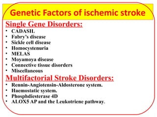
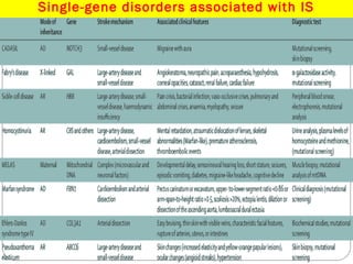
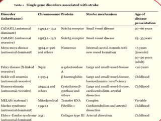
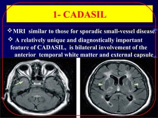
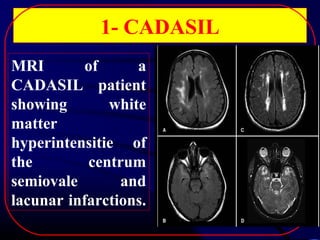
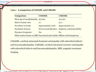
‫بسم ا‬
‫الرحمن الرحيم‬
‫قالوا سبحانك ل علم”‬

‫قالوا سبحانك ل علم”‬
‫لنا إل ما علمتنا‬
‫إنك أنت العليم‬
‫“ الحكيم‬
‫صدق ا العظيم‬
‫سورة البقرة ايه 23‬
By

Dr/ Ayman Al-malt
Assistant Lecturer Of Neurology
Faculty of medicine
. Tanta university
INTRODUCTION

Stroke, or ‘‘brain attack,’’ is defined as a
focal neurological deficit lasting for more
than 24 hours with no cause other than that
of vascular origin.
Ischemic stroke is responsible for about
80%
of
all
strokes,
intracerebral
hemorrhage for 15% and subarachnoid
hemorrhage for 5%.
 A transient ischemic attack (TIA) has the
same complex symptoms as stroke, but with
a resolution of these symptoms within 24
hours .
INTRODUCTION

Since introduction of thrombolytic therapy
in acute ischemic stroke; The American
Heart Association and American Stroke
Association (AHA/ASA) 2009 Guidelines
shift from the time-based definition of TIA to
a tissue-based definition in 2002 (3) with a new
definition for TIA as "a brief episode of
neurological dysfunction caused by focal
brain or retinal ischemia, with clinical
symptoms typically lasting less than one hour
and without evidence of acute infarction"
INTRODUCTION

 Stroke is considered to be one of the
important global health problems as 15
million people worldwide suffer a stroke
annually; of those, 5 million die and another
5 million are left permanently disabled,
placing a burden on family and
community(5).
 Stroke is the third commonest cause of
mortality after cardiac disease, cancer and
the first cause of disability(6).
Risk Factors
Non Modifiable risk
factors:
 Age
 Gender male
 Race (African-American)

Genetics ( mono or

polygenetic)

Modifiable risk factors
 High BP
 Cigarette smoking
 Alcohol intake
 Heart disease
 Atrial fibrillation
 Diabetes
 blood Cholesterol
 Sedentary lifestyle
 Obesity, Stress
Genetic factors and ischemic stroke
•
•

•
•

IS and HS have an important genetic background.
The causation of stroke is multifactorial (a
combination of environmental and genetic risk
factors) and the genetic part is very complex
(polygenic, multiple genes play a role).
Many common risk factors for stroke like DM and
hypertension are partly inherited, so many genetic
loci contribute more or less to the stroke phenotype.
Here, we will review genetic factors that play a role
in IS and HS , with a focus on monogenic forms of
stroke that can serve as a model to study the more
common phenotypes.
Genetic Factors of ischemic stroke
Single Gene Disorders:
•
•
•
•
•
•
•
•

CADASIL
Fabry’s disease
Sickle cell disease
Homocystenuria
MELAS
Moyamoya disease
Connective tissue disorders
Miscellaneous

Multifactorial Stroke Disorders:
•
•
•
•

Rennin-Angiotensin-Aldosterone system.
Haemostatic system.
Phosphdiesterase 4D
ALOX5 AP and the Leukotriene pathway.
Single-gene disorders associated with IS
1- CADASIL
Cerebral autosomal dominant arteriopathy with
subcortical infarcts and leucoencephalopathy.
1- AD small-vessel disease mutations in NOTCH3.
2- The clinical phenotype comprises migraine
recurrent strokes and TIAs, dementia, and psychiatric
disturbance with onset usually in the third to sixth
decade.
3- About a 1/3 of patients develop migraine with aura.
NOTCH3 encodes a cell-surface receptor, which has a
role in arterial development and is expressed on
vascular smooth-muscle cells. Ch19
1- CADASIL
 MRI similar to those for sporadic small-vessel disease.
 A relatively unique and diagnostically important
feature of CADASIL, is bilateral involvement of the
anterior temporal white matter and external capsule .
1- CADASIL
MRI
of
a
CADASIL patient
showing
white
matter
hyperintensitie of
the
centrum
semiovale
and
lacunar infarctions.
2- Fabry’s Disease
FD is an X-linked systemic disorder
 deficiency of lysosomal enzyme α-galactosidase A.
progressive accumulation of

glycosphingolipids,

in the myocardium, renal epithelium, skin, eye, and
vasculature.
Onset is typically in childhood or adolescence
C/P

acroparaesthesia,

angiokeratoma,

hypohidrosis ,hearing loss being common signs.

or
2- Fabry’s Disease
Systemic complications involving the kidneys, heart,
and brain usually follow in mid-adulthood.
Fabry's disease is surprisingly common in young
stroke patients. both large-artery disease and smallvessel disease (posterior circulation).
 ERT with recombinant α-galactosidase.
3- Sickle Cell Disease
SCD is the most common cause of stroke in children
ch11p15.5.
The disease can be caused by the homozygous state for
haemoglobin S (HbS) or by the compound heterozygous
state with haemoglobin C (HbC) or α-thalassaemia.
About 25% of patients with HbS/HbS (highest between 2
years and 5 years) and 10% of those with HbS/HbC will
have a stroke by the age of 45 years.
3- Sickle Cell Disease
Conversely, the risk of HS 1% is highest in 3rd
decade.
Clinically overt strokes 11% are typically due to
large-artery

disease,

characterised

by

intimal

thickening, proliferation of fibroblasts and smoothmuscle cells, and eventually thrombus formation.
 Silent small recurrent infarcts located in subcortical
regions, and attributed to small-vessel disease. ( 22%)
4- Homocystinuria
Homocystinuria encompasses a group of mostly AR
enzyme deficiencies, which cause high (>100μmol/L)
plasma

concentrations

of

homocysteine

and

homocystinuria. Ch 21
deficiency of cystathionine beta-synthase (CBS), a
key enzyme in the degradation of homocysteine.
50% of untreated patients with CBS deficiency have
a thromboembolic event by the age of 30 years
4- Homocystinuria
The disease should be considered in any child with
stroke,

mental

retardation,

a

traumatic

(mostly

downward) dislocation of the ocular lenses, or skeletal
abnormalities.
Homocystinuria must be distinguished from milder
(15–100μmol/L) hyperhomocysteinaemia, which is a
risk factor for stroke in the general population and is
associated with deficient dietary B6, B12, or folate.
4- Homocystinuria
 Homocystinuria can cause stroke (atherosclerosis
and thromboembolism but also through small-vessel
disease and arterial dissection).
Around a half of the patients with CBS deficiency
respond to B6. Those who respond tend to have a later
onset, a milder phenotype, and a better prognosis than
non-responders.
Acquired Causes Of Hyperhomocysteinemia
advanced age, tobacco use, excess coffee intake, low
dietary folate intake, and low vitamin B intake. Higher
homocysteine levels are also associated with diabetes
mellitus,

malignancies,

hypothyroidism,

lupus,

inflammatory bowel disease, and certain medication
use such as metformin, methotrexate, anticonvulsants,
theophylline, and levodopa.
5- MELAS Syndrome
The

syndrome

of

mitochondrial

myopathy,

encephalopathy, lactic acidosis, and stroke-like episodes
is associated with several mutations in mitochondrial
DNA.
MELAS is associated with various symptoms,
however, monosymptomatic cases with stroke as the
sole manifestation exist.
5- MELAS Syndrome
 The cerebral lesions underlying stroke-like episodes

in MELAS differ from typical ischaemic infarcts; the
cortex is almost invariably involved.
In many cases, lesions are not limited to vascular
territories and there are no embolic or stenotic lesions
on angiography. Also, diffusion-weighted MRI may
show an increase in the apparent diffusion coefficient
within acute lesions, suggesting vasogenic rather than
cytotoxic oedema.
5- MELAS SYNDROME
Serial diffusion-weighted MRI
obtained

during

stroke-like

episodes of a patient .
He had recurrent attacks of
throbbing headache, sparkling
phenomena,

alternating

hemiparesis and homonemous
hemianopia
6- Moyamoya disease
Moyamoya disease is a chronic progressive syndrome
that is characterised by bilateral occlusion of the
terminal carotid artery in association with
telangiectatic vessels at the base of the brain.
The disease is uncommon in non-Asian populations
whereas its prevalence in Japan 3-5/ 100 000.
The most frequent manifestations in childhood are
TIS, IS, and epileptic seizures. Rupture of
telangiectatic vessels causes ICH.
 adult type: 30 years (HS).
 Juvenile type 5 years (IS).
6- Moyamoya disease
About 10% of moyamoya cases occur as familial
cases, but the pattern of inheritance is not clear.
 autosomal dominant
Moyamoya disease has been linked to genetic loci on
chromosomes 3p, 8q, and 17q
moyamoya-like changes have been described in
association with a variety of single-gene disorders,
including sickle-cell disease, SLE, pseudoxanthoma
elasticum, and neurofibromatosis type 1.
6- Connective tissue
IS is a well-knownDisorders
complication of several heritable CT
disorders.

Marfan's syndrome

is an AD ch 15 systemic
disorder affecting the musculoskeletal system, CVS, and
the eye. The diagnosis is usually established on clinical
grounds whereas the role of genetic testing is limited.
MF is caused by mutations in a gene (FBN1)
FBN1 encodes fibrillin 1, an extracellular matrix
protein.
MF

7- Connective tissue
Disordersin many tissues,
Fibrillin 1 is expressed

including the heart and elastic arteries.
CV complications of the disease include TIAs,
IS, and subdural haematoma.
neurovascular manifestations were associated
with cardiac sources of embolism, in particular
prosthetic heart valves, mitral valve prolapse,
and AF, whereas there was no association with
aortic disease or cerebral artery dissection.
7- Connective tissue
Disorders
Ehlers-Danlos syndrome type IV, the vascular
type, is an AD disorder resulting from mutations in
COL3A1, the gene for collagen type III.
The disorder may be suspected on the basis of the
associated clinical features and can be confirmed by
mutational screening or biochemical studies on
cultured fibroblasts (synthesis of an abnormal type
III procollagen).
The mutational spectrum is broad and
neomutations are common.
 About 50% of the cases have no apparent FH.
7- Connective tissue
Disorders
complications are common and include

CV
intracranial aneurysms, arterial dissection, and
spontaneous rupture of large and mediumsized arteries.

IS has also been recognised as a complication
of
osteogenesis
imperfecta
and
pseudoxanthoma elasticum, which is associated
with stenotic lesions of the distal carotid artery
and with small-vessel disease.
8- Miscellaneous
IS can occur as a complication of several
heritable cardiomyopathies and dysrhythmias,
haemoglobinopathies, coagulopathies, and
dyslipidaemias, and vasculopathies (herdiatry
endotheliopathy
with
retinopathy
,
nephropathy and stroke).
Common Multifactorial Stroke And
Genetic Risk Factors For IS
The genetic contribution to common multifactorial
stroke seems to be polygenic.
Most likely, there are many alleles with small effect
sizes.
It is increasingly recognised that the effects of some
alleles are limited to one or few stroke subtypes and
that effect sizes may vary depending on sex and ethnic
origin.
Most previous studies investigating genetic risk
factors for human stroke have taken a candidate gene
approach using case–control methodologies.
Common Multifactorial Stroke And Genetic
Risk Factors For Ischaemic Stroke
 difficulties: sample characteristics small number and
study design, could explain much of the inconsistency
between studies.
Most common
1- Renin-angiotensin-aldosterone system
2- Inflammatory genes
3- Haemostatic system
4- Phosphodiesterase 4D
5- ALOX5AP and the leukotriene pathway
1- Renin-angiotensin-aldosterone
system
RAAS contributes to the risk of ischaemic stroke.
Among the various sequence variations in RAAS, the
insertion/deletion (I/D) polymorphism ACE is the most
extensively studied.
ACE produces angiotensin II and catabolises
bradykinin thereby affecting vascular tone, endothelial
function, and smooth-muscle-cell proliferation.
RAAS has a well-documented effect on systemic blood
pressure.
Thus, the I/D polymorphism has become a strong
candidate for cardiovascular risk.
2- Inflammatory genes
Among the most widely investigated genes are
those involved in inflammation (eg, interleukin
1, interleukin 6, TNFα, toll-like receptor 4, Pselectin and E-selectin, C-reactive protein), lipid
metabolism (eg, apolipoprotein E, paraoxonase,
epoxide hydrolase), nitric oxide release, and
extracellular

matrix

(matrix
3- Haemostatic system
Prothrombotic states, such as APC resistance and the
underlying Factor V Leiden polymorphism , are an
established risk factor for VTE but their role in IS is
still debated.
 factor V Leiden polymorphism, the prothrombin
G20210A polymorphism, and the PAI1 polymorphism
all confer a small but significant risk for IS.
Inherited Causes of Thrombosis
1- Increased levels of natural procoagulants
Factor V Leiden mutation (APC resistance)
Prothrombin 20210 mutation
FVIII, FIX, FXI, FVII, VWF
2- Decreased levels natural anticoagulants
Antithrombin (AD Ch1)
Protein C (AD Ch1)
Protein S (ADch3)
Tissue Factor Pathway Inhibitor (TFPI)
Inherited Causes of Thrombosis
3- Abnormalities of Fibrinolysis
Plasminogen deficiency Type I
Plasminogen deficiency Type II
Decreased levels of tissue plasminogen activator
(tPA)
Increased levels of plasminogen activator inhibitor
(PAI-1)
3-Haemostatic system
 prothrombotic states might be responsible for stroke
in some younger patients and in those with additional
risk factors.
However, there is less evidence for a role of
prothrombotic states in unselected patients with
common multifactorial stroke.
4- Phosphodiesterase 4D
 PDE4D gene is associated with IS in the Icelandic
population.
PDE4D ch 5q12.


PDE4D

were

associated

with

the

phenotype of cardiogenic and carotid stroke.

combined
4- Phosphodiesterase 4D
PDE4D degrades second messenger cAMP, which is a
key signal transduction molecule in multiple cell types,
including vascular endothelial, smooth muscle, and
inflammatory cells.
Since associations were limited to cardiogenic and
carotid stroke it was suggested that PDE4D acts
through atherosclerosis.
5- ALOX5AP and the leukotriene pathway
Leukotrienes are proinflammatory mediators that
are implicated in the pathogenesis and progression of
atherosclerosis.
ALOX5AP
  (arachidonate
5-lipoxygenase-activating
protein)

another gene that has been discovered through
genome-wide linkage analysis.
ALOX5AP is associated with a 1·8-fold increased risk
of MI and a 1·7-fold increased risk of IS in the Icelandic
population.
ALOX5AP encodes 5-lipoxygenase activating protein
(FLAP), an important component of the leukotriene
pathway.
Genetic Factors Of Cerebral
Hematoma
1. Hypertension
2. Coagulation
3. Amyloid Angiopathy
4. Herediatry Hgi Telangiectasia
5. Von Hippel Landau
6. Hamartomatous Tumour
7. Cm Avm
1- Hypertension
Not every hypertensive individual develops
hematoma, nor can every case of non-lobar
Hematoma be ascribed to hypertension.
The absence or presence of additional risk
factors probably determines whether the patient
will suffer from an IS, HS, or no stroke at all.
HTN is genetically determined.
1- Hypertension
Many candidate genes for hypertension have
been suggested including genes for renin ,
ACE, aldosterone, phospholipase, kallikrein,
endothelin, and adrenergic receptors.
Genome-wide linkage surveys have found
linkage for blood-pressure loci on almost all
chromosomes.
2- Coagulation :
Disturbances of coagulation – either as a
result of medication, or due to hereditary
disorders of haemostasis – are important
reasons for HS in up to 8% of patients.
Patients with cerebral amyloid angiopathy are
at a higher risk for HS after anticoagulation or
thrombolysis, especially when they are ApoE ε2
carriers.
2- Coagulation :
Hereditary disorders of haemostasis are less common
causes for HS.
HS is the leading cause of death in patients with
fibrinogen deficiencies, and the cause of death in many
patients with haemophilia A (factor VIII deficiency),
and haemophilia B (factor IX deficiency).
 Patients with factor VII, X, V, XI and XIII
deficiencies can also suffer from HS, and HS has been
described in patients with platelet disorders such as
congenital megakaryocyte hypoplasia and the Wiskott–
Aldrich syndrome
3- Amyloid Angiopathy
Amyloid is a term used to describe protein deposits
with circumscript physical characteristics: β-pleated
sheet configuration, apple green birefringence under
polarized light after Congo red staining, fibrillary
structure and high insolubility.
There are many different proteins that can
accumulate as amyloid, and there are many different
disease processes that can lead to amyloid formation.
In CAA, amyloid deposition occurs predominantly in
the cerebral blood vessels, with a preference for small
cerebral arteries and arterioles.
3- Amyloid Angiopathy
CAA can consist of amyloid-β-protein (Abeta-related
angiitis)"., cystatin C, transthyretin, or gelsolin.
Amyloid deposition in cerebral blood vessels can
have several clinical consequences, but can also remain
asymptomatic .
The vessel wall can be weakened, causing rupture
and lobar HS.
CAA can also obliterate the vessel lumen, leading to
ischemia (cerebral infarction, “incomplete” infarction,
and leukoencephalopathy).
Although CAA may contribute to the neurodegeneration of
AD, a direct causal link between the 2 disorders has not been
established
3- Amyloid Angiopathy
CAA is clearly a risk factor for HS, but there are
many patients with amyloid angiopathy, who do not
suffer from HS at all.
HS can occur in patients with cerebral CAA, but
with an enormous variety of clinical presentations (eg.
age at onset, age at death, occurrence of dementia).
In amyloid angiopathy the co-occurrence with other
risk factors such as an ApoE genotype, the presence of
hypertension, or head trauma, determines the presence
or absence of a HS.
Amyloid Angiopathy
AD ch 21 Dutch,
British, Icelandic type.
Associated with cerebral
lobar hge.
MRI of a patient with
hereditary CAA showing
multiple microbleeds and
hemorrhages.
Thank You

Genetics of stroke

  • 1.
    ‫بسم ا‬ ‫الرحمن الرحيم‬ ‫قالواسبحانك ل علم”‬ ‫قالوا سبحانك ل علم”‬ ‫لنا إل ما علمتنا‬ ‫إنك أنت العليم‬ ‫“ الحكيم‬ ‫صدق ا العظيم‬ ‫سورة البقرة ايه 23‬
  • 2.
    By Dr/ Ayman Al-malt AssistantLecturer Of Neurology Faculty of medicine . Tanta university
  • 3.
    INTRODUCTION Stroke, or ‘‘brainattack,’’ is defined as a focal neurological deficit lasting for more than 24 hours with no cause other than that of vascular origin. Ischemic stroke is responsible for about 80% of all strokes, intracerebral hemorrhage for 15% and subarachnoid hemorrhage for 5%.  A transient ischemic attack (TIA) has the same complex symptoms as stroke, but with a resolution of these symptoms within 24 hours .
  • 4.
    INTRODUCTION Since introduction ofthrombolytic therapy in acute ischemic stroke; The American Heart Association and American Stroke Association (AHA/ASA) 2009 Guidelines shift from the time-based definition of TIA to a tissue-based definition in 2002 (3) with a new definition for TIA as "a brief episode of neurological dysfunction caused by focal brain or retinal ischemia, with clinical symptoms typically lasting less than one hour and without evidence of acute infarction"
  • 5.
    INTRODUCTION  Stroke isconsidered to be one of the important global health problems as 15 million people worldwide suffer a stroke annually; of those, 5 million die and another 5 million are left permanently disabled, placing a burden on family and community(5).  Stroke is the third commonest cause of mortality after cardiac disease, cancer and the first cause of disability(6).
  • 6.
    Risk Factors Non Modifiablerisk factors:  Age  Gender male  Race (African-American) Genetics ( mono or polygenetic) Modifiable risk factors  High BP  Cigarette smoking  Alcohol intake  Heart disease  Atrial fibrillation  Diabetes  blood Cholesterol  Sedentary lifestyle  Obesity, Stress
  • 7.
    Genetic factors andischemic stroke • • • • IS and HS have an important genetic background. The causation of stroke is multifactorial (a combination of environmental and genetic risk factors) and the genetic part is very complex (polygenic, multiple genes play a role). Many common risk factors for stroke like DM and hypertension are partly inherited, so many genetic loci contribute more or less to the stroke phenotype. Here, we will review genetic factors that play a role in IS and HS , with a focus on monogenic forms of stroke that can serve as a model to study the more common phenotypes.
  • 8.
    Genetic Factors ofischemic stroke Single Gene Disorders: • • • • • • • • CADASIL Fabry’s disease Sickle cell disease Homocystenuria MELAS Moyamoya disease Connective tissue disorders Miscellaneous Multifactorial Stroke Disorders: • • • • Rennin-Angiotensin-Aldosterone system. Haemostatic system. Phosphdiesterase 4D ALOX5 AP and the Leukotriene pathway.
  • 9.
  • 11.
    1- CADASIL Cerebral autosomaldominant arteriopathy with subcortical infarcts and leucoencephalopathy. 1- AD small-vessel disease mutations in NOTCH3. 2- The clinical phenotype comprises migraine recurrent strokes and TIAs, dementia, and psychiatric disturbance with onset usually in the third to sixth decade. 3- About a 1/3 of patients develop migraine with aura. NOTCH3 encodes a cell-surface receptor, which has a role in arterial development and is expressed on vascular smooth-muscle cells. Ch19
  • 12.
    1- CADASIL  MRIsimilar to those for sporadic small-vessel disease.  A relatively unique and diagnostically important feature of CADASIL, is bilateral involvement of the anterior temporal white matter and external capsule .
  • 13.
    1- CADASIL MRI of a CADASIL patient showing white matter hyperintensitieof the centrum semiovale and lacunar infarctions.
  • 15.
    2- Fabry’s Disease FDis an X-linked systemic disorder  deficiency of lysosomal enzyme α-galactosidase A. progressive accumulation of glycosphingolipids, in the myocardium, renal epithelium, skin, eye, and vasculature. Onset is typically in childhood or adolescence C/P acroparaesthesia, angiokeratoma, hypohidrosis ,hearing loss being common signs. or
  • 16.
    2- Fabry’s Disease Systemiccomplications involving the kidneys, heart, and brain usually follow in mid-adulthood. Fabry's disease is surprisingly common in young stroke patients. both large-artery disease and smallvessel disease (posterior circulation).  ERT with recombinant α-galactosidase.
  • 17.
    3- Sickle CellDisease SCD is the most common cause of stroke in children ch11p15.5. The disease can be caused by the homozygous state for haemoglobin S (HbS) or by the compound heterozygous state with haemoglobin C (HbC) or α-thalassaemia. About 25% of patients with HbS/HbS (highest between 2 years and 5 years) and 10% of those with HbS/HbC will have a stroke by the age of 45 years.
  • 18.
    3- Sickle CellDisease Conversely, the risk of HS 1% is highest in 3rd decade. Clinically overt strokes 11% are typically due to large-artery disease, characterised by intimal thickening, proliferation of fibroblasts and smoothmuscle cells, and eventually thrombus formation.  Silent small recurrent infarcts located in subcortical regions, and attributed to small-vessel disease. ( 22%)
  • 19.
    4- Homocystinuria Homocystinuria encompassesa group of mostly AR enzyme deficiencies, which cause high (>100μmol/L) plasma concentrations of homocysteine and homocystinuria. Ch 21 deficiency of cystathionine beta-synthase (CBS), a key enzyme in the degradation of homocysteine. 50% of untreated patients with CBS deficiency have a thromboembolic event by the age of 30 years
  • 20.
    4- Homocystinuria The diseaseshould be considered in any child with stroke, mental retardation, a traumatic (mostly downward) dislocation of the ocular lenses, or skeletal abnormalities. Homocystinuria must be distinguished from milder (15–100μmol/L) hyperhomocysteinaemia, which is a risk factor for stroke in the general population and is associated with deficient dietary B6, B12, or folate.
  • 21.
    4- Homocystinuria  Homocystinuriacan cause stroke (atherosclerosis and thromboembolism but also through small-vessel disease and arterial dissection). Around a half of the patients with CBS deficiency respond to B6. Those who respond tend to have a later onset, a milder phenotype, and a better prognosis than non-responders.
  • 22.
    Acquired Causes OfHyperhomocysteinemia advanced age, tobacco use, excess coffee intake, low dietary folate intake, and low vitamin B intake. Higher homocysteine levels are also associated with diabetes mellitus, malignancies, hypothyroidism, lupus, inflammatory bowel disease, and certain medication use such as metformin, methotrexate, anticonvulsants, theophylline, and levodopa.
  • 23.
    5- MELAS Syndrome The syndrome of mitochondrial myopathy, encephalopathy,lactic acidosis, and stroke-like episodes is associated with several mutations in mitochondrial DNA. MELAS is associated with various symptoms, however, monosymptomatic cases with stroke as the sole manifestation exist.
  • 24.
    5- MELAS Syndrome The cerebral lesions underlying stroke-like episodes in MELAS differ from typical ischaemic infarcts; the cortex is almost invariably involved. In many cases, lesions are not limited to vascular territories and there are no embolic or stenotic lesions on angiography. Also, diffusion-weighted MRI may show an increase in the apparent diffusion coefficient within acute lesions, suggesting vasogenic rather than cytotoxic oedema.
  • 25.
    5- MELAS SYNDROME Serialdiffusion-weighted MRI obtained during stroke-like episodes of a patient . He had recurrent attacks of throbbing headache, sparkling phenomena, alternating hemiparesis and homonemous hemianopia
  • 26.
    6- Moyamoya disease Moyamoyadisease is a chronic progressive syndrome that is characterised by bilateral occlusion of the terminal carotid artery in association with telangiectatic vessels at the base of the brain. The disease is uncommon in non-Asian populations whereas its prevalence in Japan 3-5/ 100 000. The most frequent manifestations in childhood are TIS, IS, and epileptic seizures. Rupture of telangiectatic vessels causes ICH.  adult type: 30 years (HS).  Juvenile type 5 years (IS).
  • 27.
    6- Moyamoya disease About10% of moyamoya cases occur as familial cases, but the pattern of inheritance is not clear.  autosomal dominant Moyamoya disease has been linked to genetic loci on chromosomes 3p, 8q, and 17q moyamoya-like changes have been described in association with a variety of single-gene disorders, including sickle-cell disease, SLE, pseudoxanthoma elasticum, and neurofibromatosis type 1.
  • 28.
    6- Connective tissue ISis a well-knownDisorders complication of several heritable CT disorders. Marfan's syndrome is an AD ch 15 systemic disorder affecting the musculoskeletal system, CVS, and the eye. The diagnosis is usually established on clinical grounds whereas the role of genetic testing is limited. MF is caused by mutations in a gene (FBN1) FBN1 encodes fibrillin 1, an extracellular matrix protein.
  • 29.
    MF 7- Connective tissue Disordersinmany tissues, Fibrillin 1 is expressed including the heart and elastic arteries. CV complications of the disease include TIAs, IS, and subdural haematoma. neurovascular manifestations were associated with cardiac sources of embolism, in particular prosthetic heart valves, mitral valve prolapse, and AF, whereas there was no association with aortic disease or cerebral artery dissection.
  • 30.
    7- Connective tissue Disorders Ehlers-Danlossyndrome type IV, the vascular type, is an AD disorder resulting from mutations in COL3A1, the gene for collagen type III. The disorder may be suspected on the basis of the associated clinical features and can be confirmed by mutational screening or biochemical studies on cultured fibroblasts (synthesis of an abnormal type III procollagen). The mutational spectrum is broad and neomutations are common.  About 50% of the cases have no apparent FH.
  • 31.
    7- Connective tissue Disorders complicationsare common and include CV intracranial aneurysms, arterial dissection, and spontaneous rupture of large and mediumsized arteries. IS has also been recognised as a complication of osteogenesis imperfecta and pseudoxanthoma elasticum, which is associated with stenotic lesions of the distal carotid artery and with small-vessel disease.
  • 32.
    8- Miscellaneous IS canoccur as a complication of several heritable cardiomyopathies and dysrhythmias, haemoglobinopathies, coagulopathies, and dyslipidaemias, and vasculopathies (herdiatry endotheliopathy with retinopathy , nephropathy and stroke).
  • 33.
    Common Multifactorial StrokeAnd Genetic Risk Factors For IS The genetic contribution to common multifactorial stroke seems to be polygenic. Most likely, there are many alleles with small effect sizes. It is increasingly recognised that the effects of some alleles are limited to one or few stroke subtypes and that effect sizes may vary depending on sex and ethnic origin. Most previous studies investigating genetic risk factors for human stroke have taken a candidate gene approach using case–control methodologies.
  • 34.
    Common Multifactorial StrokeAnd Genetic Risk Factors For Ischaemic Stroke  difficulties: sample characteristics small number and study design, could explain much of the inconsistency between studies. Most common 1- Renin-angiotensin-aldosterone system 2- Inflammatory genes 3- Haemostatic system 4- Phosphodiesterase 4D 5- ALOX5AP and the leukotriene pathway
  • 35.
    1- Renin-angiotensin-aldosterone system RAAS contributesto the risk of ischaemic stroke. Among the various sequence variations in RAAS, the insertion/deletion (I/D) polymorphism ACE is the most extensively studied. ACE produces angiotensin II and catabolises bradykinin thereby affecting vascular tone, endothelial function, and smooth-muscle-cell proliferation. RAAS has a well-documented effect on systemic blood pressure. Thus, the I/D polymorphism has become a strong candidate for cardiovascular risk.
  • 36.
    2- Inflammatory genes Amongthe most widely investigated genes are those involved in inflammation (eg, interleukin 1, interleukin 6, TNFα, toll-like receptor 4, Pselectin and E-selectin, C-reactive protein), lipid metabolism (eg, apolipoprotein E, paraoxonase, epoxide hydrolase), nitric oxide release, and extracellular matrix (matrix
  • 37.
    3- Haemostatic system Prothromboticstates, such as APC resistance and the underlying Factor V Leiden polymorphism , are an established risk factor for VTE but their role in IS is still debated.  factor V Leiden polymorphism, the prothrombin G20210A polymorphism, and the PAI1 polymorphism all confer a small but significant risk for IS.
  • 38.
    Inherited Causes ofThrombosis 1- Increased levels of natural procoagulants Factor V Leiden mutation (APC resistance) Prothrombin 20210 mutation FVIII, FIX, FXI, FVII, VWF 2- Decreased levels natural anticoagulants Antithrombin (AD Ch1) Protein C (AD Ch1) Protein S (ADch3) Tissue Factor Pathway Inhibitor (TFPI)
  • 39.
    Inherited Causes ofThrombosis 3- Abnormalities of Fibrinolysis Plasminogen deficiency Type I Plasminogen deficiency Type II Decreased levels of tissue plasminogen activator (tPA) Increased levels of plasminogen activator inhibitor (PAI-1)
  • 40.
    3-Haemostatic system  prothromboticstates might be responsible for stroke in some younger patients and in those with additional risk factors. However, there is less evidence for a role of prothrombotic states in unselected patients with common multifactorial stroke.
  • 41.
    4- Phosphodiesterase 4D PDE4D gene is associated with IS in the Icelandic population. PDE4D ch 5q12.  PDE4D were associated with the phenotype of cardiogenic and carotid stroke. combined
  • 42.
    4- Phosphodiesterase 4D PDE4Ddegrades second messenger cAMP, which is a key signal transduction molecule in multiple cell types, including vascular endothelial, smooth muscle, and inflammatory cells. Since associations were limited to cardiogenic and carotid stroke it was suggested that PDE4D acts through atherosclerosis.
  • 43.
    5- ALOX5AP andthe leukotriene pathway Leukotrienes are proinflammatory mediators that are implicated in the pathogenesis and progression of atherosclerosis. ALOX5AP   (arachidonate 5-lipoxygenase-activating protein) another gene that has been discovered through genome-wide linkage analysis. ALOX5AP is associated with a 1·8-fold increased risk of MI and a 1·7-fold increased risk of IS in the Icelandic population. ALOX5AP encodes 5-lipoxygenase activating protein (FLAP), an important component of the leukotriene pathway.
  • 44.
    Genetic Factors OfCerebral Hematoma 1. Hypertension 2. Coagulation 3. Amyloid Angiopathy 4. Herediatry Hgi Telangiectasia 5. Von Hippel Landau 6. Hamartomatous Tumour 7. Cm Avm
  • 45.
    1- Hypertension Not everyhypertensive individual develops hematoma, nor can every case of non-lobar Hematoma be ascribed to hypertension. The absence or presence of additional risk factors probably determines whether the patient will suffer from an IS, HS, or no stroke at all. HTN is genetically determined.
  • 46.
    1- Hypertension Many candidategenes for hypertension have been suggested including genes for renin , ACE, aldosterone, phospholipase, kallikrein, endothelin, and adrenergic receptors. Genome-wide linkage surveys have found linkage for blood-pressure loci on almost all chromosomes.
  • 47.
    2- Coagulation : Disturbancesof coagulation – either as a result of medication, or due to hereditary disorders of haemostasis – are important reasons for HS in up to 8% of patients. Patients with cerebral amyloid angiopathy are at a higher risk for HS after anticoagulation or thrombolysis, especially when they are ApoE ε2 carriers.
  • 48.
    2- Coagulation : Hereditarydisorders of haemostasis are less common causes for HS. HS is the leading cause of death in patients with fibrinogen deficiencies, and the cause of death in many patients with haemophilia A (factor VIII deficiency), and haemophilia B (factor IX deficiency).  Patients with factor VII, X, V, XI and XIII deficiencies can also suffer from HS, and HS has been described in patients with platelet disorders such as congenital megakaryocyte hypoplasia and the Wiskott– Aldrich syndrome
  • 49.
    3- Amyloid Angiopathy Amyloidis a term used to describe protein deposits with circumscript physical characteristics: β-pleated sheet configuration, apple green birefringence under polarized light after Congo red staining, fibrillary structure and high insolubility. There are many different proteins that can accumulate as amyloid, and there are many different disease processes that can lead to amyloid formation. In CAA, amyloid deposition occurs predominantly in the cerebral blood vessels, with a preference for small cerebral arteries and arterioles.
  • 50.
    3- Amyloid Angiopathy CAAcan consist of amyloid-β-protein (Abeta-related angiitis)"., cystatin C, transthyretin, or gelsolin. Amyloid deposition in cerebral blood vessels can have several clinical consequences, but can also remain asymptomatic . The vessel wall can be weakened, causing rupture and lobar HS. CAA can also obliterate the vessel lumen, leading to ischemia (cerebral infarction, “incomplete” infarction, and leukoencephalopathy). Although CAA may contribute to the neurodegeneration of AD, a direct causal link between the 2 disorders has not been established
  • 51.
    3- Amyloid Angiopathy CAAis clearly a risk factor for HS, but there are many patients with amyloid angiopathy, who do not suffer from HS at all. HS can occur in patients with cerebral CAA, but with an enormous variety of clinical presentations (eg. age at onset, age at death, occurrence of dementia). In amyloid angiopathy the co-occurrence with other risk factors such as an ApoE genotype, the presence of hypertension, or head trauma, determines the presence or absence of a HS.
  • 52.
    Amyloid Angiopathy AD ch21 Dutch, British, Icelandic type. Associated with cerebral lobar hge. MRI of a patient with hereditary CAA showing multiple microbleeds and hemorrhages.
  • 53.