1. Dietary Balances; Regulation of Feeding; Obesity and Starvation,
Vitamins and Minerals, Body Temperature Regulation and Fever.
Energy Intake and Output
Used or stored for later use (fat)
Appropriate balanced intake (proteins, carbohydrates, fats, minerals, and vitamins)
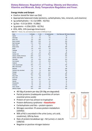
1g carbohydrates – 4.1 Cal (98% - 4(17kJ)
1g fats – 9.3 Cal (95% - 9 (38kJ)
1g proteins – 4.35kJ (92% - 4(17kJ)
45%, 40%, 15% (average Americans)
40-50g of protein per day (20-30g are degraded)
Partial proteins (inadequate quantities of certain
essential amino acids)
Protein of corn has almost no tryptophan
Protein-deficiency syndrome – Kwashiorkor
Carbohydrates and fats – protein sparers
Nitrogen excretion assess protein metabolism
(16%N2)
90% of N2 is excreted in the urine (urea, uric acid,
creatinine), 10% by feces
Rate of protein breakdown (g) – N2 (urine) x 1.1x6.25
(100/16)
Negative or positive nitrogen balance
2. “respiratory quotient” – ration of C02 production to 02 utilization (1h and more).
Fat utilization (0.7), carbohydrates (1.0), proteins (0.8)
Excess hydrogen atoms
Right after meal close to 1.0: 8-10 hr after meal about 0.7; in diabetes melitus
always about 0.7.
Regulation of Food Intake and Energy Storage
Only 27% of the energy ingested normally reaches the functional systems of the
cells
Food intake, energy expenditure and fat storage – environmental, cultural and
genetic factors + physiological controls
“epidemics” of obesity (64% & 33%)
2000 Cal daily expenditure of energy (6000-7000 Cal).
Neural Centers regulate Food intake
Sensation of hunger (rhythmical contractions of stomach and restlessness)
Appetite – desire for particular type of food
Feeling of satiety
Lateral nuclei of the hypothalamus – feeding centre (hyperphagia, inanitation)
Operates by exciting the motor drives to search for food.
Ventromedial nuclei of the hypothalamus – satiety center (aphagia, hyperphagia)
Other centers also play a major role (arcuate!), hormonal secretion (thyroid and
adrenal glands, pancreatic islet cells)
Integration of neural signals from the gastrointestinal tract (Stomach filling),
chemical signals from nutrients in the blood, signals from gastrointestinal
hormones, hormones released by adipose tissue and signals from the cerebral
cortex (sight, smell and taste)
Feeding behaviour
Orexigenic and anorexigenic substances and receptors – therapeutic sites
Feedback mechanisms for control of food
intake.
Stretch receptors in the stomach activate
sensory afferent pathways in the vagus
nerve and inhibit food intake.
Peptide YY (PYY), cholecystokinin (CCK),
and insulin are gastrointestinal hormones
that are released by the ingestion of food
and suppress further feeding.
Ghrelin is released by the stomach,
especially during fasting, and stimulates
appetite.
Leptin is a hormone produced in increasing
amounts by fat cells as they increase in
size. It inhibits food intake.
3.
4. Neurons and NTs in Hypothalamus – that stimulate or inhibit feeding
1) Pro-opiomelanocortin (POMC) neurons
a. α- MSH ( α-melanocyte-stimulating hormone)
b. CART (cocaine and amphetamine related transcript)
2) Neurons that produce orexigenic substances
a. NPY (neuropeptide Y)
b. AGRP (agouti-related protein)
Activation of POMC neurons decreases food intake and increases energy
expenditure
Activation of NPY-AGRP neurons increases food intake and reduces energy
expenditure
Major targets for:
- Leptin
- Insulin
- CCK
- Ghrelin
POMC neurons release α- MSH (acts on melanocortin receptors found especially
in neurons of the paraventricular nuclei)
Atleast 5 subtypes of melanocortin receptors
MCR-3 and MCR-4 are especially important in regulating food intake and energy
balance
Activation of these receptors reduces food intake while increasing energy
expenditure
Inhibition has an opposite effect.
MCR activation is mediated by activation of nucleus tractus solitarius
(sympathetics).
Defective signalling of the melanocortin pathway is associated with extreme
obesity
Mutations of MCR-4 – most common known monogenic (single-gene) cause of
human obesity (5-6% of early-onset severe obesity in children)
AGRP – natural antagonist of MCR-3 and MCR-4 receptors.
Role of AGRP in normal physiological control of food intake is unclear
Excessive formation of AGRP in mice and humans, due to gene mutations, is
associated with increased food intake and obesity.
NPY (arcuate nuclei) – when energy stores of the body are low – stimulates
appetite and firing of the POMC neurons is reduced – decreased activity of the
melanocortin pathway and further stimulated appetite.
5. Factors that regulate Quantity of Food intake
Short-term regulation – preventing over-eating at each meal
Long-term regulation – maintenance of normal quantities of energy
stores in the body
Short-term regulation
What turns off the eating?
1. Distending of gastrointestinal tract (stomach and the duodenum –
vagus nerve)
2. Humoral and hormonal factors
a. CCK – fat
b. Peptide YY from the ileum and colon – fat
c. Glucagon-like peptide (GLP) from intestines – enhances
glucose-dependent insulin production and secretion from the
pancreas – suppress appetite.
3. Ghrelin – oxyntic cells of the stomach and intestine,
concentrations rise during fasting, fall rapidly after a meal;
administration of ghrelin increases food intake in experimental
animals.
4. Oral receptors (experiment with oesophageal fistula; chewing,
salivation, swallowing, and tasting – shorter duration (20-40 min).
Intermediate and Long-Term Regulation
Depends on nutritional status of the body
Glucostatic, aminostatic and lipostatic theories of regulation
Glucoreceptor (increased GUK increases the rate of firing) and
glucosensitive (increased GUK decreases the firing) neurons in the
hypothalamus.
Temperature Regulation and Food Intake
Exposition to cold – increased feeding
Interaction within the hypothalmus
1. Increases metabolic rate
2. Provides increased fat for insulation
Feedback from Adipose Tissue
Hypothalamus senses energy storage through the actions of Leptin, a
peptide hormone released from adipocytes
POMC neurons of the arcuate nuclei and neurons of the paraventricular
nuclei
1. Decreased appetite stimulators (NPY I AGRP)
2. Activation of POMC neurons (α- MSH)
3. Increased substances that decrease appetite (CRH).
4. Increased sympathetic nerve activity
5. Decreased insulin secretion by the pancreatic β cells
6. Mice or humans with mutations that render their fat cells unable to
produce leptin or mutations that cause defective leptin receptors in the
hypothalamus – marked by hyperphagia and morbid obesity.
In most obese humans no deficiency of leptin production
Many other mechanisms, questionable summary
Obesity – excess of body fat
BMI = mass (kg)/height2
(m2
)
25-30 overweight, more than 30 – obesity
Measurement of total body fat (skin-fold thickness, bioelectrical
impedance, or underwater weighing; 25% & 35%)
Obesity results from greater energy intake than energy expenditure.
For each 9.3 Cal (38.9 kJ) of excess energy – 1 gram of fat is stored
1/3 energy used each day by the average person goes into muscular
activity (2/3)
Increase in physical activity
Psychological factors
Three meals a day and that each meal must be filling
During or after stressful situations (Death of a parent, a severe illness, or
even mental depression)
Eating can be a means of releasing tension.
7. Childhood over-nutrition
Rate of formation of new fat cells
Number of fat cells in obese children is often as three times that in normal
children
Hyperplastic and hypertrophic obesity
New adipocytes can differentiate from fibroblast-like preadipocytes at any period
of life.
Neurogenic Abnormalities
Lesions in the ventromedial nuclei of the hypothalamus – tumours
Functional organization of the hypothalamic or other neurogenic feeding
centers in obese individuals may be different
Abnormalities of neurotransmitters or receptor mechanisms
Genetic Factors
Obesity definitely runs in families
Identical twins mass is usually within 1.5, or 2.5kg
20-25% of cases of obesity may be caused by genetic factors
1. Mutations of MCR-4
2. Congenital leptin deficiency
3. Mutations of the leptin receptor
Treatment of Obesity
Reducing energy intake or/and increasing energy expenditure
Large quantities of “bulk” (non-nutritive cellulose substances, distention).
Prevent vitamin deficiencies
Amphetamines, sibutramine – dangerous overexcite sympathetic nervous system
and raise pressure, addiction
Altering lipid metabolism
Orilistat (a lipase inhibitor) – reduces the intestinal digestion of fat
Loss of fat – soluble vitamins in the fees
Increase in physical activity
Various surgical procedures (gastric bypass surgery and gastric banding surgery)
Inanition
Lack of food or
Psychological and hypothalamic disorders
Anorexia nervosa – reduction in food intake caused primarily by diminished
appetite, nauseated by food
Cachexia – weight loss greater than that caused by reduced food intake alone
(tumors, AIDS).
8. Starvation
Tissues preferentially use carbohydrates for energy
Protein depletion: rapid depletion at first, then greatly slowed depletion, and
finally rapid depletion again shortly before death
Gluconeogenesis decreases to 1/5
State of ketosis (β – hydroxybutyrate – brain)
9.
10.
11.
12.
13. Body Temperature Regulation and Fever
Normal Body Temperature
“Core” temperature = 0.6 °C (1.1 °F).
(nude person exposed to air temperatures 10-55°C)
Skin temperature rises and falls with the temperature of the
surroundings (ability to lose heat to the surroundings)
Normal Core Temperature
Range of normal temperature (36-37.5°C)
Average normal core temperature – 36.5-37°C (measured orally; rectally
0.5°C higher).
Regulatory mechanisms are not perfect: temperature increases during
exercise and varies with temperature extremes of the surroundings
Balance between heat production and heat loss
Heat production
Heat – principal by-product of metabolism
Metabolic rate of the body:
1. Basal rate of metabolism
2. Muscle activity
3. Effect of thyroxine, (hGH, testosterone)
4. Effect of sympathetic stimulation
5. Increased chemical activity in the cells
6. Thermogenic effect of food
14. Heat Loss
Heat is generated in deep organs
- Liver
- Brain
- Heart
- Skeletal muscles
Heat is lost to the air via skin
Rate at which heat is lost:
1. How rapidly heat can be conducted from where it is produced to the
skin
2. How rapidly heat can then be transferred from the skin to the
surroundings
Insulator system of the body
Skin subcutaneous tissues (fat) – insulator
Conduction of heat through fat = 1/3 conduction through other tissues
Insulator properties of female body are better than male body
Blood flow to the skin from the Body core
Enables heat to be conducted from the core of the body to the skin
Especially important is a continuous venous plexus
Rate of blood flow into the skin venous plexus can vary tremendously (0-
30% CO)
Skin is an effective controlled “heat radiator” system
Flow of blood to the skin is a most effective mechanism for heat transfer
from the body core to the skin
Vasoconstriction of the arterioles and the arteriovenous anastomoses
that supply blood to the venous plexus of the skin is controlled almost
entirely by the sympathetic nervous system.
15. Basic Physics of how heat is lost from the skin surface:
Radiation (about 60%, infrared heat rays, a type of electromagnetic
wave (5-20um), in all directions)
Conduction (about 3% direct conduction from to solid objects, about
15% to air – convection (currents), suspension in water!)
Evaporation (evaporation of 1g water – 0.58 Cal (2.5kJ) heat, insensibly
and evaporation of sweat, necessary cooling mechanism at very high air
temperatures.
Effect of clothing
Increasing the thickness of the so-called private zone of air+decreasing
air currents
Rate of heat loss from the body by conduction and convection (to ½, or
1/6 arctic-type clothing)
Coating the inside of clothing with a thin layer of gold – reflects radiant
heat back
Extreme caution against allowing the clothing to become wet
Sweating
Starts by stimulation of the anterior hypothalamus-preoptic area in the
brain by electricity or by excess heat
Nerve impulses are transmitted in the autonomic pathways to the spinal
cord and then through sympathetic outflow to the skin everywhere in
the body.
Sweat glands are innervated by cholinergic nerve fibers (but that run in
the sympathetic nerves along with the adrenergic fibers)
They can also be stimulated by epinephrine or norepinephrine
circulating in the blood.
16. Mechanism of sweat secretion
1. Deep subdermal coiled portion – secretes the sweat (primary or
precursor secretion)
2. Duct portion (modify concentrations of constituents)
Primary secretion
Active secretory product of the epithelial cells
Composition is similar to that of plasma (Na+ = 142mmol/L, a CL- =
104mmol/L), does not contain plasma proteins
Reabsorption of ions
Slight stimulation – most of Na+ and Cl- are reabsorbed (concentration
of each falls to as low as 5 mmol/L)
This reduces the osmotic pressure of the sweat fluid to such a low level
that most of the water is also reabsorbed, which concentrates most of
the other consituents (urea, K+, lactic acid)
Strong stimulation – Na+ and Cl- are reabsorbed to concentrations of 50-
60 mmol/L, little of the water is reabsorbed – significant loss of NaCl
17. Acclimatization, Role of Aldosterone
Normal unacclimatized person – 1L/h sweat
After 1-6 weeks – 2-3L/h sweat
Removing 10x more heat from the body
Change in the internal sweat gland cells to increase their sweating
capability
Better conservation of body salt – increased secretion of aldosterone
(decreases loses from 15-30 g/day to 3-5 g/day)
Loss of Heat by panting
Substitute mechanism due to:
1. Surfaces often covered with fur
2. Skin of most lower animals is not supplied with sweat glands
Panting center is associated with pneumotaxic respiratory center in the
pons
Evaporation of saliva from the tongue, w/o increase in alveolar
ventilation
Role of the hypothalamus
Experiments with use of a thermode
Principal areas in the brain for temperature control are the preoptic and
anterior hypothalamic nuclei of the hypothalamus
Large numbers of heat-sensitive neurons
About 1/3rd
as many cold-sensitive neurons
Heating of preoptic area – profuse sweating and vasodilation in the skin
18. Detection of temperature
Temperature receptors in skin and in a few specific deep tissues (spinal
cord, abdominal viscera, around the great veins)
In the skin: cold receptors (far more) and warmth receptors
In deep tissues: function differently from the skin receptors because
they are exposed to the body core temperature, they detect mainly cold.
Integration of the central and Peripheral Temperature Signals
Area of the hypothalamus that is located bilaterally in the posterior
hypothalamus approximately at the level of the mammilarry bodies
combination and integration of signals from the preoptic area and from
elsewhere in the body
Temperature-Decreasing Mechanisms
vasodilation
in the skin (inhibition of the sympathetic centers in the posterior
sweating
evaporation
decrease in heat production
inhibition of shivering and chemical thermogenesis
Temperature-Increasing Mechanisms
vasoconstriction
in the skin (stimulation of the posterior hypothalamic sympathetic centers)
piloerection
hairs "standing on end", not important in humans, thick layer of "insulator air"
increase in thermogenesis
promoting shivering, sympathetic excitation of heat production, and thyroxine
secretion
Hypothalamic Stimulation of Shivering
primary motor center for shivering located in the dorsomedial portion of the
posterior hypothalamus near wall of the 3rd ventricle
normally inhibited by signals from the heat center in anterior preoptic area
cold signals from the skin and spinal cord
body heat production can rise 4-5x normal
Transmits signals to anterior motor neurons
signals are nonrhythmical and do not cause the actual muscle shaking
they increase the tone of the skeletal muscles throughout the body
when the tone rises above a certain critical level, shivering begins
results from feedback oscillation of the muscle spindle stretch reflex mechanism
19. Sympathetic "Chemical" Excitation
ability of norepinephrine and epinephrine to uncouple oxidative
phosphorylation
foodstuffs are oxidized but do not cause ATP to be formed – release of heat
directly proportional to the amount of brown fat (acclimatization)
adults do not have brown fat (increased rate of heat production 10-15%, in
infants100%)
Increased Thyroxine Output
cooling preoptic area – increases production of TRH increased TSH
increased tiroksina activates uncoupling protein
yet another mechanism of chemical thermogenesis
requires several weeks' exposure to cold
humans seldom allow themselves to be exposed to the same degree of
cold
Concept of a "Set-Point"
critical body core temperature37,1 °C
called the "set-point" of the temperature control mechanism
feedback gain of the temperature control system = (ratio of the change
in environmental temperature to the change in body core temperature)
– 1
changes about 1°C for each 25° to 30°C change in environmental temperature
(~27)
extremely high gain (baroreceptor feedback gain< 2)
Skin Temperature Can Slightly Alter the Set-Point
decrease in skin temperature – increase in set-point for sweating
decrease in skin temperature– increase in set-pointfor shivering
Behavioral Control
even more potent
person makes appropriate environmental adjustments to re-establish comfort
there are local skin temperature reflexes
after cutting the spinal cord in the neck above the sympathetic outflow from the
cord regulation becomes extremely poor
20. Fever
body temperature above the usual range of normal
1. abnormalities in the brain itself
2. toxic substances that affect the temperature-regulating centers
Resetting the Hypothalamic Temperature-Regulating Center
many proteins, breakdown products of proteins, lipopolysaccharide toxins
released from bacterial cell membranes – pyrogens
some pyrogens act directly and immediately
other pyrogens function indirectly and may require several hours of latency
(endotoxins from gram-negative bacteria)
phagocytizion of bacteria – release of interleukin-1 (IL1, leukocyte or endogenous
pyrogen)
IL1 in 8-10 min significantly increases temperature (in nanograms)
IL1 inducing formation of prostaglandin E2
drugs that impedes the formation of prostaglandins from arachidonic acid –
antipyretics (aspirin)
Fever Caused by Brain Lesions
• almost always after surgery in the region of the hypothalamus
• compression of the hypothalamus by a brain tumor
Characteristics of Febrile Conditions
• chills – extremely cold feeling, vasoconstriction in the skin, shivers
• crisis or “flush”– after factor is removed, intense sweating and the hot skin
• heatstrokebody temperature rises beyond a critical temperature – 40-42 °C (105° to
108°F, dizziness, abdominal distress, vomiting, delirium, loss of consciousness)
local hemorrhages and parenchymatous degeneration of cells
• especially in the brain, but also liver and kidneys
• acclimatization
Exposure of the Body to Extreme Cold
person exposed to ice water for 20 to 30 minutes ordinarily dies because of heart
standstill or heart fibrillation
once the body temperature has fallen below about 85°F (30 °C), the ability of the
hypothalamus to regulate temperature is lost
sleepiness, coma– depresses the activity of the central nervous system
frostbites (lobes of the ears and in the digits of the hands and feet) – formation of
ice crystals– permanent damage– gangrene
artificial hypothermia (heart surgery)