SlideShare a Scribd company logo
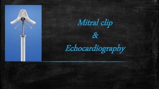
Mitral clip
&
Echocardiography
Denton Arthur Cooley
(August 22, 1920 – November 18, 2016)
Introduction
▪ Only technique available to alter the mitral valve (MV) morphology,
annulus diameter, and reduce MR.
▪ Alfieri in 1991, surgically sutured the free edges of the mid-portions of
the anterior (A2) and posterior (P2) MV scallops.
▪ Maisano et al. demonstrated that with the Alfieri technique 90 % of
patients with both functional and degenerative MR are free from the
combined endpoint of recurrent MR ≥2+ and re-operation after 5
years.
Echo & Mitral Clip
▪ Echocardiography is the essential imaging modality for
MitraClip treatment.
(i) Patient selection
(ii) Guidance of the procedure
(iii) Identification as well as the assessment of complications
(iv) The evaluation of the final result after clip implantation
(v) The assessment at followup of MR severity, left
ventricular (LV) chamber size and function as well as
pulmonary artery pressures.
Patient selection Grading of MR severity
▪ MR needs to be moderate to severe (3+) or severe ( 4+).
▪ Chronic severe MR is generally associated with left atrial (LA) and LV
enlargement.
▪ Severe MR as timed by CW Doppler is generally holosystolic.
▪ ‘E’-wave in the mitral inflow is taller than the ‘A’-wave
▪ An ‘E’-wave taller than 1.2–1.5 m/s usually indicates severe
MITRAL VALVE ANATOMY ON TEE
Determination Of The Morphology
▪ TTE vsTEE
▪ Morphology
▪ Eccentric jet
▪ All pulmonary veins
▪ Detailed anatomy of mitral apparatus
▪ Description of scallops and commissures
10
Anatomic Measurements
▪ Degenerative Mitral Regurgitation (DMR)
–Flail Gap
–FlailWidth
▪ Functional Mitral Regurgitation (FMR)
–Coaptation Length
–Coaptation Depth
Posterior Leaflet Length
Ideal Valves for Mitral clip
▪ Central origination of MR (A2/P2 segments)
▪ No severe calcification in the grasping area
▪ PML should be long enough to allow secure capturing by the Clip.
▪ No excessive flail width and an excessive flail gap
▪ Some residual coaptation length should exist in functional MR
Intermediate suitability
it has been reported that in isolated patients with anatomy outside these
criteria, successful treatment with the MitraClip may be achieved.These
include
–Pathology in the P1 or P3 segment
–MVA 4 cm2 and 3 cm2 with good leaflet mobility
–Mobile posterior leaflet length between 7 and 10 mm, leaflet
restriction in systole
Main procedural steps
1.Transseptal puncture
2. Introduction of the Steerable Guide Catheter (SGC) into the left atrium
3. Advancement of the Clip Delivery System (CDS) into the left atrium
4. Steering and positioning of the MitraClip above the mitral valve
5. Advancing the MitraClip into the left ventricle
6. Grasping of the leaflets and assessment of proper leaflet insertion
7. Clip detachment
Using 3D TEE Before procedure
▪ Benefits of Orthogonal views
▪ Detailed MV anatomy and function and all components of the mitral
apparatus
▪ Scallop Identification
▪ Identification of clefts, gaps, and perforations
▪ The mitral annulus visulaization
▪ Delivery catheters, wires, devices, and target structures can be
visualized in one single view with 3DTEE
▪ MitraClip positioning perpendicular to the line of coaptation in the
middle segments of the MV.
▪ Should be combination of 2D and 3D
▪ Biner et al, demonstrated that the usage of 2D and 3DTEE in
combination is associated with a remarkable 28% reduction in
procedure times.
Using 3D TEE during procedure
Transseptal Puncture
▪ Most important aspects of the MitraClip procedure.
▪ Optimal puncture site is located superiorly and posteriorly
in the interatrial septum (IAS )
▪ ThreeTEE planes are used to determine the correct site:
– Short-axis view at the base for anterior–posterior orientation
– Bi-caval view for superior - inferior orientation
– Four-chamber view to direct the height above the MV.
Transseptal puncture
▪ What is tenting ‘tenting’
▪ Height above mitral annulus
– Degenerative 4-5 cm
– Functional 3.5 cm
▪ PFO and ASDs should be avoided
Introduction of the Steerable Guide
Catheter (SGC)
3. Continous monitoring to avoid injury
4. Dilator is retrieved first, then extra stiff wire
1. Wire in Pulmonary vein
2. Identify Dilator, SGC
Advancement of the Clip Delivery System
(CDS) into the left atrium
▪ The CDS Advanced and then dipped down
▪ Slow advancement to avoid injury
▪ 3DTEE and x-plane views are used to visualize the distance of the CDS from the LA wall
Steering and positioning of the MitraClip
above the mitral valve
▪ Medial– lateral Clip adjustments ---intercommissural view
▪ Anterior–posterior adjustments ---long-axis (LVOT) view
▪ Mitral Clip should be perpendicular to the line of coaptation
▪ Opened Clip can be visualized in full length in the longaxis view
▪ No Clip arms should be seen in the intercommissural view
▪ The MitraClip should split the regurgitation jet in both orthogonal views
▪ A single 3D enface-view
X Plane
Advancing the MitraClip into the left
ventricle
▪ Maintain same position as previous
▪ Passage across the MV in open state.
▪ Reassess in LV as might rotate
▪ 2DTEE (intercommissural and LVOT)
and 3DTEE views are used
Grasping Of The Leaflets And Assessment
Of Proper Leaflet Insertion
▪ After suitable position grasping done in LVOT
view (2D)
▪ 3D has no clear benefit in this part of the
procedure
▪ Longer loop during the grasping
▪ Initial partial closure followed by full
▪ Multiple planes are useful
▪ Insertion of the posterior leaflet is commonly
best seen in the LVOT view
▪ Insertion of the anterior leaflet in the four-
chamber view.
Assessment Of Result And Mitraclip Release
▪ Grade of regurgitation
▪ stenosis of the MV
▪ Complications
▪ Effects ofAnesthesia on assessment
▪ Post clip quantitative Doppler evaluation is problematic due to multiple
orifice
▪ Potential overestimation of residualMR in patients with multiple jets.
▪ Artefacts caused by the MitraClip may also alter the findings
▪ Small persistent colour jets, even if multiple, are certainly congruent with
mild MR.
Assessment Of Result And Mitraclip Release
▪ Pulmonary vein flow estimation
▪ The PISA method is not validated for multiple MR jets
▪ Regurgitation volume can be calculated
▪ 3D-acquisition of the LV volumes is preferable for the calculation when
available.
▪ Summation of 2D measured venae contracta is not reliable in the presence of
multiple jets.
▪ Measurement of the vena contracta area in 3D echocardiography shows
potential for the quantification of MR with irregularly shaped vena contracta
areas, although currently no reference data are available.
Assessment Of Result And Mitraclip Release
Additional parameters
In addition to echocardiography, some other parameters may be
helpful for the evaluation of the severity of residual MR:
▪ LA pressure measurements (V-wave). A decrease or even a
normalization of LA pressures may be seen in case of effective MR
treatment.
▪ Stroke–volume measurements via right heart catheterization
▪ LV angiography to assess the amount of regurgitation by judging the
density and the expansion of the MR jet in the LA.
▪ Mean gradient of up to 5 mmHg is considered acceptable.
▪ Planimetry of the two orifices should be performed, ideally with 3DTEE,
alternatively with 2DTEE in the transgastric short-axis view.
▪ In the EVEREST I and II studies, a planimetric MVA of 1.5 cm2 was
considered criteria for clinically significant mitral stenosis.
▪ Once in place, it has been demonstrated that the MitraClip device did not
result in clinically significant MV stenosis during 2 years followup
Additional parameters
Additional MitraClip implantation
▪ Same positioning of clip in LA.
▪ Clip should be Closed during advancement into LV to avoid
entaglement
▪ Fluoroscopy is more helpful thanTEE for the positioning of a
second MitraClip
▪ It should be aligned as parallel as possible to the first Clip.
Post-procedural follow-up
For post-procedural follow-up,TTE is usually sufficient. current guidelines
includes
▪ Colour flow Doppler of the MR
▪ MV inflow gradient
▪ Assessment of pulmonary vein flow
▪ Residual ASD evaluation
▪ Systolic pulmonary artery pressure
▪ LV size and volume measurements in systole and diastole
▪ LVEF
Thanks for your patience
Future Perspectives
Role of 3D echocardiography in the assessment of MR severity may increase in the
future
Major advantage being the ability of this modality to assess vena contracta area
which is most commonly non-circular especially in patients suffering from functional
MR.
Usually, the vena contracta area is quantified in a post-processing step using
planimetry of 3D colour regurgitation jets in orthogonal views.
The 3D derived vena contracta area correlated better with effective regurgitant
orifice area derived from Doppler measurements than 2D vena contracta diameter
measurements.
Real-time three-dimensional volume colour flow Doppler was feasible, accurate, and reproducible for mitral inflow and
aortic stroke volume measurements.
During interventional procedures including the guidance of a MitraClip implantation, there is an increasing reliance on 3D
TEE.
A newly developed EchoNavigator system (Philips Healthcare) may facilitate procedural guidance by matching
echocardiographic and fluoroscopic images.
Although only limited clinical data are available at this time, the new features of the EchoNavigator system may support
the understanding of the spatial relation between the Echo and X-ray image, thus makingTissue bridge created by the
MitraClip. Specular
isosceles triangles indicate uniform and symmetrical tension of
the Clip on both leaflets.
946 N. C.Wunderlich and R. J. Siegel
Downloaded from http://ehjcimaging.oxfordjournals.org/ by guest on November 5, 2016

More Related Content

What's hot

Strategies of handling side branch during pci
Strategies of handling side branch during pciStrategies of handling side branch during pci
Strategies of handling side branch during pciManjunath D
 
TAVI procedure review with cases
TAVI procedure review with cases TAVI procedure review with cases
TAVI procedure review with cases Abdelkader Almanfi
 
PERCUTANEOUS TREATMENT STRATEGIES OF VALVULAR HEART DISEASE
PERCUTANEOUS TREATMENT STRATEGIES OF VALVULAR HEART DISEASEPERCUTANEOUS TREATMENT STRATEGIES OF VALVULAR HEART DISEASE
PERCUTANEOUS TREATMENT STRATEGIES OF VALVULAR HEART DISEASEPraveen Nagula
 
Mitral valve repair//TRANSCATHETER MITRAL VALVE REPAIR/ TRANSCATHETER MITRAL ...
Mitral valve repair//TRANSCATHETER MITRAL VALVE REPAIR/ TRANSCATHETER MITRAL ...Mitral valve repair//TRANSCATHETER MITRAL VALVE REPAIR/ TRANSCATHETER MITRAL ...
Mitral valve repair//TRANSCATHETER MITRAL VALVE REPAIR/ TRANSCATHETER MITRAL ...Dr Virbhan Balai
 
Localization of WPW( accessory Pathway) by surface ECG
Localization of WPW( accessory Pathway) by surface ECGLocalization of WPW( accessory Pathway) by surface ECG
Localization of WPW( accessory Pathway) by surface ECGRaghu Kishore Galla
 
Tte and tee assessment for asd closure 2
Tte and tee assessment for asd closure 2Tte and tee assessment for asd closure 2
Tte and tee assessment for asd closure 2Rahul Chalwade
 
Rotablation - Dr Hafeesh Fazulu - PIMS May 5th 2021
Rotablation - Dr Hafeesh Fazulu - PIMS May 5th 2021Rotablation - Dr Hafeesh Fazulu - PIMS May 5th 2021
Rotablation - Dr Hafeesh Fazulu - PIMS May 5th 2021Hafeesh Fazulu
 
ECHOCARDIOGRAPHIC EVALUATION OF MITRAL VALVE DISEASE
ECHOCARDIOGRAPHIC EVALUATION OF MITRAL VALVE DISEASEECHOCARDIOGRAPHIC EVALUATION OF MITRAL VALVE DISEASE
ECHOCARDIOGRAPHIC EVALUATION OF MITRAL VALVE DISEASEPraveen Nagula
 
Mitral stenosis - Echocardiography
Mitral stenosis - EchocardiographyMitral stenosis - Echocardiography
Mitral stenosis - EchocardiographyAnkur Gupta
 
TAVI - Transcatheter Aortic Valve Implantation
TAVI - Transcatheter Aortic Valve ImplantationTAVI - Transcatheter Aortic Valve Implantation
TAVI - Transcatheter Aortic Valve ImplantationSrikanthK120
 
LVAD - Left Ventricular Assist Device
LVAD - Left Ventricular Assist DeviceLVAD - Left Ventricular Assist Device
LVAD - Left Ventricular Assist DeviceVishal Vanani
 
How to echo... tricuspid regurgitation.ppt
How to echo... tricuspid regurgitation.pptHow to echo... tricuspid regurgitation.ppt
How to echo... tricuspid regurgitation.pptVinayak Vadgaonkar
 
Bifurcation lesions
Bifurcation lesionsBifurcation lesions
Bifurcation lesionsAhmed Kamel
 
Mitral valve surgery chordal preservation
Mitral valve surgery  chordal preservationMitral valve surgery  chordal preservation
Mitral valve surgery chordal preservationJyotindra Singh
 
Coronary Ostial stenting techniques:Current status
Coronary Ostial stenting techniques:Current statusCoronary Ostial stenting techniques:Current status
Coronary Ostial stenting techniques:Current statusPawan Ola
 

What's hot (20)

Septal puncure ppt
Septal puncure pptSeptal puncure ppt
Septal puncure ppt
 
Strategies of handling side branch during pci
Strategies of handling side branch during pciStrategies of handling side branch during pci
Strategies of handling side branch during pci
 
TAVI procedure review with cases
TAVI procedure review with cases TAVI procedure review with cases
TAVI procedure review with cases
 
PERCUTANEOUS TREATMENT STRATEGIES OF VALVULAR HEART DISEASE
PERCUTANEOUS TREATMENT STRATEGIES OF VALVULAR HEART DISEASEPERCUTANEOUS TREATMENT STRATEGIES OF VALVULAR HEART DISEASE
PERCUTANEOUS TREATMENT STRATEGIES OF VALVULAR HEART DISEASE
 
Mitral valve repair//TRANSCATHETER MITRAL VALVE REPAIR/ TRANSCATHETER MITRAL ...
Mitral valve repair//TRANSCATHETER MITRAL VALVE REPAIR/ TRANSCATHETER MITRAL ...Mitral valve repair//TRANSCATHETER MITRAL VALVE REPAIR/ TRANSCATHETER MITRAL ...
Mitral valve repair//TRANSCATHETER MITRAL VALVE REPAIR/ TRANSCATHETER MITRAL ...
 
M mode lecture
M mode lectureM mode lecture
M mode lecture
 
Localization of WPW( accessory Pathway) by surface ECG
Localization of WPW( accessory Pathway) by surface ECGLocalization of WPW( accessory Pathway) by surface ECG
Localization of WPW( accessory Pathway) by surface ECG
 
Tte and tee assessment for asd closure 2
Tte and tee assessment for asd closure 2Tte and tee assessment for asd closure 2
Tte and tee assessment for asd closure 2
 
Rotablation - Dr Hafeesh Fazulu - PIMS May 5th 2021
Rotablation - Dr Hafeesh Fazulu - PIMS May 5th 2021Rotablation - Dr Hafeesh Fazulu - PIMS May 5th 2021
Rotablation - Dr Hafeesh Fazulu - PIMS May 5th 2021
 
ECHOCARDIOGRAPHIC EVALUATION OF MITRAL VALVE DISEASE
ECHOCARDIOGRAPHIC EVALUATION OF MITRAL VALVE DISEASEECHOCARDIOGRAPHIC EVALUATION OF MITRAL VALVE DISEASE
ECHOCARDIOGRAPHIC EVALUATION OF MITRAL VALVE DISEASE
 
Mitral stenosis - Echocardiography
Mitral stenosis - EchocardiographyMitral stenosis - Echocardiography
Mitral stenosis - Echocardiography
 
Vascular closure devices
Vascular closure devicesVascular closure devices
Vascular closure devices
 
Bifurcation stenting
Bifurcation stentingBifurcation stenting
Bifurcation stenting
 
TAVI - Transcatheter Aortic Valve Implantation
TAVI - Transcatheter Aortic Valve ImplantationTAVI - Transcatheter Aortic Valve Implantation
TAVI - Transcatheter Aortic Valve Implantation
 
LVAD - Left Ventricular Assist Device
LVAD - Left Ventricular Assist DeviceLVAD - Left Ventricular Assist Device
LVAD - Left Ventricular Assist Device
 
How to echo... tricuspid regurgitation.ppt
How to echo... tricuspid regurgitation.pptHow to echo... tricuspid regurgitation.ppt
How to echo... tricuspid regurgitation.ppt
 
Bifurcation lesions
Bifurcation lesionsBifurcation lesions
Bifurcation lesions
 
Ebstein's anomaly echocardiogram
Ebstein's anomaly echocardiogramEbstein's anomaly echocardiogram
Ebstein's anomaly echocardiogram
 
Mitral valve surgery chordal preservation
Mitral valve surgery  chordal preservationMitral valve surgery  chordal preservation
Mitral valve surgery chordal preservation
 
Coronary Ostial stenting techniques:Current status
Coronary Ostial stenting techniques:Current statusCoronary Ostial stenting techniques:Current status
Coronary Ostial stenting techniques:Current status
 

Similar to Mitral clip (Dr.Azam)

ECHOCARDIOGRAPHY IN INTERVENTIONS
ECHOCARDIOGRAPHY IN INTERVENTIONSECHOCARDIOGRAPHY IN INTERVENTIONS
ECHOCARDIOGRAPHY IN INTERVENTIONSPraveen Nagula
 
TEE Acquisition Guide
TEE Acquisition GuideTEE Acquisition Guide
TEE Acquisition Guidelspage15
 
Thorax cardio pre procedure ct s cheung
Thorax cardio pre procedure ct s cheungThorax cardio pre procedure ct s cheung
Thorax cardio pre procedure ct s cheungJFIM
 
Echocardiography of Mitral regurgitation
Echocardiography of Mitral regurgitationEchocardiography of Mitral regurgitation
Echocardiography of Mitral regurgitationDr. Muhammad AzAm Shah
 
Percutaneous mitral valve interventions (repair)
Percutaneous mitral valve interventions (repair) Percutaneous mitral valve interventions (repair)
Percutaneous mitral valve interventions (repair) ajay pratap singh
 
TTE Acquisition Guide
TTE Acquisition GuideTTE Acquisition Guide
TTE Acquisition Guidelspage15
 
Percutaneous Transcatheter Mitral Valve Replacement
Percutaneous Transcatheter Mitral Valve ReplacementPercutaneous Transcatheter Mitral Valve Replacement
Percutaneous Transcatheter Mitral Valve ReplacementShadab Ahmad
 
Anesthesia for robotic cardiac surgery kuwait experience
Anesthesia for robotic cardiac surgery kuwait experienceAnesthesia for robotic cardiac surgery kuwait experience
Anesthesia for robotic cardiac surgery kuwait experienceEslam Elsayed
 
LV systolic function.pptx
LV systolic function.pptxLV systolic function.pptx
LV systolic function.pptxTaher salman
 
Tricuspid interventions
Tricuspid interventionsTricuspid interventions
Tricuspid interventionsdrsrb
 
ASSESSMENT OF RV FUNCTION.pptx
ASSESSMENT OF RV FUNCTION.pptxASSESSMENT OF RV FUNCTION.pptx
ASSESSMENT OF RV FUNCTION.pptxAlAmin837379
 
[123doc] - bai-giang-sieu-am-tim-3d-trong-danh-gia-va-can-thiep-cac-benh-ly-v...
[123doc] - bai-giang-sieu-am-tim-3d-trong-danh-gia-va-can-thiep-cac-benh-ly-v...[123doc] - bai-giang-sieu-am-tim-3d-trong-danh-gia-va-can-thiep-cac-benh-ly-v...
[123doc] - bai-giang-sieu-am-tim-3d-trong-danh-gia-va-can-thiep-cac-benh-ly-v...Thọ Văn
 
Multivalvular disease
Multivalvular diseaseMultivalvular disease
Multivalvular diseaseAmit Verma
 
Mitral valve repair and related aspects
Mitral valve repair and related aspectsMitral valve repair and related aspects
Mitral valve repair and related aspectsDheeraj Sharma
 

Similar to Mitral clip (Dr.Azam) (20)

ECHOCARDIOGRAPHY IN INTERVENTIONS
ECHOCARDIOGRAPHY IN INTERVENTIONSECHOCARDIOGRAPHY IN INTERVENTIONS
ECHOCARDIOGRAPHY IN INTERVENTIONS
 
TEE Acquisition Guide
TEE Acquisition GuideTEE Acquisition Guide
TEE Acquisition Guide
 
Thorax cardio pre procedure ct s cheung
Thorax cardio pre procedure ct s cheungThorax cardio pre procedure ct s cheung
Thorax cardio pre procedure ct s cheung
 
Echocardiography of Mitral regurgitation
Echocardiography of Mitral regurgitationEchocardiography of Mitral regurgitation
Echocardiography of Mitral regurgitation
 
Pbmv dibyasundar mahanta
Pbmv dibyasundar mahantaPbmv dibyasundar mahanta
Pbmv dibyasundar mahanta
 
Percutaneous mitral valve interventions (repair)
Percutaneous mitral valve interventions (repair) Percutaneous mitral valve interventions (repair)
Percutaneous mitral valve interventions (repair)
 
TTE Acquisition Guide
TTE Acquisition GuideTTE Acquisition Guide
TTE Acquisition Guide
 
Percutaneous Transcatheter Mitral Valve Replacement
Percutaneous Transcatheter Mitral Valve ReplacementPercutaneous Transcatheter Mitral Valve Replacement
Percutaneous Transcatheter Mitral Valve Replacement
 
Anesthesia for robotic cardiac surgery kuwait experience
Anesthesia for robotic cardiac surgery kuwait experienceAnesthesia for robotic cardiac surgery kuwait experience
Anesthesia for robotic cardiac surgery kuwait experience
 
LV systolic function.pptx
LV systolic function.pptxLV systolic function.pptx
LV systolic function.pptx
 
Tricuspid interventions
Tricuspid interventionsTricuspid interventions
Tricuspid interventions
 
ASSESSMENT OF RV FUNCTION.pptx
ASSESSMENT OF RV FUNCTION.pptxASSESSMENT OF RV FUNCTION.pptx
ASSESSMENT OF RV FUNCTION.pptx
 
[123doc] - bai-giang-sieu-am-tim-3d-trong-danh-gia-va-can-thiep-cac-benh-ly-v...
[123doc] - bai-giang-sieu-am-tim-3d-trong-danh-gia-va-can-thiep-cac-benh-ly-v...[123doc] - bai-giang-sieu-am-tim-3d-trong-danh-gia-va-can-thiep-cac-benh-ly-v...
[123doc] - bai-giang-sieu-am-tim-3d-trong-danh-gia-va-can-thiep-cac-benh-ly-v...
 
Transcatheter therapy for mitral regurgitation
Transcatheter therapy for mitral regurgitationTranscatheter therapy for mitral regurgitation
Transcatheter therapy for mitral regurgitation
 
Tessitore E - AIMRADIAL 2014 - Sheathless
Tessitore E - AIMRADIAL 2014 - SheathlessTessitore E - AIMRADIAL 2014 - Sheathless
Tessitore E - AIMRADIAL 2014 - Sheathless
 
RT in Ca esophagus
RT in Ca esophagusRT in Ca esophagus
RT in Ca esophagus
 
Multivalvular disease
Multivalvular diseaseMultivalvular disease
Multivalvular disease
 
Mitral valve repair and related aspects
Mitral valve repair and related aspectsMitral valve repair and related aspects
Mitral valve repair and related aspects
 
Aminian A - AIMRADIAL 2013 - Glidesheath slender
Aminian A - AIMRADIAL 2013 - Glidesheath slenderAminian A - AIMRADIAL 2013 - Glidesheath slender
Aminian A - AIMRADIAL 2013 - Glidesheath slender
 
Echo of repaired tof
Echo of repaired tofEcho of repaired tof
Echo of repaired tof
 

More from Dr. Muhammad AzAm Shah

More from Dr. Muhammad AzAm Shah (7)

MR Tawfeeq.pptx
MR Tawfeeq.pptxMR Tawfeeq.pptx
MR Tawfeeq.pptx
 
Heart failure (Azam).pptx
Heart failure (Azam).pptxHeart failure (Azam).pptx
Heart failure (Azam).pptx
 
Basics/Physics of Echocardiography
Basics/Physics of EchocardiographyBasics/Physics of Echocardiography
Basics/Physics of Echocardiography
 
3D Echocardiography
3D Echocardiography3D Echocardiography
3D Echocardiography
 
Right Ventricle Echocardiography
Right Ventricle EchocardiographyRight Ventricle Echocardiography
Right Ventricle Echocardiography
 
Echocardiography Mitral stenosis
Echocardiography Mitral stenosis Echocardiography Mitral stenosis
Echocardiography Mitral stenosis
 
Echocardiography of Aortic stenosis
Echocardiography of Aortic stenosis Echocardiography of Aortic stenosis
Echocardiography of Aortic stenosis
 

Recently uploaded

The Normal Electrocardiogram - Part I of II
The Normal Electrocardiogram - Part I of IIThe Normal Electrocardiogram - Part I of II
The Normal Electrocardiogram - Part I of IIMedicoseAcademics
 
ANATOMY OF THE LOWER URINARY TRACT AND MALE [Autosaved] [Autosaved].pptx
ANATOMY OF THE LOWER URINARY TRACT AND MALE [Autosaved] [Autosaved].pptxANATOMY OF THE LOWER URINARY TRACT AND MALE [Autosaved] [Autosaved].pptx
ANATOMY OF THE LOWER URINARY TRACT AND MALE [Autosaved] [Autosaved].pptxBright Chipili
 
Arterial health throughout cancer treatment and exercise rehabilitation in wo...
Arterial health throughout cancer treatment and exercise rehabilitation in wo...Arterial health throughout cancer treatment and exercise rehabilitation in wo...
Arterial health throughout cancer treatment and exercise rehabilitation in wo...Catherine Liao
 
How to Give Better Lectures: Some Tips for Doctors
How to Give Better Lectures: Some Tips for DoctorsHow to Give Better Lectures: Some Tips for Doctors
How to Give Better Lectures: Some Tips for DoctorsLanceCatedral
 
New Directions in Targeted Therapeutic Approaches for Older Adults With Mantl...
New Directions in Targeted Therapeutic Approaches for Older Adults With Mantl...New Directions in Targeted Therapeutic Approaches for Older Adults With Mantl...
New Directions in Targeted Therapeutic Approaches for Older Adults With Mantl...i3 Health
 
Surat @ℂall @Girls ꧁❤8527049040❤꧂@ℂall @Girls Service Vip Top Model Safe
Surat @ℂall @Girls ꧁❤8527049040❤꧂@ℂall @Girls Service Vip Top Model SafeSurat @ℂall @Girls ꧁❤8527049040❤꧂@ℂall @Girls Service Vip Top Model Safe
Surat @ℂall @Girls ꧁❤8527049040❤꧂@ℂall @Girls Service Vip Top Model SafeSavita Shen $i11
 
PT MANAGEMENT OF URINARY INCONTINENCE.pptx
PT MANAGEMENT OF URINARY INCONTINENCE.pptxPT MANAGEMENT OF URINARY INCONTINENCE.pptx
PT MANAGEMENT OF URINARY INCONTINENCE.pptxdrtabassum4
 
Mastering Wealth: A Path to Financial Freedom
Mastering Wealth: A Path to Financial FreedomMastering Wealth: A Path to Financial Freedom
Mastering Wealth: A Path to Financial FreedomFatimaMary4
 
The History of Diagnostic Medical imaging
The History of Diagnostic Medical imagingThe History of Diagnostic Medical imaging
The History of Diagnostic Medical imagingYahye Mohamed
 
The POPPY STUDY (Preconception to post-partum cardiovascular function in prim...
The POPPY STUDY (Preconception to post-partum cardiovascular function in prim...The POPPY STUDY (Preconception to post-partum cardiovascular function in prim...
The POPPY STUDY (Preconception to post-partum cardiovascular function in prim...Catherine Liao
 
Retinal consideration in cataract surgery
Retinal consideration in cataract surgeryRetinal consideration in cataract surgery
Retinal consideration in cataract surgeryKafrELShiekh University
 
Impact of cancers therapies on the loss in cardiac function, myocardial fffic...
Impact of cancers therapies on the loss in cardiac function, myocardial fffic...Impact of cancers therapies on the loss in cardiac function, myocardial fffic...
Impact of cancers therapies on the loss in cardiac function, myocardial fffic...Catherine Liao
 
CURRENT HEALTH PROBLEMS AND ITS SOLUTION BY AYURVEDA.pptx
CURRENT HEALTH PROBLEMS AND ITS SOLUTION BY AYURVEDA.pptxCURRENT HEALTH PROBLEMS AND ITS SOLUTION BY AYURVEDA.pptx
CURRENT HEALTH PROBLEMS AND ITS SOLUTION BY AYURVEDA.pptxDr KHALID B.M
 
Temporal, Infratemporal & Pterygopalatine BY Dr.RIG.pptx
Temporal, Infratemporal & Pterygopalatine BY Dr.RIG.pptxTemporal, Infratemporal & Pterygopalatine BY Dr.RIG.pptx
Temporal, Infratemporal & Pterygopalatine BY Dr.RIG.pptxDr. Rabia Inam Gandapore
 
Alcohol_Dr. Jeenal Mistry MD Pharmacology.pdf
Alcohol_Dr. Jeenal Mistry MD Pharmacology.pdfAlcohol_Dr. Jeenal Mistry MD Pharmacology.pdf
Alcohol_Dr. Jeenal Mistry MD Pharmacology.pdfDr Jeenal Mistry
 
For Better Surat #ℂall #Girl Service ❤85270-49040❤ Surat #ℂall #Girls
For Better Surat #ℂall #Girl Service ❤85270-49040❤ Surat #ℂall #GirlsFor Better Surat #ℂall #Girl Service ❤85270-49040❤ Surat #ℂall #Girls
For Better Surat #ℂall #Girl Service ❤85270-49040❤ Surat #ℂall #GirlsSavita Shen $i11
 
Scientificity and feasibility study of non-invasive central arterial pressure...
Scientificity and feasibility study of non-invasive central arterial pressure...Scientificity and feasibility study of non-invasive central arterial pressure...
Scientificity and feasibility study of non-invasive central arterial pressure...Catherine Liao
 
TEST BANK For Williams' Essentials of Nutrition and Diet Therapy, 13th Editio...
TEST BANK For Williams' Essentials of Nutrition and Diet Therapy, 13th Editio...TEST BANK For Williams' Essentials of Nutrition and Diet Therapy, 13th Editio...
TEST BANK For Williams' Essentials of Nutrition and Diet Therapy, 13th Editio...kevinkariuki227
 
Final CAPNOCYTOPHAGA INFECTION by Gauri Gawande.pptx
Final CAPNOCYTOPHAGA INFECTION by Gauri Gawande.pptxFinal CAPNOCYTOPHAGA INFECTION by Gauri Gawande.pptx
Final CAPNOCYTOPHAGA INFECTION by Gauri Gawande.pptxgauripg8
 
Non-Invasive assessment of arterial stiffness in advanced heart failure patie...
Non-Invasive assessment of arterial stiffness in advanced heart failure patie...Non-Invasive assessment of arterial stiffness in advanced heart failure patie...
Non-Invasive assessment of arterial stiffness in advanced heart failure patie...Catherine Liao
 

Recently uploaded (20)

The Normal Electrocardiogram - Part I of II
The Normal Electrocardiogram - Part I of IIThe Normal Electrocardiogram - Part I of II
The Normal Electrocardiogram - Part I of II
 
ANATOMY OF THE LOWER URINARY TRACT AND MALE [Autosaved] [Autosaved].pptx
ANATOMY OF THE LOWER URINARY TRACT AND MALE [Autosaved] [Autosaved].pptxANATOMY OF THE LOWER URINARY TRACT AND MALE [Autosaved] [Autosaved].pptx
ANATOMY OF THE LOWER URINARY TRACT AND MALE [Autosaved] [Autosaved].pptx
 
Arterial health throughout cancer treatment and exercise rehabilitation in wo...
Arterial health throughout cancer treatment and exercise rehabilitation in wo...Arterial health throughout cancer treatment and exercise rehabilitation in wo...
Arterial health throughout cancer treatment and exercise rehabilitation in wo...
 
How to Give Better Lectures: Some Tips for Doctors
How to Give Better Lectures: Some Tips for DoctorsHow to Give Better Lectures: Some Tips for Doctors
How to Give Better Lectures: Some Tips for Doctors
 
New Directions in Targeted Therapeutic Approaches for Older Adults With Mantl...
New Directions in Targeted Therapeutic Approaches for Older Adults With Mantl...New Directions in Targeted Therapeutic Approaches for Older Adults With Mantl...
New Directions in Targeted Therapeutic Approaches for Older Adults With Mantl...
 
Surat @ℂall @Girls ꧁❤8527049040❤꧂@ℂall @Girls Service Vip Top Model Safe
Surat @ℂall @Girls ꧁❤8527049040❤꧂@ℂall @Girls Service Vip Top Model SafeSurat @ℂall @Girls ꧁❤8527049040❤꧂@ℂall @Girls Service Vip Top Model Safe
Surat @ℂall @Girls ꧁❤8527049040❤꧂@ℂall @Girls Service Vip Top Model Safe
 
PT MANAGEMENT OF URINARY INCONTINENCE.pptx
PT MANAGEMENT OF URINARY INCONTINENCE.pptxPT MANAGEMENT OF URINARY INCONTINENCE.pptx
PT MANAGEMENT OF URINARY INCONTINENCE.pptx
 
Mastering Wealth: A Path to Financial Freedom
Mastering Wealth: A Path to Financial FreedomMastering Wealth: A Path to Financial Freedom
Mastering Wealth: A Path to Financial Freedom
 
The History of Diagnostic Medical imaging
The History of Diagnostic Medical imagingThe History of Diagnostic Medical imaging
The History of Diagnostic Medical imaging
 
The POPPY STUDY (Preconception to post-partum cardiovascular function in prim...
The POPPY STUDY (Preconception to post-partum cardiovascular function in prim...The POPPY STUDY (Preconception to post-partum cardiovascular function in prim...
The POPPY STUDY (Preconception to post-partum cardiovascular function in prim...
 
Retinal consideration in cataract surgery
Retinal consideration in cataract surgeryRetinal consideration in cataract surgery
Retinal consideration in cataract surgery
 
Impact of cancers therapies on the loss in cardiac function, myocardial fffic...
Impact of cancers therapies on the loss in cardiac function, myocardial fffic...Impact of cancers therapies on the loss in cardiac function, myocardial fffic...
Impact of cancers therapies on the loss in cardiac function, myocardial fffic...
 
CURRENT HEALTH PROBLEMS AND ITS SOLUTION BY AYURVEDA.pptx
CURRENT HEALTH PROBLEMS AND ITS SOLUTION BY AYURVEDA.pptxCURRENT HEALTH PROBLEMS AND ITS SOLUTION BY AYURVEDA.pptx
CURRENT HEALTH PROBLEMS AND ITS SOLUTION BY AYURVEDA.pptx
 
Temporal, Infratemporal & Pterygopalatine BY Dr.RIG.pptx
Temporal, Infratemporal & Pterygopalatine BY Dr.RIG.pptxTemporal, Infratemporal & Pterygopalatine BY Dr.RIG.pptx
Temporal, Infratemporal & Pterygopalatine BY Dr.RIG.pptx
 
Alcohol_Dr. Jeenal Mistry MD Pharmacology.pdf
Alcohol_Dr. Jeenal Mistry MD Pharmacology.pdfAlcohol_Dr. Jeenal Mistry MD Pharmacology.pdf
Alcohol_Dr. Jeenal Mistry MD Pharmacology.pdf
 
For Better Surat #ℂall #Girl Service ❤85270-49040❤ Surat #ℂall #Girls
For Better Surat #ℂall #Girl Service ❤85270-49040❤ Surat #ℂall #GirlsFor Better Surat #ℂall #Girl Service ❤85270-49040❤ Surat #ℂall #Girls
For Better Surat #ℂall #Girl Service ❤85270-49040❤ Surat #ℂall #Girls
 
Scientificity and feasibility study of non-invasive central arterial pressure...
Scientificity and feasibility study of non-invasive central arterial pressure...Scientificity and feasibility study of non-invasive central arterial pressure...
Scientificity and feasibility study of non-invasive central arterial pressure...
 
TEST BANK For Williams' Essentials of Nutrition and Diet Therapy, 13th Editio...
TEST BANK For Williams' Essentials of Nutrition and Diet Therapy, 13th Editio...TEST BANK For Williams' Essentials of Nutrition and Diet Therapy, 13th Editio...
TEST BANK For Williams' Essentials of Nutrition and Diet Therapy, 13th Editio...
 
Final CAPNOCYTOPHAGA INFECTION by Gauri Gawande.pptx
Final CAPNOCYTOPHAGA INFECTION by Gauri Gawande.pptxFinal CAPNOCYTOPHAGA INFECTION by Gauri Gawande.pptx
Final CAPNOCYTOPHAGA INFECTION by Gauri Gawande.pptx
 
Non-Invasive assessment of arterial stiffness in advanced heart failure patie...
Non-Invasive assessment of arterial stiffness in advanced heart failure patie...Non-Invasive assessment of arterial stiffness in advanced heart failure patie...
Non-Invasive assessment of arterial stiffness in advanced heart failure patie...
 

Mitral clip (Dr.Azam)

  • 2.
  • 3. Denton Arthur Cooley (August 22, 1920 – November 18, 2016)
  • 4. Introduction ▪ Only technique available to alter the mitral valve (MV) morphology, annulus diameter, and reduce MR. ▪ Alfieri in 1991, surgically sutured the free edges of the mid-portions of the anterior (A2) and posterior (P2) MV scallops. ▪ Maisano et al. demonstrated that with the Alfieri technique 90 % of patients with both functional and degenerative MR are free from the combined endpoint of recurrent MR ≥2+ and re-operation after 5 years.
  • 5. Echo & Mitral Clip ▪ Echocardiography is the essential imaging modality for MitraClip treatment. (i) Patient selection (ii) Guidance of the procedure (iii) Identification as well as the assessment of complications (iv) The evaluation of the final result after clip implantation (v) The assessment at followup of MR severity, left ventricular (LV) chamber size and function as well as pulmonary artery pressures.
  • 6. Patient selection Grading of MR severity ▪ MR needs to be moderate to severe (3+) or severe ( 4+). ▪ Chronic severe MR is generally associated with left atrial (LA) and LV enlargement. ▪ Severe MR as timed by CW Doppler is generally holosystolic. ▪ ‘E’-wave in the mitral inflow is taller than the ‘A’-wave ▪ An ‘E’-wave taller than 1.2–1.5 m/s usually indicates severe
  • 7.
  • 9. Determination Of The Morphology ▪ TTE vsTEE ▪ Morphology ▪ Eccentric jet ▪ All pulmonary veins ▪ Detailed anatomy of mitral apparatus ▪ Description of scallops and commissures
  • 10. 10 Anatomic Measurements ▪ Degenerative Mitral Regurgitation (DMR) –Flail Gap –FlailWidth ▪ Functional Mitral Regurgitation (FMR) –Coaptation Length –Coaptation Depth
  • 11.
  • 12.
  • 14.
  • 15.
  • 16. Ideal Valves for Mitral clip ▪ Central origination of MR (A2/P2 segments) ▪ No severe calcification in the grasping area ▪ PML should be long enough to allow secure capturing by the Clip. ▪ No excessive flail width and an excessive flail gap ▪ Some residual coaptation length should exist in functional MR
  • 17. Intermediate suitability it has been reported that in isolated patients with anatomy outside these criteria, successful treatment with the MitraClip may be achieved.These include –Pathology in the P1 or P3 segment –MVA 4 cm2 and 3 cm2 with good leaflet mobility –Mobile posterior leaflet length between 7 and 10 mm, leaflet restriction in systole
  • 18. Main procedural steps 1.Transseptal puncture 2. Introduction of the Steerable Guide Catheter (SGC) into the left atrium 3. Advancement of the Clip Delivery System (CDS) into the left atrium 4. Steering and positioning of the MitraClip above the mitral valve 5. Advancing the MitraClip into the left ventricle 6. Grasping of the leaflets and assessment of proper leaflet insertion 7. Clip detachment
  • 19. Using 3D TEE Before procedure ▪ Benefits of Orthogonal views ▪ Detailed MV anatomy and function and all components of the mitral apparatus ▪ Scallop Identification ▪ Identification of clefts, gaps, and perforations ▪ The mitral annulus visulaization
  • 20. ▪ Delivery catheters, wires, devices, and target structures can be visualized in one single view with 3DTEE ▪ MitraClip positioning perpendicular to the line of coaptation in the middle segments of the MV. ▪ Should be combination of 2D and 3D ▪ Biner et al, demonstrated that the usage of 2D and 3DTEE in combination is associated with a remarkable 28% reduction in procedure times. Using 3D TEE during procedure
  • 21. Transseptal Puncture ▪ Most important aspects of the MitraClip procedure. ▪ Optimal puncture site is located superiorly and posteriorly in the interatrial septum (IAS ) ▪ ThreeTEE planes are used to determine the correct site: – Short-axis view at the base for anterior–posterior orientation – Bi-caval view for superior - inferior orientation – Four-chamber view to direct the height above the MV.
  • 22. Transseptal puncture ▪ What is tenting ‘tenting’ ▪ Height above mitral annulus – Degenerative 4-5 cm – Functional 3.5 cm ▪ PFO and ASDs should be avoided
  • 23. Introduction of the Steerable Guide Catheter (SGC) 3. Continous monitoring to avoid injury 4. Dilator is retrieved first, then extra stiff wire 1. Wire in Pulmonary vein 2. Identify Dilator, SGC
  • 24. Advancement of the Clip Delivery System (CDS) into the left atrium ▪ The CDS Advanced and then dipped down ▪ Slow advancement to avoid injury ▪ 3DTEE and x-plane views are used to visualize the distance of the CDS from the LA wall
  • 25. Steering and positioning of the MitraClip above the mitral valve ▪ Medial– lateral Clip adjustments ---intercommissural view ▪ Anterior–posterior adjustments ---long-axis (LVOT) view ▪ Mitral Clip should be perpendicular to the line of coaptation ▪ Opened Clip can be visualized in full length in the longaxis view ▪ No Clip arms should be seen in the intercommissural view ▪ The MitraClip should split the regurgitation jet in both orthogonal views ▪ A single 3D enface-view X Plane
  • 26.
  • 27. Advancing the MitraClip into the left ventricle ▪ Maintain same position as previous ▪ Passage across the MV in open state. ▪ Reassess in LV as might rotate ▪ 2DTEE (intercommissural and LVOT) and 3DTEE views are used
  • 28. Grasping Of The Leaflets And Assessment Of Proper Leaflet Insertion ▪ After suitable position grasping done in LVOT view (2D) ▪ 3D has no clear benefit in this part of the procedure ▪ Longer loop during the grasping ▪ Initial partial closure followed by full ▪ Multiple planes are useful ▪ Insertion of the posterior leaflet is commonly best seen in the LVOT view ▪ Insertion of the anterior leaflet in the four- chamber view.
  • 29.
  • 30. Assessment Of Result And Mitraclip Release ▪ Grade of regurgitation ▪ stenosis of the MV ▪ Complications ▪ Effects ofAnesthesia on assessment ▪ Post clip quantitative Doppler evaluation is problematic due to multiple orifice ▪ Potential overestimation of residualMR in patients with multiple jets. ▪ Artefacts caused by the MitraClip may also alter the findings ▪ Small persistent colour jets, even if multiple, are certainly congruent with mild MR.
  • 31. Assessment Of Result And Mitraclip Release
  • 32. ▪ Pulmonary vein flow estimation ▪ The PISA method is not validated for multiple MR jets ▪ Regurgitation volume can be calculated ▪ 3D-acquisition of the LV volumes is preferable for the calculation when available. ▪ Summation of 2D measured venae contracta is not reliable in the presence of multiple jets. ▪ Measurement of the vena contracta area in 3D echocardiography shows potential for the quantification of MR with irregularly shaped vena contracta areas, although currently no reference data are available. Assessment Of Result And Mitraclip Release
  • 33. Additional parameters In addition to echocardiography, some other parameters may be helpful for the evaluation of the severity of residual MR: ▪ LA pressure measurements (V-wave). A decrease or even a normalization of LA pressures may be seen in case of effective MR treatment. ▪ Stroke–volume measurements via right heart catheterization ▪ LV angiography to assess the amount of regurgitation by judging the density and the expansion of the MR jet in the LA.
  • 34. ▪ Mean gradient of up to 5 mmHg is considered acceptable. ▪ Planimetry of the two orifices should be performed, ideally with 3DTEE, alternatively with 2DTEE in the transgastric short-axis view. ▪ In the EVEREST I and II studies, a planimetric MVA of 1.5 cm2 was considered criteria for clinically significant mitral stenosis. ▪ Once in place, it has been demonstrated that the MitraClip device did not result in clinically significant MV stenosis during 2 years followup Additional parameters
  • 35. Additional MitraClip implantation ▪ Same positioning of clip in LA. ▪ Clip should be Closed during advancement into LV to avoid entaglement ▪ Fluoroscopy is more helpful thanTEE for the positioning of a second MitraClip ▪ It should be aligned as parallel as possible to the first Clip.
  • 36.
  • 37. Post-procedural follow-up For post-procedural follow-up,TTE is usually sufficient. current guidelines includes ▪ Colour flow Doppler of the MR ▪ MV inflow gradient ▪ Assessment of pulmonary vein flow ▪ Residual ASD evaluation ▪ Systolic pulmonary artery pressure ▪ LV size and volume measurements in systole and diastole ▪ LVEF
  • 38. Thanks for your patience
  • 39. Future Perspectives Role of 3D echocardiography in the assessment of MR severity may increase in the future Major advantage being the ability of this modality to assess vena contracta area which is most commonly non-circular especially in patients suffering from functional MR. Usually, the vena contracta area is quantified in a post-processing step using planimetry of 3D colour regurgitation jets in orthogonal views. The 3D derived vena contracta area correlated better with effective regurgitant orifice area derived from Doppler measurements than 2D vena contracta diameter measurements.
  • 40. Real-time three-dimensional volume colour flow Doppler was feasible, accurate, and reproducible for mitral inflow and aortic stroke volume measurements. During interventional procedures including the guidance of a MitraClip implantation, there is an increasing reliance on 3D TEE. A newly developed EchoNavigator system (Philips Healthcare) may facilitate procedural guidance by matching echocardiographic and fluoroscopic images. Although only limited clinical data are available at this time, the new features of the EchoNavigator system may support the understanding of the spatial relation between the Echo and X-ray image, thus makingTissue bridge created by the MitraClip. Specular isosceles triangles indicate uniform and symmetrical tension of the Clip on both leaflets. 946 N. C.Wunderlich and R. J. Siegel Downloaded from http://ehjcimaging.oxfordjournals.org/ by guest on November 5, 2016