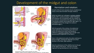
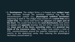
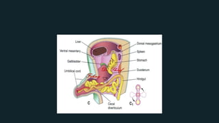
1. The midgut forms a U-shaped loop that herniates through the umbilical ring and undergoes two rotations to return to the abdominal cavity, positioning the appendix on the right.
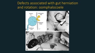
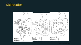
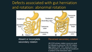
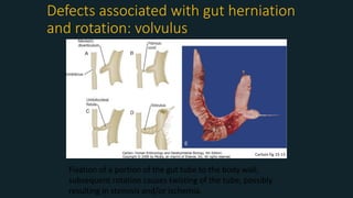
2. Failure of the midgut to return or complete rotation can result in defects like omphalocele, malrotation, intestinal atresia or stenosis.
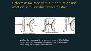
3. Common midgut defects include Meckel's diverticulum from persistence of the vitelline duct and intestinal malrotation from incomplete rotation.
Kidney development - embryology of urinary systemSaachiGupta4
Part of the development of the urinary system
The ascent of the kidney, stages of development of kidney
discussion about pronephros, mesonephros, metanephros.
05.28.09(b): Development of the Urinary SystemOpen.Michigan
Slideshow is from the University of Michigan Medical
School's M1 Embryology sequence
View additional course materials on Open.Michigan:
openmi.ch/med-M1Embryology
In this presentation the development of Small intestine and Pancreas has been discussed. The viewer would be able to understand the concept of physiological herniation and rotation of the Primary intestinal loop with in the connecting stalk.
Here is a presentation made by MBChB level 3 students for the lecture series on GIT Pathology. Hope it helps you. Few typos but better will come.It includes Hirshsprung's disease, Diveticulosis, Colitis, Colorectal Carcinoma among others
The small intestine is the part of the alimentary canal that is continuous with the stomach at the pyloric orifice and leads into the large intestine through the iliocaecal valve. It is the part where the chemical digestion of food is completed and most of the absorption of nutrients take place.
It extends from the ileum to the anus.
It reabsorbs water converting liquid chyme into semi solid stools.
It consists of the following parts: 1)Caecum and vermiformis appendix. 2)Ascending colon and hepatic flexure. 3) Transverse colon and splenic flexure 4)Descending colon 5)Sigmoid colon 6) Rectum and 7) Anal canal.
The proximal half as far as the splenic flexure – reabsorbs water and electrolytes from fluid chyme .
The distal colon beyond the splenic flexure-stores formed faeces until they are excreted.
Safalta Digital marketing institute in Noida, provide complete applications that encompass a huge range of virtual advertising and marketing additives, which includes search engine optimization, virtual communication advertising, pay-per-click on marketing, content material advertising, internet analytics, and greater. These university courses are designed for students who possess a comprehensive understanding of virtual marketing strategies and attributes.Safalta Digital Marketing Institute in Noida is a first choice for young individuals or students who are looking to start their careers in the field of digital advertising. The institute gives specialized courses designed and certification.
for beginners, providing thorough training in areas such as SEO, digital communication marketing, and PPC training in Noida. After finishing the program, students receive the certifications recognised by top different universitie, setting a strong foundation for a successful career in digital marketing.
The French Revolution, which began in 1789, was a period of radical social and political upheaval in France. It marked the decline of absolute monarchies, the rise of secular and democratic republics, and the eventual rise of Napoleon Bonaparte. This revolutionary period is crucial in understanding the transition from feudalism to modernity in Europe.
For more information, visit-www.vavaclasses.com
Macroeconomics- Movie Location
This will be used as part of your Personal Professional Portfolio once graded.
Objective:
Prepare a presentation or a paper using research, basic comparative analysis, data organization and application of economic information. You will make an informed assessment of an economic climate outside of the United States to accomplish an entertainment industry objective.
Unit 8 - Information and Communication Technology (Paper I).pdfThiyagu K
This slides describes the basic concepts of ICT, basics of Email, Emerging Technology and Digital Initiatives in Education. This presentations aligns with the UGC Paper I syllabus.
Normal Labour/ Stages of Labour/ Mechanism of LabourWasim Ak
Normal labor is also termed spontaneous labor, defined as the natural physiological process through which the fetus, placenta, and membranes are expelled from the uterus through the birth canal at term (37 to 42 weeks
Introduction to AI for Nonprofits with Tapp NetworkTechSoup
Dive into the world of AI! Experts Jon Hill and Tareq Monaur will guide you through AI's role in enhancing nonprofit websites and basic marketing strategies, making it easy to understand and apply.
Biological screening of herbal drugs: Introduction and Need for
Phyto-Pharmacological Screening, New Strategies for evaluating
Natural Products, In vitro evaluation techniques for Antioxidants, Antimicrobial and Anticancer drugs. In vivo evaluation techniques
for Anti-inflammatory, Antiulcer, Anticancer, Wound healing, Antidiabetic, Hepatoprotective, Cardio protective, Diuretics and
Antifertility, Toxicity studies as per OECD guidelines
June 3, 2024 Anti-Semitism Letter Sent to MIT President Kornbluth and MIT Cor...Levi Shapiro
Letter from the Congress of the United States regarding Anti-Semitism sent June 3rd to MIT President Sally Kornbluth, MIT Corp Chair, Mark Gorenberg
Dear Dr. Kornbluth and Mr. Gorenberg,
The US House of Representatives is deeply concerned by ongoing and pervasive acts of antisemitic
harassment and intimidation at the Massachusetts Institute of Technology (MIT). Failing to act decisively to ensure a safe learning environment for all students would be a grave dereliction of your responsibilities as President of MIT and Chair of the MIT Corporation.
This Congress will not stand idly by and allow an environment hostile to Jewish students to persist. The House believes that your institution is in violation of Title VI of the Civil Rights Act, and the inability or
unwillingness to rectify this violation through action requires accountability.
Postsecondary education is a unique opportunity for students to learn and have their ideas and beliefs challenged. However, universities receiving hundreds of millions of federal funds annually have denied
students that opportunity and have been hijacked to become venues for the promotion of terrorism, antisemitic harassment and intimidation, unlawful encampments, and in some cases, assaults and riots.
The House of Representatives will not countenance the use of federal funds to indoctrinate students into hateful, antisemitic, anti-American supporters of terrorism. Investigations into campus antisemitism by the Committee on Education and the Workforce and the Committee on Ways and Means have been expanded into a Congress-wide probe across all relevant jurisdictions to address this national crisis. The undersigned Committees will conduct oversight into the use of federal funds at MIT and its learning environment under authorities granted to each Committee.
• The Committee on Education and the Workforce has been investigating your institution since December 7, 2023. The Committee has broad jurisdiction over postsecondary education, including its compliance with Title VI of the Civil Rights Act, campus safety concerns over disruptions to the learning environment, and the awarding of federal student aid under the Higher Education Act.
• The Committee on Oversight and Accountability is investigating the sources of funding and other support flowing to groups espousing pro-Hamas propaganda and engaged in antisemitic harassment and intimidation of students. The Committee on Oversight and Accountability is the principal oversight committee of the US House of Representatives and has broad authority to investigate “any matter” at “any time” under House Rule X.
• The Committee on Ways and Means has been investigating several universities since November 15, 2023, when the Committee held a hearing entitled From Ivory Towers to Dark Corners: Investigating the Nexus Between Antisemitism, Tax-Exempt Universities, and Terror Financing. The Committee followed the hearing with letters to those institutions on January 10, 202
2. DISTAL DUODENUM
• Distal or lower duodenum arises from the cranial most portion of the
midgut and is served by anterior and posterior branches of the
inferior pancreaticoduodenal artery, which is a branch of the superior
mesentery artery.
• As with the rest of the duodenum, becomes secondarily
retroperitoneal as with the rest of the entire gi tract, the lumen is
obliterated transiently during development and then recanalizes.
• Failure to recanalize the duodenum can result in stenosis (narrowing)
or atresia (complete blockage)
3. Development of the midgut and colon
Herniation and rotation:
• Growth of the GI tract exceeds volume of abdominal
cavity so the tube herniates through umbilicus
• While herniated, gut undergoes a primary rotation
(fig B) of 90° “counterclockwise” (when looking at
the embryo); this corresponds with the rotation of
the stomach, and positions the appendix on the left.
The primary rotation also brings the right vagus n. to
the FRONT (hence the change in its name to
ANTERIOR vagus n.
• With the growth of the embryo, the abdominal
cavity expands thus drawing the gut tube back
within the abdominal cavity and causing an
additional, secondary rotation (fig C) of 180° CCW
(positioning the appendix on the RIGHT)
• Once in the abdominal cavity, the colon continues to
grow in length, pushing the appendix to its final
position in the lower right quadrant.
• Note the attachment of the vitelline duct to the gut
at the region of the ileum. The duct normally
regresses during development, but not always….
4. • 1. Development. The midgut forms a U-shaped loop (midgut loop)
that herniates through the primitive umbilical ring into the
extraembryonic coelom (i.e., physiological umbilical herniation)
beginning at week 6. The midgut loop consists of a cranial limb and a
caudal limb. The cranial limb forms the jejunum and upper part of
the ileum. The caudal limb forms the cecal diverticulum, from which
the cecum and appendix develop; the rest of the caudal limb forms
the lower part of the ileum, ascending colon, and proximal two
thirds of the transverse colon. The midgut loop rotates a total of
270_ counterclockwise around the superior mesenteric artery as it
returns to the abdominal cavity, thus reducing the physiological
herniation, around week 12.
5.
6. • 2. Sources. Simple columnar absorptive cells lining midgut
derivatives, goblet cells, Paneth cells, and enteroendocrine cells
comprising the intestinal glands are derived from endoderm.
• The lamina propria, muscularis mucosae, submucosa, and inner
circular and outer longitudinal smooth muscle of the muscularis
externa and serosa are derived from visceral mesoderm.
7. • 3. Clinical considerations
• a. Omphalocele: occurs when abdominal contents herniates through
the umbilical ring and persists outside the body, covered variably by a
translucent peritoneal membrane sac (a light gray, shiny sac)
protruding from the base of the umbilical cord.
• Large omphaloceles may contain stomach, liver, and intestines. Small
omphaloceles contain only intestines. Omphaloceles are
• usually associated with other congenital anomalies (e.g., trisomy 13,
trisomy 18, or Beckwith-Wiedemann syndrome) and with
• increased levels of alfa-fetoprotein.
8.
9. • b. Gastroschisis: occurs when there is a defect in the ventral
abdominal wall, usually to the right of the umbilical ring, through
which there is a massive evisceration of intestines (other organs may
also be involved). The intestines are not covered by a peritoneal
membrane, are directly exposed to amniotic fluid, are thickened, and
are covered with adhesions.
10.
11. • c. Ileal diverticulum (Meckel’s diverticulum): occurs when a remnant
of the vitelline duct persists, thereby forming an outpouching located
on the antimesenteric border of the ileum. The outpouching may
connect to the umbilicus via a fibrous cord or fistula. A Meckel’s
diverticulum is usually located about 30 cm proximal to the ileocecal
valve in infants and varies in length from 2 to 15 cm. Heterotopic
gastric mucosa may be present, which leads to ulceration,
perforation, or gastrointestinal bleeding, especially if a large number
of parietal cells are present. It is associated clinically with symptoms
resembling appendicitis and bright-red or dark-red stools (bloody).
12. • d. Nonrotation of the midgut loop: occurs when the midgut loop rotates only
90_ counterclockwise, thereby positioning the small intestine entirely on the right
side and the large intestine entirely on the left side, with the cecum located
either in the left upper quadrant or the left iliac fossa.
• Note the small intestines (SI) on the right side and the large intestines (LI) on the
left side.
• e. Malrotation of the midgut loop: occurs when the midgut loop undergoes only
partial counterclockwise rotation.
• This results in the cecum and appendix lying in a subpyloric or subhepatic
location and the small intestine being suspended by only a vascular pedicle (i.e.,
not a broad mesentery). A major clinical complication of malrotation is volvulus
(twisting of the small intestines around the vascular pedicle), which may cause
necrosis due to compromised blood supply. (Note: The abnormal position of the
appendix due to malrotation of the midgut should be considered when
diagnosing appendicitis).
13. • g. Intestinal atresia and stenosis: Atresia occurs when the lumen of
the intestines is completely occluded, whereas stenosis occurs when
the lumen of the intestines is narrowed.
• The causes of these conditions seem to be both failed recanalization
and/or an ischemic intrauterine event (“vascular accident”).
14. • i. Intussusception: occurs when a segment of bowel invaginates or
telescopes into an adjacent bowel segment, leading to obstruction or
ischemia. This is one of the most common causes of obstruction in
children younger than 2 years of age, is most often idiopathic, and is
most commonly involves the ileum and colon (i.e., ileocolic). It is
associated clinically with acute onset of intermittent abdominal pain,
vomiting, bloody stools, diarrhea, and somnolence.
15.
16. • j. Retrocecal and retrocolic appendix occurs when the appendix is located
on the posterior side of the cecum or colon, respectively. These anomalies
are very common and important to remember during appendectomies.
Note: The appendix is normally found on the medial side of the cecum.
• Derivatives of the hindgut are supplied by the inferior mesenteric artery.
• A. Distal one third of the transverse colon, descending colon, sigmoid
colon.
• 1. Development. The cranial end of the hindgut develops into the distal
one third of the transverse colon, descending colon, and sigmoid colon.
The terminal end of the hindgut is an endoderm-lined pouch called the
cloaca, which contacts the surface ectoderm of the proctodeum to form
the cloacal membrane.
17. Defects associated with gut herniation and
rotation: vitelline duct abnormalities
Vitelline duct abnormalities of some sort occur in ~2% of all live
births. Note that these aberrant structures are almost always
found along the ileal portion of the GI tract.
Langman’s fig 14-32
19. Midgut Anomalies
• Clinical correlation –
• Duodenal atresia is due to failed canalization.
• Omphalocele results from failure of the midgut loop to return to the
abdomen.
• Meckel’s diverticulum occurs when a remnant of the yolk sac (Vitelline duct)
persists.
• Malrotation occurs if the midgut does not complete the rotation prior to
returning to the abdomen.
21. Defects associated with gut herniation
and rotation: abnormal rotation
Absent or incomplete
secondary rotation
Reversed secondary rotation
(90 CCW primary rotation occurs as usual
but followed by abnormal 180 CW rotation.
Net rotation is 90° CW; viscera are in their
normal location, but note that the duodenum
is anterior to the transverse colon)
Langman’s fig 14-33
22. Defects associated with gut herniation
and rotation: volvulus
Fixation of a portion of the gut tube to the body wall;
subsequent rotation causes twisting of the tube, possibly
resulting in stenosis and/or ischemia.
Carlson fig 15-13
Editor's Notes
Development of Colonic Midgut
Physiologic umbilical herniation of midgut out of abdominal cavity between the 6th and 10th week. The midgut rotates 90 degrees counterclockwise around the superior mesenteric artery axis leaving the caudal midgut to left. As the umbilical hernia reduces the colon returns after the small intestine and does an additional 180 degree counterclockwise rotation. This is followed by fixation of the colon.
A cecal diverticulum presents at the 6th week and develops into the cecum and appendix. The appendix is initially located at the caudal midgut loop and by birth it is located at the distal end of cecum. Unequal cecal growth results in the appendix being medial to the cecum.
Different types of malrotation:
Malrotation of the intestines results in a pedacle that can twist causing either Midgut and cecal volvulus. Intestinal malrotation may occur as an isolated event or in association with other types of congenital anomalies.
Rotational abnormalities may be grouped by the developmental stage at which they occur.
1. Arrest during the bowel herniation phase (Omphalocele)
2. Arrest or abnormalities of rotation (Malrotation, Gastroschisis) Nonrotation is the most common and leads to the small intestine remaining entirely to the right of the artery, with the cecum at or near the midline and the colon in the left abdomen. The proximal jejunum and colon pass very closely to the SMA, leaving a narrow pedicle as the base of the mesentery. In malrotation the midgut rotation is incomplete, the duodenal-jejunal loop remains to the right of the SMA, and the ileocecal loop comes to lie in the right upper quadrant, anterior to the SMA and closely related to the duodenum. The entire midgut is attached by a very narrow pedicle, SMA and SMV are in this pedicle. A volvulus of this pedicle may occur.
3. Arrest in the last phase resulting in a mobile cecum, an unattached duodenum, or an unattached small bowel mesentery, which allows cecal volvulus.
Sigmoid volvulus is more common in adults, especially African’s who often have a redundant colon. It can occur in children and is thought to be related to intestinal malrotation, omphalomesenteric abnormalities, Hirschsprung’s disease or anal stenosis. There is also a connection with diabetes mellitus.