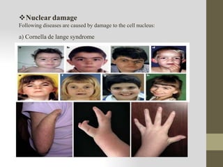
BASIC PRINCIPLES
OF CELL INJURY
AND ADAPTATIONS
Definition:
Cell injury is defined as a variety of stresses in cell
that encounters as a result of changes in its internal and
external environment.
The causes of cell injury, reversible or irreversible
may be broadly classified into two large groups:
a)Genetic causes
b)Acquired causes
1. Hypoxia and Ischemia
2. PhysicalAgents
3. Chemicals and drugs
4. MicrobialAgents
5. Immunological agents:
6. Psychological factors
Causes of cell injury
a) Genetic causes: Genetic defects may results in pathological changes as seen
in Sickle cell anemia
b)Acquired causes: Based on the underlying agents the acquired causes of cell
injury may be further classified as under:
1. Hypoxia and Ischemia:
Cells require oxygen to generate energy and carry out metabolic functions.
Deficiency of oxygen (hypoxia) results in failure to carry out these activities by
the cells.
 Thus hypoxia is the most common cause of cell injury. The causes of hypoxia
are as under:
a) Reduced supply of blood to cells i.e Ischemia
b) Other causes include:Anemia, carbon monoxide poisioning.
2. PhysicalAgents:
These are
 Mechanical trauma (e.g: Road accidents)
 Thermal trauma (e.g: By heat and cold)
 Electricity
 Radiations (e.g: Ultraviolet and ionizing)
 Rapid changes in atmospheric pressure.
3. Chemicals and drugs: These includes:
 Chemical poisons (e.g: cyanide, arsenic, mercury)
 Strong acids and alkalis
 Environmental pollutants
 Insecticides and pesticides
 Hypertonic glucose and salts
 Oxygen at higher concentration
 Social agents (e.g:Alcohol, Narcotic drugs)
These agents may cause severe damage at cellular level by altering
membrane permeability, osmotic homeostasis or integrity of an enzyme or
cofactor resulting in death of the cell.
4.Microbial Agents: Injuries by microbes includes infections caused by
bacteria, viruses, fungi, rickettsiae, protozoa, metazoan and other parasites.
5. Immunological agents: It protects the host against various injurious
agents it may also turn lethal and cause cell injury.
e.g: Hypersensitivity reaction, anaphylactic reaction, autoimmune reaction.
6. Nutritional imbalance: Nutritional deficiency (protein deficiency
Kwashiorkor, marasmus) or an excess of nutrients (e.g obesity, atherosclerosis,
hypertension) may result in nutritional imbalance which causes cell injury.
7. Psychological factors: There are no specific biochemical or morphological
changes that occurs in common mental diseases. However, problem of drug
addiction, alcoholism and smoking results in various organic diseases such as
liver damage, chronic bronchitis, lung cancer, peptic ulcer, hypertension,
Ischemic heart diseases etc.
Pathogenesis
 Cell membrane damage
 Mitochondrial Damage
 Ribosomal damage
 Nuclear damage
 Cell membrane damage
Cell membrane diseases are life threatning disorders
which are genetic in nature.
They usually work against proteins in the body which
are important for ion channels and receptors within
the membrane.
Following are some of the important disorders related to cell membrane:
a) Hyaline membrane disease
It is commonly associated with pre-term infants. It affects the
lungs at the time of birth causing respiratory distress. As a result, lungs
require abnormal levels of oxygen and carbon dioxide exchange after
birth.
b) Alzheimer’s disease
It is a progressive
disease that destroys memory and
other important mental functions.
The oxidative stress caused by
Alzheimer’s disease in the brain
results in phospholipid alterations.
These alterations disrupt functions
of the brain cells.
c) Cystic fibrosis:
It is a disease which brings about an
excessive production of fluid in the
lungs due to a defective calcium ion
channel. when the channel mutate
leading to alteration in proteins, it
causes the mucus to build up in the
lungs, creating difficulty in breathing.
d) Duchenne muscular dystrophy:
This disease affects dystrophin in the muscle cell. Dystrophin allows the
muscle cell wall to connect with the intracellular section. In absence of
dystrophin, the cell membrane is incapable of repairing itself, destroying it
and causing muscular dystrophy.
 Mitochondrial damage
Mitochondrial damage can lead to diseases which are together termed as
mitochondrial diseases.
 Following are the examples of mitochondrial diseases:
a) Diabetes mellitus and deafness (DAD):
It is subtype of diabetes which is caused from a point mutation at position
3243 in human mitochondrial DNA and it is characterized by diabtes and
sensorineural hearing loss.
b)Leber’s hereditary optic
neuropathy (LHON):
It is a mitochondrially inherited
(transmitted from mother to
offspring) dengeration of retinal
ganglion cells (RGCs) and their
axons that leads to an acute or
subacute loss of central vision;
this affects predominantly young
adult males.
 RIBOSOMALDAMAGE
 Damage to ribosomes can lead to diseases which termed as
Ribosomopathy.
 Ribosomopathies are caused by alterations in the structure or
function of ribosomal.
 Name of some these disease are listed below.
 Affected individuals may also have an
opening in the roof of the mouth (cleft
palate) with or without a split in the
upper lip (cleft lip).
 They may have a short, webbed neck;
shoulder blades that are smaller and
higher than usual; and abnormalities
a) Diamond-blackfan anemia: It is characterized normocytic or macrocytic
anemia (low RBC counts) with decreased erythroid progenitor cells in the bone
marrow.
of their hands, most commonly
malformed or absent thumbs.
b)Dyskeratosis congenital (DKC): The entity was classically defined
by the abnormal skin pigmentation, nail dystrophy ie abnormal
changes in the shape, color, texture, and growth of the fingernails
or toenails. Nail dystrophy is often caused by infection or injury to
the nail. Leukoplakia of the oral mucosa ie thick, white patches on
the inside surfaces of your mouth caused by chronic irritation, such
as from tobacco use, including smoking and chewing. It is
characterized by short telomeres nothing but region of repetitive
nucleotide sequences associated with specialized proteins at the
ends of linear chromosomes..
b)Shwachman-Diamond syndrome: It is characterized by bone
marrow dysfunction, skeletal abnormalities and short stature.
c) 5q-myelodysplastic syndrome: It is type of bone marrow
disorder.
d)Treacher Collins syndrome: A inherited condition in which
bones and tissues in face aren’t developed.
Nuclear damage
Following diseases are caused by damage to the cell nucleus:
a) Cornella de lange syndrome
 It is a rare genetic disorder present from birth.
 Some of the important symptoms of the disease are as follows:
•Low birth weight
•Delayed growth and small stature
•Development delay
•Missing limbs or portions of limbs
•Microcephaly: small head size
•Excessive body hair
•Hearing impairment and vision abnormalities
•Partial joining of second and third toes
•Seizures, heart defects.
b) Revesz syndrome:
 It is a fatal disease which causes retinopathy ie disease of the retina which results in impairment or
loss of vision and bone marrow failure.
 Other symptoms include:
•severe aplastic anemia (Aplastic anemia is a condition that occurs when your body stops producing
enough new blood cells.)
•intrauterine growth retardation
•fine sparse hair (hair that is less dense; may be due to reduced follicle numbers or due to the inability
to retain some of the hairs of the coat over time at older age older age, due to slower hair growth)
•skin pigmentation
•ataxia (group of disorders that affect co-ordination, balance and speech during walking, speaking,
swallowing due to cerebellar hypoplasia (lack of cells in tissues or organs that affects their
functioning.).
•It is genetic disease thought to be caused by short telomeres.
c) Schinzel giedon
 It is congenital, neurogenerative terminal
syndrome.
 It exhibits severe midface retraction, skull
abnormalities, renal anomalies.
 Babies with the syndrome have severe mental
retardation, growth retardation and global
development delay.
d) Spinal muscular atrophy
 It is characterized by loss of motor neurons and progressively muscle
wasting, often leading to early death.
 It is caused by a genetic defect in the SMN1 gene, which encodes SMN,
a protein which is needed for survival of motor neurons.
 Muscular atrophy first affects proximal muscles and lung muscles.
MORPHOLOGY OF CELL INJURY
1. Morphology of reversible cell injury
 Cellular swelling
 fatty changes
2. Morphology of irreversible cell injury
 Necrosis
 Apoptosis
3. Intracellular acculmulations
 Acculmulations of lipids, proteins, carbohydrates.
 Abnormal substances e.g: as in storage disease
 Accumulations of pigments e.g: endogenous pigment, exogenous pigment.
4. Subcellular alterations in cell injury
ATROPHY
Muscle atrophy
Definition: Reduction of the number and size of the
parenchyma cells of an organ or its an organ or its part
which was once normal is called as atrophy.
Atrophy is a partial or complete wasting away of a part
of body.
 It includes mutations, which can destroy the gene to
build up the organ, poor nourishment, poor
circulation, loss of hormonal support and loss of
nerve supply to the target organ.
e.g: Atrophy as a part of normal development include
shrinkage and involution of the thymus in early
childhood and the tonsils in adolescence. In old age it
may include loss teeth, hair, weakening of muscles or
loss of weight in organ and sluggish mental activity.
 Muscle atrophies: Disuse atrophy of muscle and
bones, with loss of mass and strength, can occur
after prolonged immobility, such as extended bed
rest, or having a body part in a cast.
e.g: darkness for the eye or being bed ridden for the
legs.
HYPERTROPHY
Definition: It is the increase in the volume of
an organ or tissue due to enlargement of its
component cells.
 Eccentric hypertrophy is observed where the
walls and chamber of a hollow organ
undergo growth in which the overall size
and volume are enlarged.
 Physiologic hypertrophy:
e.g: enlarged size of the uterus in pregnancy.
 Pathologic hypertrophy:
e.g i) hypertrophy of cardiac muscles in
systemic hypertension
ii) hypertrophy of smooth muscle e.g:
pyloric stenosis (in stomach)
HYPERPLASIA
Definition:: It is an increase in the amount of organic tissue which results
from cell proliferation which may lead to gross enlargement of an organ
and may be confused with benign neoplasia/ tumour.
Gross changes: Enlargement of affected organ or tissue.
Microscopical changes: There is increase in the the number of the cells
which is due to the rate of DNAsynthesis and hence mitosis of cells.
It is common pre-neoplastic response to stimulus. Microscopically, cells
resemble normal cells but are increased in numbers.
Physiologic hyperplasia:
e.g: 1) hormonal hyperplasia i.e hyperplasia due to hormonal stimulation.
E.g-hyperplasia of female breasts at puberty, during pregnancy and
lactation.
Pathologic hyperplasia:
e.g: 1) Endometrial hyperplasia following oestrogen excess.
2) In wound healing there is formation of granulation tissue due to
proliferation of fibroblast and endothelial cells.
Definition: It is defined as reversible change of one of epithelial or
mesenchymal cells, usually in response to abnormal stimuli and often reverts
back to normal on removal of stimulus.
Metaplasia is broadly divided into two types:
a) Epithelial Metaplasia: Metastatic change may be patchy or diffuse and
usually results in replacement by stronger but less well specialized epithelium.
Some common types of epithelial metaplasia are:
Squamous metaplasia
e.g: i. In bronchous of chronic smokers
ii. In vitamin A deficiency
Columnar metaplasia
e.g: intestinal metaplasia in healed chronic gastric ulcer.
b) Mesenchymal metaplasia: In this case transformation of one adult type of
mesenchymal tissue to another.
METAPLASIA
DYSPLASIA
Definition: It refers to an abnormality of development
or an epithelial anamoly of growth and differentiation
i.e. epithelial dysplasia.
The term dysplasia is typically used when the cellular
abnormality is restricted to the originating tissue, as in
case of an early, in situ neoplasm.
Examples of dysplasia include epithelial dysplasia of
the cervix.
In cervical intra-epithelial neoplasia an increased
population of immature cells, restricted to the mucosal
surface do not invade through the basement membrane
to the deeper soft tissues.
Dysplasia is characterized by 4 major pathological
microscopical changes:
1. Anisocytosis: cells of unequal size.
2. Poikilocytosis: abnormally shaped cells.
3. Hyperchromatism: excessive pigmentation.
4.Presence of mitotic figures: An unusual number of
cells which are currently dividing.
The most notable components of the cell which are targets of cell damage are
the DNA and the cell membrane. There are two types of damages are as
follows:
1) Sub-lethal (reversible) changes:
They are of two types: cellular swelling and fatty change.
a)Cellular swelling: two basic morphological changes manifested by sub-
lethally injured cells are cell swelling and fatty change.
b)Fatty changes: sometimes the cells are damaged to such an extent that they
are unable to metabolize fat adequately. In such cases small vacuoles of fat
accumulate and become dispersed within cytoplasm.
2) Lethal changes:
 Necrosis:
Definition: it is characterized by cytoplasmic
swelling, irreversible damage to the plasma
membrane, and organelle breakdown leading
to cell death.
Types of cell damage
The stages of cellular necrosis include following:
a)Pyknosis : clumping of chromosomes and shrinking to the nmucleus of the
cell.
b)Karyorrhexis: fragmentation of the nucleus and breakup of the chromatin
into unstructural granules.
c) Karyolysis: dissolution of the cell nucleus.
 Apoptosis:
Definition: it is form of coordinated and internally programmed cell death which
is of significance in a variety of physiologic and pathologic conditions.
Biochemical changes:
Proteolysis of cytoskeletal proteins
Proteins-protein cross linking
Fragmentation of nuclear chromatin by activation of nuclease.
Appearance of phosphatidyl serine on the outer surface of cell membrane.
Intracellular Accumulation
One of the manifestations of metabolic dearrangements in cells is the intracellular
accumulations of abnormal amounts of various substances.
It falls into two main categories:
 A Normal cellular constituent like water, lipids, proteins and carbohydrates,
which accumulate in excess.
 An abnormal substance, either exogenous like products of infectious agents
of endogenous such as product of abnormal synthesis or metabolism.
Abnormal accumulation of some major components and diseases related to them
are mentioned below:
 Lipids:
All major classes of lipids can accumulate in cells: triglycerides, cholesterol and
phospholipids.
Some of the diseases related to lipids are mentioned below:
a) Steatosis (fatty changes): It is an abnormal accumulation of triglycerides
within parenchymal cells.
b)Atherosclerosis: In atherosclerotic plaques, smooth muscle cells and macrophages
within the intimal layer of the aorta and large arteries are filled with lipid vacuoles,
most of which are made up of cholesterol and cholesterol esters.
c)Xanthomas: Intracellular accumulation of cholesterol within macrophages is a
characterisistic of acquired and hereditary hyperlipidemic states.
d) Cholesterolosis: it refers to the focal accumulation of cholesterol-laden
macrophages in the lamina propria of the gall bladder.
 Proteins
Intracellular accumulation of proteins appears as rounded, eosinophilic droplets,
vacuoles or aggregates in the cytoplasm. Excess of proteins within the cells sufficient
to cause morphologically visible accumulation have different causes as follows:
a) Reabsorption: Droplets in proximal renal tubules are seen in renal diseases
associated with proteinuria.
b)Accumulation of cytoskeletal proteins: There are several types of cytoskeletal
proteins.
e.g: Microtubules (20-25 nm in diameter)
c) Aggregation of abnormal proteins: Abnormal or misfolded proteins may deposit
in tissues and can interfere with normal functions.
 Hyaline change:
Hyaline refers to an alteration within cells or in the extracellular space which gives a
homogeneous, glassy, pink appearance in routine histologic sections stained with
hematoxylin and eosin.
e.g: Reabsorption, Russell bodies, alcoholic hyaline are e.g of intracellular hyaline
deposits.
 Glycogen: Glycogen accumulates within cells in a group of related genetic disordes
which are collectively reffered to as the glycogen storage disease or glycogenosis.
 Calcification: It is accumulation of calcium salts in a body tissue. It normally
occurs in formation of bone, but calcium can be deposited abnormally in soft tissue,
causing it to harden.
e.g: Calcification of soft tissues like arteries, cartilage and heart valves can be caused by
vitamin K2 deficiency.Calcification can manifest in many ways in the body. Some of
them may be caused by poor calcium absorption.
The symptoms are as follows:
 Kidney stones
 Gall stones
 Tartar on teeth
Calcification:
• It is accumulation of calcium salts in a body tissue. It normally
occurs in formation of bone, but calcium can be deposited
abnormally in soft tissue, causing it to harden.
• e.g: Calcification of soft tissues like arteries, cartilage and heart
valves can be caused by vitamin K2 deficiency.
• Calcification can manifest in many ways in the body. Some of
them may be caused by poor calcium absorption.
• The symptoms are as follows:
 Kidney stones
 Gall stones
 Tartar on teeth
Enzyme leakage and death death:
The major cellular organelle containing digestive enzymes are lysosomes.
Following enzymes are present in lysosomes:
 Acid phosphatoses for phosphate esters.
 Nucleases: DNAase, RNAase for breakdown of nucleic acids.
 Protein digesting enzmes: collagenase, cathepsins etc.
 Carbohydrate digesting enzymes: beta-glycosidases, hexosaminidaseAetc.
 Lipid digesting enzymes: sphingomyelinase, esterases.
 Silicosis: It may lead to tuberculosis.
 Pompe disease (alpha-glucosidase).
 Gaucher disease.
 Sandhoff disease.
Acidosis and alkalosis
 Acidosis:
It is an increased acidity in the blood and other body tissue. Usually it refers to
acidity of blood plasma. Usually it refers to acidity of blood plasma. Acidosis is
said to occur when arterial pH falls below 7.35, except in the foetus.
There is another term called acidemia, which describes the state of low blood pH.
Acidosis is used to describe the processes leading to these states.
 Metabolic acidosis: It may results from either increased production of
metabolic acids, like lactic acid or disturbances in the ability to excrete acid via
kidney
Lactic acidosis may occur from one of the followng causes:
 Severe hypoxemia
 Hypoperfusion
 Respiratory acidosis: in respiratory acidosis, carbon dioxide is increased
while the bicarbonate is normal or increased. It results from a build up of
carbon dioxide in blood, termed as hypercapnia, due to hypoventilation.
Sign and symptoms of acidosis:
 Headaches
 confusion
 feeling tired
 sleepiness
 dysfunction of cerebrum which may progress to coma.
 Alkalosis:
Alkalosis is a condition in which pH of tissue is elevated beyond the normal
range of 7.35 to 7.55. It is a result of decresed hydrogen ion concentration,
leading to increased bicarbonate or alternatively a direct result of increased
bicarbonate concentration.
Alkalosis refers to a process by which the pH is increased. Alkalemia refers
to pH which is higher than normal, specifically in the blood.The causes of
metabolic alkalosis can divided into 2 categories, depending on urine
chloride levels.
Causes of alkalosis:
a) Chloride responsive alkalosis:
It caused by following reasons:
 Loss of hydrogens ions: caused by vomiting or kidney failure.
 Congenital chloride diarrhoea-a rare condition
 Cystic fibrosis
 Contraction alkalosis: caused by dehydration/use of diuretics.
b) Chloride resistant alkalosis:
It caused by following reasons:
 Retention of bicarbonate
 Shift of hydrogen ions into intracellular space: seen in hypokalemia.
 Alkalotic agents like bicarbonate
 Liddle syndrome characterized by hypertension and hypoaldosteronism.
 Aminoglycoside toxicity can induce a hypokakalemic metabolic acidosis.
Electrolyte Imbalance
 Electrolytes play a vital role in maintain homeostasis within the body.
 They help to regulate heart and neurological function, fluid balance, oxygen
delivery, acid-base balance etc.
 Electrolyte imbalances can develop by one the following mechanisms:
 Excessive ingestion
 Diminished elimination
 Diminished ingestion
 Excessive elimination
 The most serious electrolyte disturbances involve abnormalities in the level of
sodium, potassium, or calcium.
 Chronic laxatives abuse, severe diarrhoea or vomiting can cause electrolyte
disturbances along with dehydration.
 Electrolytes are important because cells like nerve, heart and muscle use them
to maintain voltages across their cell membranes and to carry electrical
impulses across themselves and to other cells.
 Electrolytes are loss through sweat particulary in the form of sodium and
potassium. Excessive sweating in summer causes weakness due to loss of
electroytes. Electrolytes must be replaces to keep their concentration in body
fluids constant.
SELF STUDY PART AS PER
REDUCED SYLLABUS
Introduction & definitions of Homeostasis,
Components and Types of Feedback systems.

1.1cell injury-1.pptx

  • 1.
    BASIC PRINCIPLES OF CELLINJURY AND ADAPTATIONS
  • 2.
    Definition: Cell injury isdefined as a variety of stresses in cell that encounters as a result of changes in its internal and external environment.
  • 4.
    The causes ofcell injury, reversible or irreversible may be broadly classified into two large groups: a)Genetic causes b)Acquired causes 1. Hypoxia and Ischemia 2. PhysicalAgents 3. Chemicals and drugs 4. MicrobialAgents 5. Immunological agents: 6. Psychological factors Causes of cell injury
  • 5.
    a) Genetic causes:Genetic defects may results in pathological changes as seen in Sickle cell anemia b)Acquired causes: Based on the underlying agents the acquired causes of cell injury may be further classified as under: 1. Hypoxia and Ischemia: Cells require oxygen to generate energy and carry out metabolic functions. Deficiency of oxygen (hypoxia) results in failure to carry out these activities by the cells.  Thus hypoxia is the most common cause of cell injury. The causes of hypoxia are as under: a) Reduced supply of blood to cells i.e Ischemia b) Other causes include:Anemia, carbon monoxide poisioning. 2. PhysicalAgents: These are  Mechanical trauma (e.g: Road accidents)  Thermal trauma (e.g: By heat and cold)  Electricity  Radiations (e.g: Ultraviolet and ionizing)  Rapid changes in atmospheric pressure.
  • 6.
    3. Chemicals anddrugs: These includes:  Chemical poisons (e.g: cyanide, arsenic, mercury)  Strong acids and alkalis  Environmental pollutants  Insecticides and pesticides  Hypertonic glucose and salts  Oxygen at higher concentration  Social agents (e.g:Alcohol, Narcotic drugs) These agents may cause severe damage at cellular level by altering membrane permeability, osmotic homeostasis or integrity of an enzyme or cofactor resulting in death of the cell. 4.Microbial Agents: Injuries by microbes includes infections caused by bacteria, viruses, fungi, rickettsiae, protozoa, metazoan and other parasites. 5. Immunological agents: It protects the host against various injurious agents it may also turn lethal and cause cell injury. e.g: Hypersensitivity reaction, anaphylactic reaction, autoimmune reaction.
  • 7.
    6. Nutritional imbalance:Nutritional deficiency (protein deficiency Kwashiorkor, marasmus) or an excess of nutrients (e.g obesity, atherosclerosis, hypertension) may result in nutritional imbalance which causes cell injury. 7. Psychological factors: There are no specific biochemical or morphological changes that occurs in common mental diseases. However, problem of drug addiction, alcoholism and smoking results in various organic diseases such as liver damage, chronic bronchitis, lung cancer, peptic ulcer, hypertension, Ischemic heart diseases etc.
  • 8.
    Pathogenesis  Cell membranedamage  Mitochondrial Damage  Ribosomal damage  Nuclear damage
  • 9.
     Cell membranedamage Cell membrane diseases are life threatning disorders which are genetic in nature. They usually work against proteins in the body which are important for ion channels and receptors within the membrane.
  • 10.
    Following are someof the important disorders related to cell membrane: a) Hyaline membrane disease It is commonly associated with pre-term infants. It affects the lungs at the time of birth causing respiratory distress. As a result, lungs require abnormal levels of oxygen and carbon dioxide exchange after birth. b) Alzheimer’s disease It is a progressive disease that destroys memory and other important mental functions. The oxidative stress caused by Alzheimer’s disease in the brain results in phospholipid alterations. These alterations disrupt functions of the brain cells.
  • 11.
    c) Cystic fibrosis: Itis a disease which brings about an excessive production of fluid in the lungs due to a defective calcium ion channel. when the channel mutate leading to alteration in proteins, it causes the mucus to build up in the lungs, creating difficulty in breathing. d) Duchenne muscular dystrophy: This disease affects dystrophin in the muscle cell. Dystrophin allows the muscle cell wall to connect with the intracellular section. In absence of dystrophin, the cell membrane is incapable of repairing itself, destroying it and causing muscular dystrophy.
  • 12.
     Mitochondrial damage Mitochondrialdamage can lead to diseases which are together termed as mitochondrial diseases.
  • 13.
     Following arethe examples of mitochondrial diseases: a) Diabetes mellitus and deafness (DAD): It is subtype of diabetes which is caused from a point mutation at position 3243 in human mitochondrial DNA and it is characterized by diabtes and sensorineural hearing loss. b)Leber’s hereditary optic neuropathy (LHON): It is a mitochondrially inherited (transmitted from mother to offspring) dengeration of retinal ganglion cells (RGCs) and their axons that leads to an acute or subacute loss of central vision; this affects predominantly young adult males.
  • 14.
     RIBOSOMALDAMAGE  Damageto ribosomes can lead to diseases which termed as Ribosomopathy.  Ribosomopathies are caused by alterations in the structure or function of ribosomal.  Name of some these disease are listed below.
  • 15.
     Affected individualsmay also have an opening in the roof of the mouth (cleft palate) with or without a split in the upper lip (cleft lip).  They may have a short, webbed neck; shoulder blades that are smaller and higher than usual; and abnormalities a) Diamond-blackfan anemia: It is characterized normocytic or macrocytic anemia (low RBC counts) with decreased erythroid progenitor cells in the bone marrow. of their hands, most commonly malformed or absent thumbs. b)Dyskeratosis congenital (DKC): The entity was classically defined by the abnormal skin pigmentation, nail dystrophy ie abnormal changes in the shape, color, texture, and growth of the fingernails or toenails. Nail dystrophy is often caused by infection or injury to the nail. Leukoplakia of the oral mucosa ie thick, white patches on the inside surfaces of your mouth caused by chronic irritation, such as from tobacco use, including smoking and chewing. It is characterized by short telomeres nothing but region of repetitive nucleotide sequences associated with specialized proteins at the ends of linear chromosomes..
  • 16.
    b)Shwachman-Diamond syndrome: Itis characterized by bone marrow dysfunction, skeletal abnormalities and short stature. c) 5q-myelodysplastic syndrome: It is type of bone marrow disorder. d)Treacher Collins syndrome: A inherited condition in which bones and tissues in face aren’t developed.
  • 17.
    Nuclear damage Following diseasesare caused by damage to the cell nucleus: a) Cornella de lange syndrome
  • 18.
     It isa rare genetic disorder present from birth.  Some of the important symptoms of the disease are as follows: •Low birth weight •Delayed growth and small stature •Development delay •Missing limbs or portions of limbs •Microcephaly: small head size •Excessive body hair •Hearing impairment and vision abnormalities •Partial joining of second and third toes •Seizures, heart defects. b) Revesz syndrome:  It is a fatal disease which causes retinopathy ie disease of the retina which results in impairment or loss of vision and bone marrow failure.  Other symptoms include: •severe aplastic anemia (Aplastic anemia is a condition that occurs when your body stops producing enough new blood cells.) •intrauterine growth retardation •fine sparse hair (hair that is less dense; may be due to reduced follicle numbers or due to the inability to retain some of the hairs of the coat over time at older age older age, due to slower hair growth) •skin pigmentation •ataxia (group of disorders that affect co-ordination, balance and speech during walking, speaking, swallowing due to cerebellar hypoplasia (lack of cells in tissues or organs that affects their functioning.). •It is genetic disease thought to be caused by short telomeres.
  • 19.
    c) Schinzel giedon It is congenital, neurogenerative terminal syndrome.  It exhibits severe midface retraction, skull abnormalities, renal anomalies.  Babies with the syndrome have severe mental retardation, growth retardation and global development delay. d) Spinal muscular atrophy  It is characterized by loss of motor neurons and progressively muscle wasting, often leading to early death.  It is caused by a genetic defect in the SMN1 gene, which encodes SMN, a protein which is needed for survival of motor neurons.  Muscular atrophy first affects proximal muscles and lung muscles.
  • 20.
    MORPHOLOGY OF CELLINJURY 1. Morphology of reversible cell injury  Cellular swelling  fatty changes 2. Morphology of irreversible cell injury  Necrosis  Apoptosis 3. Intracellular acculmulations  Acculmulations of lipids, proteins, carbohydrates.  Abnormal substances e.g: as in storage disease  Accumulations of pigments e.g: endogenous pigment, exogenous pigment. 4. Subcellular alterations in cell injury
  • 21.
    ATROPHY Muscle atrophy Definition: Reductionof the number and size of the parenchyma cells of an organ or its an organ or its part which was once normal is called as atrophy. Atrophy is a partial or complete wasting away of a part of body.  It includes mutations, which can destroy the gene to build up the organ, poor nourishment, poor circulation, loss of hormonal support and loss of nerve supply to the target organ. e.g: Atrophy as a part of normal development include shrinkage and involution of the thymus in early childhood and the tonsils in adolescence. In old age it may include loss teeth, hair, weakening of muscles or loss of weight in organ and sluggish mental activity.  Muscle atrophies: Disuse atrophy of muscle and bones, with loss of mass and strength, can occur after prolonged immobility, such as extended bed rest, or having a body part in a cast. e.g: darkness for the eye or being bed ridden for the legs.
  • 22.
    HYPERTROPHY Definition: It isthe increase in the volume of an organ or tissue due to enlargement of its component cells.  Eccentric hypertrophy is observed where the walls and chamber of a hollow organ undergo growth in which the overall size and volume are enlarged.  Physiologic hypertrophy: e.g: enlarged size of the uterus in pregnancy.  Pathologic hypertrophy: e.g i) hypertrophy of cardiac muscles in systemic hypertension ii) hypertrophy of smooth muscle e.g: pyloric stenosis (in stomach)
  • 24.
    HYPERPLASIA Definition:: It isan increase in the amount of organic tissue which results from cell proliferation which may lead to gross enlargement of an organ and may be confused with benign neoplasia/ tumour. Gross changes: Enlargement of affected organ or tissue. Microscopical changes: There is increase in the the number of the cells which is due to the rate of DNAsynthesis and hence mitosis of cells. It is common pre-neoplastic response to stimulus. Microscopically, cells resemble normal cells but are increased in numbers. Physiologic hyperplasia: e.g: 1) hormonal hyperplasia i.e hyperplasia due to hormonal stimulation. E.g-hyperplasia of female breasts at puberty, during pregnancy and lactation. Pathologic hyperplasia: e.g: 1) Endometrial hyperplasia following oestrogen excess. 2) In wound healing there is formation of granulation tissue due to proliferation of fibroblast and endothelial cells.
  • 25.
    Definition: It isdefined as reversible change of one of epithelial or mesenchymal cells, usually in response to abnormal stimuli and often reverts back to normal on removal of stimulus. Metaplasia is broadly divided into two types: a) Epithelial Metaplasia: Metastatic change may be patchy or diffuse and usually results in replacement by stronger but less well specialized epithelium. Some common types of epithelial metaplasia are: Squamous metaplasia e.g: i. In bronchous of chronic smokers ii. In vitamin A deficiency Columnar metaplasia e.g: intestinal metaplasia in healed chronic gastric ulcer. b) Mesenchymal metaplasia: In this case transformation of one adult type of mesenchymal tissue to another. METAPLASIA
  • 26.
    DYSPLASIA Definition: It refersto an abnormality of development or an epithelial anamoly of growth and differentiation i.e. epithelial dysplasia. The term dysplasia is typically used when the cellular abnormality is restricted to the originating tissue, as in case of an early, in situ neoplasm. Examples of dysplasia include epithelial dysplasia of the cervix. In cervical intra-epithelial neoplasia an increased population of immature cells, restricted to the mucosal surface do not invade through the basement membrane to the deeper soft tissues. Dysplasia is characterized by 4 major pathological microscopical changes: 1. Anisocytosis: cells of unequal size. 2. Poikilocytosis: abnormally shaped cells. 3. Hyperchromatism: excessive pigmentation. 4.Presence of mitotic figures: An unusual number of cells which are currently dividing.
  • 27.
    The most notablecomponents of the cell which are targets of cell damage are the DNA and the cell membrane. There are two types of damages are as follows: 1) Sub-lethal (reversible) changes: They are of two types: cellular swelling and fatty change. a)Cellular swelling: two basic morphological changes manifested by sub- lethally injured cells are cell swelling and fatty change. b)Fatty changes: sometimes the cells are damaged to such an extent that they are unable to metabolize fat adequately. In such cases small vacuoles of fat accumulate and become dispersed within cytoplasm. 2) Lethal changes:  Necrosis: Definition: it is characterized by cytoplasmic swelling, irreversible damage to the plasma membrane, and organelle breakdown leading to cell death. Types of cell damage
  • 28.
    The stages ofcellular necrosis include following: a)Pyknosis : clumping of chromosomes and shrinking to the nmucleus of the cell. b)Karyorrhexis: fragmentation of the nucleus and breakup of the chromatin into unstructural granules. c) Karyolysis: dissolution of the cell nucleus.  Apoptosis: Definition: it is form of coordinated and internally programmed cell death which is of significance in a variety of physiologic and pathologic conditions. Biochemical changes: Proteolysis of cytoskeletal proteins Proteins-protein cross linking Fragmentation of nuclear chromatin by activation of nuclease. Appearance of phosphatidyl serine on the outer surface of cell membrane.
  • 29.
    Intracellular Accumulation One ofthe manifestations of metabolic dearrangements in cells is the intracellular accumulations of abnormal amounts of various substances. It falls into two main categories:  A Normal cellular constituent like water, lipids, proteins and carbohydrates, which accumulate in excess.  An abnormal substance, either exogenous like products of infectious agents of endogenous such as product of abnormal synthesis or metabolism. Abnormal accumulation of some major components and diseases related to them are mentioned below:  Lipids: All major classes of lipids can accumulate in cells: triglycerides, cholesterol and phospholipids. Some of the diseases related to lipids are mentioned below: a) Steatosis (fatty changes): It is an abnormal accumulation of triglycerides within parenchymal cells.
  • 30.
    b)Atherosclerosis: In atheroscleroticplaques, smooth muscle cells and macrophages within the intimal layer of the aorta and large arteries are filled with lipid vacuoles, most of which are made up of cholesterol and cholesterol esters. c)Xanthomas: Intracellular accumulation of cholesterol within macrophages is a characterisistic of acquired and hereditary hyperlipidemic states. d) Cholesterolosis: it refers to the focal accumulation of cholesterol-laden macrophages in the lamina propria of the gall bladder.  Proteins Intracellular accumulation of proteins appears as rounded, eosinophilic droplets, vacuoles or aggregates in the cytoplasm. Excess of proteins within the cells sufficient to cause morphologically visible accumulation have different causes as follows: a) Reabsorption: Droplets in proximal renal tubules are seen in renal diseases associated with proteinuria. b)Accumulation of cytoskeletal proteins: There are several types of cytoskeletal proteins. e.g: Microtubules (20-25 nm in diameter) c) Aggregation of abnormal proteins: Abnormal or misfolded proteins may deposit in tissues and can interfere with normal functions.
  • 31.
     Hyaline change: Hyalinerefers to an alteration within cells or in the extracellular space which gives a homogeneous, glassy, pink appearance in routine histologic sections stained with hematoxylin and eosin. e.g: Reabsorption, Russell bodies, alcoholic hyaline are e.g of intracellular hyaline deposits.  Glycogen: Glycogen accumulates within cells in a group of related genetic disordes which are collectively reffered to as the glycogen storage disease or glycogenosis.  Calcification: It is accumulation of calcium salts in a body tissue. It normally occurs in formation of bone, but calcium can be deposited abnormally in soft tissue, causing it to harden. e.g: Calcification of soft tissues like arteries, cartilage and heart valves can be caused by vitamin K2 deficiency.Calcification can manifest in many ways in the body. Some of them may be caused by poor calcium absorption. The symptoms are as follows:  Kidney stones  Gall stones  Tartar on teeth
  • 32.
    Calcification: • It isaccumulation of calcium salts in a body tissue. It normally occurs in formation of bone, but calcium can be deposited abnormally in soft tissue, causing it to harden. • e.g: Calcification of soft tissues like arteries, cartilage and heart valves can be caused by vitamin K2 deficiency. • Calcification can manifest in many ways in the body. Some of them may be caused by poor calcium absorption. • The symptoms are as follows:  Kidney stones  Gall stones  Tartar on teeth
  • 33.
    Enzyme leakage anddeath death: The major cellular organelle containing digestive enzymes are lysosomes. Following enzymes are present in lysosomes:  Acid phosphatoses for phosphate esters.  Nucleases: DNAase, RNAase for breakdown of nucleic acids.  Protein digesting enzmes: collagenase, cathepsins etc.  Carbohydrate digesting enzymes: beta-glycosidases, hexosaminidaseAetc.  Lipid digesting enzymes: sphingomyelinase, esterases.  Silicosis: It may lead to tuberculosis.  Pompe disease (alpha-glucosidase).  Gaucher disease.  Sandhoff disease.
  • 34.
    Acidosis and alkalosis Acidosis: It is an increased acidity in the blood and other body tissue. Usually it refers to acidity of blood plasma. Usually it refers to acidity of blood plasma. Acidosis is said to occur when arterial pH falls below 7.35, except in the foetus. There is another term called acidemia, which describes the state of low blood pH. Acidosis is used to describe the processes leading to these states.  Metabolic acidosis: It may results from either increased production of metabolic acids, like lactic acid or disturbances in the ability to excrete acid via kidney Lactic acidosis may occur from one of the followng causes:  Severe hypoxemia  Hypoperfusion  Respiratory acidosis: in respiratory acidosis, carbon dioxide is increased while the bicarbonate is normal or increased. It results from a build up of carbon dioxide in blood, termed as hypercapnia, due to hypoventilation.
  • 35.
    Sign and symptomsof acidosis:  Headaches  confusion  feeling tired  sleepiness  dysfunction of cerebrum which may progress to coma.  Alkalosis: Alkalosis is a condition in which pH of tissue is elevated beyond the normal range of 7.35 to 7.55. It is a result of decresed hydrogen ion concentration, leading to increased bicarbonate or alternatively a direct result of increased bicarbonate concentration. Alkalosis refers to a process by which the pH is increased. Alkalemia refers to pH which is higher than normal, specifically in the blood.The causes of metabolic alkalosis can divided into 2 categories, depending on urine chloride levels.
  • 36.
    Causes of alkalosis: a)Chloride responsive alkalosis: It caused by following reasons:  Loss of hydrogens ions: caused by vomiting or kidney failure.  Congenital chloride diarrhoea-a rare condition  Cystic fibrosis  Contraction alkalosis: caused by dehydration/use of diuretics. b) Chloride resistant alkalosis: It caused by following reasons:  Retention of bicarbonate  Shift of hydrogen ions into intracellular space: seen in hypokalemia.  Alkalotic agents like bicarbonate  Liddle syndrome characterized by hypertension and hypoaldosteronism.  Aminoglycoside toxicity can induce a hypokakalemic metabolic acidosis.
  • 37.
    Electrolyte Imbalance  Electrolytesplay a vital role in maintain homeostasis within the body.  They help to regulate heart and neurological function, fluid balance, oxygen delivery, acid-base balance etc.  Electrolyte imbalances can develop by one the following mechanisms:  Excessive ingestion  Diminished elimination  Diminished ingestion  Excessive elimination  The most serious electrolyte disturbances involve abnormalities in the level of sodium, potassium, or calcium.  Chronic laxatives abuse, severe diarrhoea or vomiting can cause electrolyte disturbances along with dehydration.  Electrolytes are important because cells like nerve, heart and muscle use them to maintain voltages across their cell membranes and to carry electrical impulses across themselves and to other cells.  Electrolytes are loss through sweat particulary in the form of sodium and potassium. Excessive sweating in summer causes weakness due to loss of electroytes. Electrolytes must be replaces to keep their concentration in body fluids constant.
  • 38.
    SELF STUDY PARTAS PER REDUCED SYLLABUS Introduction & definitions of Homeostasis, Components and Types of Feedback systems.