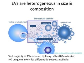
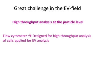
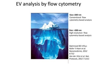
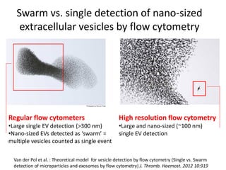
Flow cytometric analysis of
individual extracellular vesicles
Marca Wauben
Utrecht University
Dept. Biochemistry & Cell Biology
Fac. Veterinary Medicine
The Netherlands
Extracellular vesicles have changed the way we
look at communication in and between
biological systems
EV offer tremendous opportunities for clinical applications
ranging from biomarkers for diagnosis or prognosis
to therapeutic application of EV or mimics for drug delivery
Therapeutic application of
extracellular vesicles or mimics
Next hurdle to take: Large scale preparation and isolation of
well-defined vesicles
Quality control: Quantitative & qualitative analysis
Multiparameter analysis of individual vesicles
Vesicle-based biomarkers: A novel class
between small molecule and cellular
biomarkers
High potential biomarkers
BUT………
Major technical problem is the analysis of specific subsets (rare
events) of vesicles in complex body fluids
EVs are heterogeneous in size &
composition
Vast majority of EVs released by living cells <200nm in size
NO unique markers for different EV-subsets available
Cellular and Molecular Life Sciences 2011; 68(16):2667-88
Great challenge in the EV-field
• Cargo incorporation into EVs is dynamic
Individual EV analysis
can discriminate
• Mixed population of EVs
Bulk-based analysis methods e.g. Western-blotting,
proteomics, (bead)capture assays, ELISA
To monitor quantitative and qualitative changes in
EV-subsets
Great challenge in the EV-field
High throughput analysis at the particle level
Flow cytometer  Designed for high throughput analysis
of cells applied for EV analysis
EV analysis by flow cytometry
Size:>300 nm
Conventional flow
cytometry-based analysis
Size: <300 nm
High resolution flow
cytometry-based analysis
Optimized BD Influx:
Nolte-’t Hoen et al.
Nanomedicine, 2012
8:712
Van der Vlist et al. Nat.
Protocols, 2012 7:1311
Trigger signal
• Uniform parameter to detect all EVs of
interest
• Light scatter is useful as a trigger parameter
for cells and large EVs
• Small EVs (<300nm)  background problems
Scatter-based thresholding
Detection of fluorescent nano-sized beads
FSC-based
thresholding
10
0
10
1
10
2
10
3
10
410
0
10
1
10
2
10
3
10
4
threshold
SSC
Reduced wide-angle FSC
Nolte-’t Hoen et al. Nanomedicine, 2012 8:712
Van der Vlist et al. Nat. Protocols, 2012 7:1311
100 nm
200 nm
Fluorescence-based thresholding
Detection of fluorescent nano-sized beads
10
0
10
1
10
2
10
3
10
410
0
10
1
10
2
10
3
10
4
threshold
10
0
10
1
10
2
10
3
10
410
0
10
1
10
2
10
3
10
4
threshold
100 nm
beads
200 nm
beads
Reduced wide-angle FSC
Fluorescence
noise
Nolte-’t Hoen et al. Nanomedicine, 2012 8:712
Van der Vlist et al. Nat. Protocols, 2012 7:1311
Vesicle isolation:
Cell culture supernatant

2x 200g

2x 500g

1x 10,000g

1x 100,000g
Pelleted vesicles
Generic label PKH-67
Collection of
density
gradient
fractions
Flow cytometry
(Generic) fluorescent labeling of cell-derived vesicles
Specific protein
labeling
(FL-Ab)
Nolte-’t Hoen et al. Nanomedicine, 2012 8:712
Van der Vlist et al. Nat. Protocols, 2012 7:1311
High-resolution flow cytometric
analysis of nano-sized EVs
Reduced wide-angle FSC
10
0
10
1
10
2
10
3
10
410
0
10
1
10
2
10
3
10
4
PKH67Fluorescence
threshold 0
10000
20000
30000
40000
50000
60000
1,26
1,24
1,22
1,20
1,18
1,15
1,11
1,08
1,07
1,06
DC
Numberofevents
Density (g/ml)
QuantificationDetection
Nolte-’t Hoen et al., Nanomedicine 2012; Van der Vlist/Nolte-’t Hoen et al., Nature Protocols 2012; Van der Vlist et al., J Extracellular Vesicles 2012
MFG-E8 (B-PE)
MHCII(APC)
10
0
10
1
10
2
10
3
10
410
0
10
1
10
2
10
3
10
4
10
0
10
1
10
2
10
3
10
410
0
10
1
10
2
10
3
10
4
vesicles
act-DC
vesicles
non-act-DC
Reduced wide-angle FSC
PHK67fluorescence
10
0
10
1
10
2
10
3
10
410
0
10
1
10
2
10
3
10
4
1
3
2
Characterization
Proteins
T cell extracellular vesicles
Light scattering
Any flow cytometer can measure something
when concentrations are high enough…….
BUT what does the signal mean?
Van der Pol et al. : Theoretical model for vesicle detection by flow cytometry (Single vs. Swarm
detection of microparticles and exosomes by flow cytometry) J. Thromb. Haemost. 2012 10:919
Swarm vs. single detection of nano-sized
extracellular vesicles by flow cytometry
Regular flow cytometers
•Large single EV detection (>300 nm)
•Nano-sized EVs detected as ‘swarm’ =
multiple vesicles counted as single event
High resolution flow cytometry
•Large and nano-sized (~100 nm)
single EV detection
Swarm detection influences quantitative and
qualitative flow cytometric analysis of nano-
sized EVs
• Regular flow cytometers can be used for swarm detection of
nano-sized EVs  ‘Bulk-based’ analysis (no information on EV-
subsets, no quantitative analysis)
• For high resolution flow cytometry proper concentrations
should be used for genuine single nano-sized vesicle-based
analysis
-Reproducibility and comparison of results
-Development and evaluation of (novel)techniques
No gold standard technique available
Need for EV-like standards to calibrate and compare
Need for sample preparation guidelines
Need for sample analysis by several techniques
Need for standardization of high
throughput EV-analysis
Need for comprehensive reporting
of well-controlled experiments

ISEV2014 - Introduction to flow cytometry analysis of EV's (M. Wauben)

  • 1.
    Flow cytometric analysisof individual extracellular vesicles Marca Wauben Utrecht University Dept. Biochemistry & Cell Biology Fac. Veterinary Medicine The Netherlands
  • 2.
    Extracellular vesicles havechanged the way we look at communication in and between biological systems EV offer tremendous opportunities for clinical applications ranging from biomarkers for diagnosis or prognosis to therapeutic application of EV or mimics for drug delivery
  • 3.
    Therapeutic application of extracellularvesicles or mimics Next hurdle to take: Large scale preparation and isolation of well-defined vesicles Quality control: Quantitative & qualitative analysis Multiparameter analysis of individual vesicles
  • 4.
    Vesicle-based biomarkers: Anovel class between small molecule and cellular biomarkers High potential biomarkers BUT……… Major technical problem is the analysis of specific subsets (rare events) of vesicles in complex body fluids
  • 5.
    EVs are heterogeneousin size & composition Vast majority of EVs released by living cells <200nm in size NO unique markers for different EV-subsets available Cellular and Molecular Life Sciences 2011; 68(16):2667-88
  • 6.
    Great challenge inthe EV-field • Cargo incorporation into EVs is dynamic Individual EV analysis can discriminate • Mixed population of EVs Bulk-based analysis methods e.g. Western-blotting, proteomics, (bead)capture assays, ELISA To monitor quantitative and qualitative changes in EV-subsets
  • 7.
    Great challenge inthe EV-field High throughput analysis at the particle level Flow cytometer  Designed for high throughput analysis of cells applied for EV analysis
  • 8.
    EV analysis byflow cytometry Size:>300 nm Conventional flow cytometry-based analysis Size: <300 nm High resolution flow cytometry-based analysis Optimized BD Influx: Nolte-’t Hoen et al. Nanomedicine, 2012 8:712 Van der Vlist et al. Nat. Protocols, 2012 7:1311
  • 9.
    Trigger signal • Uniformparameter to detect all EVs of interest • Light scatter is useful as a trigger parameter for cells and large EVs • Small EVs (<300nm)  background problems
  • 10.
    Scatter-based thresholding Detection offluorescent nano-sized beads FSC-based thresholding 10 0 10 1 10 2 10 3 10 410 0 10 1 10 2 10 3 10 4 threshold SSC Reduced wide-angle FSC Nolte-’t Hoen et al. Nanomedicine, 2012 8:712 Van der Vlist et al. Nat. Protocols, 2012 7:1311 100 nm 200 nm
  • 11.
    Fluorescence-based thresholding Detection offluorescent nano-sized beads 10 0 10 1 10 2 10 3 10 410 0 10 1 10 2 10 3 10 4 threshold 10 0 10 1 10 2 10 3 10 410 0 10 1 10 2 10 3 10 4 threshold 100 nm beads 200 nm beads Reduced wide-angle FSC Fluorescence noise Nolte-’t Hoen et al. Nanomedicine, 2012 8:712 Van der Vlist et al. Nat. Protocols, 2012 7:1311
  • 12.
    Vesicle isolation: Cell culturesupernatant  2x 200g  2x 500g  1x 10,000g  1x 100,000g Pelleted vesicles Generic label PKH-67 Collection of density gradient fractions Flow cytometry (Generic) fluorescent labeling of cell-derived vesicles Specific protein labeling (FL-Ab) Nolte-’t Hoen et al. Nanomedicine, 2012 8:712 Van der Vlist et al. Nat. Protocols, 2012 7:1311
  • 13.
    High-resolution flow cytometric analysisof nano-sized EVs Reduced wide-angle FSC 10 0 10 1 10 2 10 3 10 410 0 10 1 10 2 10 3 10 4 PKH67Fluorescence threshold 0 10000 20000 30000 40000 50000 60000 1,26 1,24 1,22 1,20 1,18 1,15 1,11 1,08 1,07 1,06 DC Numberofevents Density (g/ml) QuantificationDetection Nolte-’t Hoen et al., Nanomedicine 2012; Van der Vlist/Nolte-’t Hoen et al., Nature Protocols 2012; Van der Vlist et al., J Extracellular Vesicles 2012 MFG-E8 (B-PE) MHCII(APC) 10 0 10 1 10 2 10 3 10 410 0 10 1 10 2 10 3 10 4 10 0 10 1 10 2 10 3 10 410 0 10 1 10 2 10 3 10 4 vesicles act-DC vesicles non-act-DC Reduced wide-angle FSC PHK67fluorescence 10 0 10 1 10 2 10 3 10 410 0 10 1 10 2 10 3 10 4 1 3 2 Characterization Proteins T cell extracellular vesicles Light scattering
  • 14.
    Any flow cytometercan measure something when concentrations are high enough……. BUT what does the signal mean?
  • 15.
    Van der Polet al. : Theoretical model for vesicle detection by flow cytometry (Single vs. Swarm detection of microparticles and exosomes by flow cytometry) J. Thromb. Haemost. 2012 10:919 Swarm vs. single detection of nano-sized extracellular vesicles by flow cytometry Regular flow cytometers •Large single EV detection (>300 nm) •Nano-sized EVs detected as ‘swarm’ = multiple vesicles counted as single event High resolution flow cytometry •Large and nano-sized (~100 nm) single EV detection
  • 16.
    Swarm detection influencesquantitative and qualitative flow cytometric analysis of nano- sized EVs • Regular flow cytometers can be used for swarm detection of nano-sized EVs  ‘Bulk-based’ analysis (no information on EV- subsets, no quantitative analysis) • For high resolution flow cytometry proper concentrations should be used for genuine single nano-sized vesicle-based analysis
  • 17.
    -Reproducibility and comparisonof results -Development and evaluation of (novel)techniques No gold standard technique available Need for EV-like standards to calibrate and compare Need for sample preparation guidelines Need for sample analysis by several techniques Need for standardization of high throughput EV-analysis Need for comprehensive reporting of well-controlled experiments