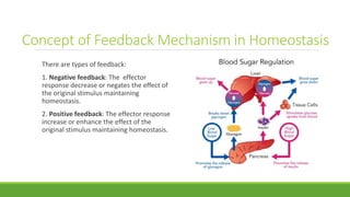
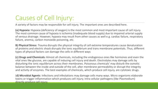
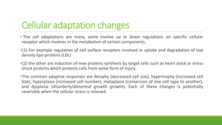
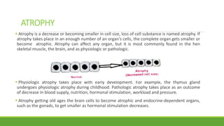
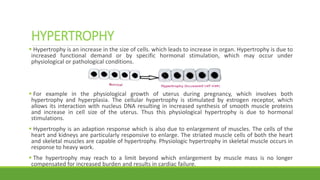
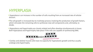
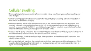
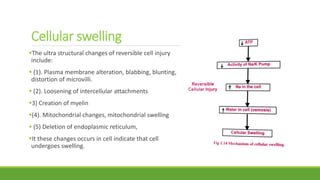
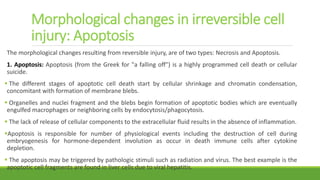
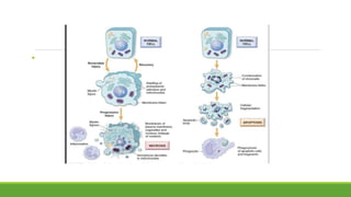
This is a presentation on the topic of Adaptations, Cell injury and cell death, prepared by Dr Ashish Jawarkar, he is MD in pathology and a teacher at Parul institute of Medical sciences and research Vadodara.
Difference between Apoptosis versus Necrosis and Types of Necrosis.pptxRukhshanda Ramzaan
Apoptosis Versus Nercosis
Apoptosis Necrosis
Predefined cell suicide or programmed cell death. Natural physiological Process. Involve one cell at a time. Cell shrinkage (Dense eosinophilic cytoplasm) Pyknosis (Condensation) and Karyorrhexis (fragmentation) of nuclear material Formation of membrane blebs and apoptotic bodies
Phagocytosis of apoptotic bodies by Macrophages
Caspase dependent pathway
No Inflammation (no immune response) Premature, unprogrammed cell death always pathological. Involve many cells Cell Swelling (Swelling of endoplasmic reticulum and mitochondria) and membrane blebs Pyknosis (condensation), Karyorrhexis (Fragmentation) and Karyolysis (lysis)of the nucleus. Breakdown of the plasma membrane, organelles (enzymatic digestion), leakage of cellular contents
Increased eosinophilia, Accumulation of Myelin figures (whorled precipitated Phospholipids)
Initiate Inflammation (Strong immune response)
The study of the blood flow is called hemodynamics.
Thus hemodynamics deals with the dynamics of blood flow. The circulatory system is controlled by homeostatic mechanisms, much as hydraulic circuits and are controlled by control systems.
Hemodynamic response continuously monitors and adjusts to conditions in the body and its environment. Thus hemodynamics explains the physical laws that govern the flow of blood in the blood vessels.
Cellular adaptations, injury and death.. Lecture 1Ashish Jawarkar
This is a series of lectures on general pathology useful for undergraduate and postgraduate pathology students. The ppts here have are enriched with explanatory pictures as well as useful video links.. hope you find them useful
Basic principles of Cell injury and AdaptationAkshayYadav176
Basic principles of Cell injury and Adaptation:
(As per new syllabus of PCI)
Introduction, definitions, Homeostasis, Components and Types of Feedback systems, Causes of cellular injury,Pathogenesis (Cell membrane damage, Mitochondrial damage, Ribosome damage, Nuclear damage),Morphology of cell injury – Adaptive changes (Atrophy, Hypertrophy, hyperplasia, Metaplasia, Dysplasia),Cell swelling, Intra cellular accumulation, Calcification, Enzyme leakage and Cell Death Acidosis & Alkalosis,Electrolyte imbalance.
This ppt on basic Introduction of General Pathology including the etiology and pathogenesis and their factors .
General Pathology into simple terms for you to understand and use in your exam.
These simplified slides by Dr. Sidra Arshad present an overview of the non-respiratory functions of the respiratory tract.
Learning objectives:
1. Enlist the non-respiratory functions of the respiratory tract
2. Briefly explain how these functions are carried out
3. Discuss the significance of dead space
4. Differentiate between minute ventilation and alveolar ventilation
5. Describe the cough and sneeze reflexes
Study Resources:
1. Chapter 39, Guyton and Hall Textbook of Medical Physiology, 14th edition
2. Chapter 34, Ganong’s Review of Medical Physiology, 26th edition
3. Chapter 17, Human Physiology by Lauralee Sherwood, 9th edition
4. Non-respiratory functions of the lungs https://academic.oup.com/bjaed/article/13/3/98/278874
micro teaching on communication m.sc nursing.pdfAnurag Sharma
Microteaching is a unique model of practice teaching. It is a viable instrument for the. desired change in the teaching behavior or the behavior potential which, in specified types of real. classroom situations, tends to facilitate the achievement of specified types of objectives.
ARTIFICIAL INTELLIGENCE IN HEALTHCARE.pdfAnujkumaranit
Artificial intelligence (AI) refers to the simulation of human intelligence processes by machines, especially computer systems. It encompasses tasks such as learning, reasoning, problem-solving, perception, and language understanding. AI technologies are revolutionizing various fields, from healthcare to finance, by enabling machines to perform tasks that typically require human intelligence.
Lung Cancer: Artificial Intelligence, Synergetics, Complex System Analysis, S...Oleg Kshivets
RESULTS: Overall life span (LS) was 2252.1±1742.5 days and cumulative 5-year survival (5YS) reached 73.2%, 10 years – 64.8%, 20 years – 42.5%. 513 LCP lived more than 5 years (LS=3124.6±1525.6 days), 148 LCP – more than 10 years (LS=5054.4±1504.1 days).199 LCP died because of LC (LS=562.7±374.5 days). 5YS of LCP after bi/lobectomies was significantly superior in comparison with LCP after pneumonectomies (78.1% vs.63.7%, P=0.00001 by log-rank test). AT significantly improved 5YS (66.3% vs. 34.8%) (P=0.00000 by log-rank test) only for LCP with N1-2. Cox modeling displayed that 5YS of LCP significantly depended on: phase transition (PT) early-invasive LC in terms of synergetics, PT N0—N12, cell ratio factors (ratio between cancer cells- CC and blood cells subpopulations), G1-3, histology, glucose, AT, blood cell circuit, prothrombin index, heparin tolerance, recalcification time (P=0.000-0.038). Neural networks, genetic algorithm selection and bootstrap simulation revealed relationships between 5YS and PT early-invasive LC (rank=1), PT N0—N12 (rank=2), thrombocytes/CC (3), erythrocytes/CC (4), eosinophils/CC (5), healthy cells/CC (6), lymphocytes/CC (7), segmented neutrophils/CC (8), stick neutrophils/CC (9), monocytes/CC (10); leucocytes/CC (11). Correct prediction of 5YS was 100% by neural networks computing (area under ROC curve=1.0; error=0.0).
CONCLUSIONS: 5YS of LCP after radical procedures significantly depended on: 1) PT early-invasive cancer; 2) PT N0--N12; 3) cell ratio factors; 4) blood cell circuit; 5) biochemical factors; 6) hemostasis system; 7) AT; 8) LC characteristics; 9) LC cell dynamics; 10) surgery type: lobectomy/pneumonectomy; 11) anthropometric data. Optimal diagnosis and treatment strategies for LC are: 1) screening and early detection of LC; 2) availability of experienced thoracic surgeons because of complexity of radical procedures; 3) aggressive en block surgery and adequate lymph node dissection for completeness; 4) precise prediction; 5) adjuvant chemoimmunoradiotherapy for LCP with unfavorable prognosis.
The prostate is an exocrine gland of the male mammalian reproductive system
It is a walnut-sized gland that forms part of the male reproductive system and is located in front of the rectum and just below the urinary bladder
Function is to store and secrete a clear, slightly alkaline fluid that constitutes 10-30% of the volume of the seminal fluid that along with the spermatozoa, constitutes semen
A healthy human prostate measures (4cm-vertical, by 3cm-horizontal, 2cm ant-post ).
It surrounds the urethra just below the urinary bladder. It has anterior, median, posterior and two lateral lobes
It’s work is regulated by androgens which are responsible for male sex characteristics
Generalised disease of the prostate due to hormonal derangement which leads to non malignant enlargement of the gland (increase in the number of epithelial cells and stromal tissue)to cause compression of the urethra leading to symptoms (LUTS
New Drug Discovery and Development .....NEHA GUPTA
The "New Drug Discovery and Development" process involves the identification, design, testing, and manufacturing of novel pharmaceutical compounds with the aim of introducing new and improved treatments for various medical conditions. This comprehensive endeavor encompasses various stages, including target identification, preclinical studies, clinical trials, regulatory approval, and post-market surveillance. It involves multidisciplinary collaboration among scientists, researchers, clinicians, regulatory experts, and pharmaceutical companies to bring innovative therapies to market and address unmet medical needs.
Ethanol (CH3CH2OH), or beverage alcohol, is a two-carbon alcohol
that is rapidly distributed in the body and brain. Ethanol alters many
neurochemical systems and has rewarding and addictive properties. It
is the oldest recreational drug and likely contributes to more morbidity,
mortality, and public health costs than all illicit drugs combined. The
5th edition of the Diagnostic and Statistical Manual of Mental Disorders
(DSM-5) integrates alcohol abuse and alcohol dependence into a single
disorder called alcohol use disorder (AUD), with mild, moderate,
and severe subclassifications (American Psychiatric Association, 2013).
In the DSM-5, all types of substance abuse and dependence have been
combined into a single substance use disorder (SUD) on a continuum
from mild to severe. A diagnosis of AUD requires that at least two of
the 11 DSM-5 behaviors be present within a 12-month period (mild
AUD: 2–3 criteria; moderate AUD: 4–5 criteria; severe AUD: 6–11 criteria).
The four main behavioral effects of AUD are impaired control over
drinking, negative social consequences, risky use, and altered physiological
effects (tolerance, withdrawal). This chapter presents an overview
of the prevalence and harmful consequences of AUD in the U.S.,
the systemic nature of the disease, neurocircuitry and stages of AUD,
comorbidities, fetal alcohol spectrum disorders, genetic risk factors, and
pharmacotherapies for AUD.
Couples presenting to the infertility clinic- Do they really have infertility...Sujoy Dasgupta
Dr Sujoy Dasgupta presented the study on "Couples presenting to the infertility clinic- Do they really have infertility? – The unexplored stories of non-consummation" in the 13th Congress of the Asia Pacific Initiative on Reproduction (ASPIRE 2024) at Manila on 24 May, 2024.
Pulmonary Thromboembolism - etilogy, types, medical- Surgical and nursing man...VarunMahajani
Disruption of blood supply to lung alveoli due to blockage of one or more pulmonary blood vessels is called as Pulmonary thromboembolism. In this presentation we will discuss its causes, types and its management in depth.
- Video recording of this lecture in English language: https://youtu.be/lK81BzxMqdo
- Video recording of this lecture in Arabic language: https://youtu.be/Ve4P0COk9OI
- Link to download the book free: https://nephrotube.blogspot.com/p/nephrotube-nephrology-books.html
- Link to NephroTube website: www.NephroTube.com
- Link to NephroTube social media accounts: https://nephrotube.blogspot.com/p/join-nephrotube-on-social-media.html
Acute scrotum is a general term referring to an emergency condition affecting the contents or the wall of the scrotum.
There are a number of conditions that present acutely, predominantly with pain and/or swelling
A careful and detailed history and examination, and in some cases, investigations allow differentiation between these diagnoses. A prompt diagnosis is essential as the patient may require urgent surgical intervention
Testicular torsion refers to twisting of the spermatic cord, causing ischaemia of the testicle.
Testicular torsion results from inadequate fixation of the testis to the tunica vaginalis producing ischemia from reduced arterial inflow and venous outflow obstruction.
The prevalence of testicular torsion in adult patients hospitalized with acute scrotal pain is approximately 25 to 50 percent
HOT NEW PRODUCT! BIG SALES FAST SHIPPING NOW FROM CHINA!! EU KU DB BK substit...GL Anaacs
Contact us if you are interested:
Email / Skype : kefaya1771@gmail.com
Threema: PXHY5PDH
New BATCH Ku !!! MUCH IN DEMAND FAST SALE EVERY BATCH HAPPY GOOD EFFECT BIG BATCH !
Contact me on Threema or skype to start big business!!
Hot-sale products:
NEW HOT EUTYLONE WHITE CRYSTAL!!
5cl-adba precursor (semi finished )
5cl-adba raw materials
ADBB precursor (semi finished )
ADBB raw materials
APVP powder
5fadb/4f-adb
Jwh018 / Jwh210
Eutylone crystal
Protonitazene (hydrochloride) CAS: 119276-01-6
Flubrotizolam CAS: 57801-95-3
Metonitazene CAS: 14680-51-4
Payment terms: Western Union,MoneyGram,Bitcoin or USDT.
Deliver Time: Usually 7-15days
Shipping method: FedEx, TNT, DHL,UPS etc.Our deliveries are 100% safe, fast, reliable and discreet.
Samples will be sent for your evaluation!If you are interested in, please contact me, let's talk details.
We specializes in exporting high quality Research chemical, medical intermediate, Pharmaceutical chemicals and so on. Products are exported to USA, Canada, France, Korea, Japan,Russia, Southeast Asia and other countries.
Prix Galien International 2024 Forum ProgramLevi Shapiro
June 20, 2024, Prix Galien International and Jerusalem Ethics Forum in ROME. Detailed agenda including panels:
- ADVANCES IN CARDIOLOGY: A NEW PARADIGM IS COMING
- WOMEN’S HEALTH: FERTILITY PRESERVATION
- WHAT’S NEW IN THE TREATMENT OF INFECTIOUS,
ONCOLOGICAL AND INFLAMMATORY SKIN DISEASES?
- ARTIFICIAL INTELLIGENCE AND ETHICS
- GENE THERAPY
- BEYOND BORDERS: GLOBAL INITIATIVES FOR DEMOCRATIZING LIFE SCIENCE TECHNOLOGIES AND PROMOTING ACCESS TO HEALTHCARE
- ETHICAL CHALLENGES IN LIFE SCIENCES
- Prix Galien International Awards Ceremony
Recomendações da OMS sobre cuidados maternos e neonatais para uma experiência pós-natal positiva.
Em consonância com os ODS – Objetivos do Desenvolvimento Sustentável e a Estratégia Global para a Saúde das Mulheres, Crianças e Adolescentes, e aplicando uma abordagem baseada nos direitos humanos, os esforços de cuidados pós-natais devem expandir-se para além da cobertura e da simples sobrevivência, de modo a incluir cuidados de qualidade.
Estas diretrizes visam melhorar a qualidade dos cuidados pós-natais essenciais e de rotina prestados às mulheres e aos recém-nascidos, com o objetivo final de melhorar a saúde e o bem-estar materno e neonatal.
Uma “experiência pós-natal positiva” é um resultado importante para todas as mulheres que dão à luz e para os seus recém-nascidos, estabelecendo as bases para a melhoria da saúde e do bem-estar a curto e longo prazo. Uma experiência pós-natal positiva é definida como aquela em que as mulheres, pessoas que gestam, os recém-nascidos, os casais, os pais, os cuidadores e as famílias recebem informação consistente, garantia e apoio de profissionais de saúde motivados; e onde um sistema de saúde flexível e com recursos reconheça as necessidades das mulheres e dos bebês e respeite o seu contexto cultural.
Estas diretrizes consolidadas apresentam algumas recomendações novas e já bem fundamentadas sobre cuidados pós-natais de rotina para mulheres e neonatos que recebem cuidados no pós-parto em unidades de saúde ou na comunidade, independentemente dos recursos disponíveis.
É fornecido um conjunto abrangente de recomendações para cuidados durante o período puerperal, com ênfase nos cuidados essenciais que todas as mulheres e recém-nascidos devem receber, e com a devida atenção à qualidade dos cuidados; isto é, a entrega e a experiência do cuidado recebido. Estas diretrizes atualizam e ampliam as recomendações da OMS de 2014 sobre cuidados pós-natais da mãe e do recém-nascido e complementam as atuais diretrizes da OMS sobre a gestão de complicações pós-natais.
O estabelecimento da amamentação e o manejo das principais intercorrências é contemplada.
Recomendamos muito.
Vamos discutir essas recomendações no nosso curso de pós-graduação em Aleitamento no Instituto Ciclos.
Esta publicação só está disponível em inglês até o momento.
Prof. Marcus Renato de Carvalho
www.agostodourado.com
2. There are two separate field involve in pathophysiology
First is Physiology: the study of body and its function
Second is Pathology: the study of disease and its impact on body.
When combine, pathophysiology concern with study of mechanism
responsible for initiation, development, treatment of pathological
process in humans and animals and provide appropriate care to the
patient.
Pathophysiology
3. Basic principals of Cell Injury & Adaptation
Introduction, Definitions,
Homeostasis,
Components and Types of Feedback systems,
Causes of cellular injury,
Pathogenesis (Cell membrane damage, Mitochondrial damage, Ribosome damage, Nuclear
damage)
Morphology of cell injury – Adaptive changes (Atrophy, Hypertrophy, hyperplasia, Metaplasia,
Dysplasia),Cell swelling,
Intra cellular accumulation, Calcification, Enzyme leakage and Cell Death Acidosis & Alkalosis,
Electrolyte imbalance.
4. Introduction
Cells are the basic units of tissues, which form organs and systems in the human body.
Traditionally, body cells are divided into two main types: epithelial and mesenchymal cells
In 1859, Virchow first published a cellular theory of disease, bringing in the concept that
diseases occur due to peculiarities at the level of cells
Thus, most forms diseases begin with cell injury followed by consequent loss of cellular
function.
Cell injury is defined as a variety of stresses a cell encounters as a result of changes in internal
and external environment.
In general, cells of the body have the inbuilt mechanism to deal with changes in the
environment to an extent.
The environment of a living cells frequently changing. Thus, the availability of nutrient en,
blood supply and regulatory substances like hormones is not the same at all time. Homeostatic
mechanisms try to keep these changes within physiological range.
5. The cellular response to stress
The cellular response to stress may vary and depends upon the
following variables:
i) Type of cell and tissue involved
ii) Extent and type of cell injury
Various forms of cellular responses to cell injury may be as
follows:
1. When there is improved working demand, the cell may
adapt to the changes which are revealed morphologically and
then revert back to normal after stress is removed (Cellular
adaptation)
2. If the stress is mild to moderate, the injured cell may recover
(reversible cell injury) while when the damage is persistent cell
death may occur (Irreversible cell injury)
6. Homeostasis refers to stability, balance or equilibrium in the body's internal environment due to constant interaction of
body's many regulatory processes.
Maintaining a stable internal environment requires constant monitoring and adjustments as conditions change.
Components Homeostatic mechanism is maintained by control system or feedback system, having three components;
1. Receptor (sensor): It is a body structure that monitors changes in a controlled condition and sends input to a control
center (integrator). The input is in the form of nerve impulses or chemical signals. Example: Certain nerve endings in the
skin sense temperature and detect changes.
2. Control centre (integrator): It integrates (puts together) data from sensor provides commands to an effector but this
time the effector produces a physiological response that adds to the initial changes in the controlled condition. Example-
the brain, sets the range values within which a controlled condition should be maintained, evaluates the input it receives
from receptors and generates output commands when they are needed. The output from the control center occurs as
nerve impulses, or hormones and other chemical signals.
3. Effectors: It is the mechanism that has a body structure that receives output from the control center and produces a
response or effect that changes the controlled condition. Nearly every organ or tissue in the body can behave as an
effector
Homeostasis
7. Homeostasis
Example: If the temperature of a body is decreased from set point, the receptor detects the
change in temperature and signals the thermostat (effector). Result in shivering which generate
heat and raises body temperature. Similarly, it the temperature is increased from set point,
thermo receptor are involved in the detection of temperature change . Once stimulated, it act
on effectors for cooling (sweat gland) or heating (e.g. muscles) the body to reverse the change to
set point.
8. Concept of Feedback Mechanism in Homeostasis
There are types of feedback:
1. Negative feedback: The effector
response decrease or negates the effect of
the original stimulus maintaining
homeostasis.
2. Positive feedback: The effector response
increase or enhance the effect of the
original stimulus maintaining homeostasis.
9.
10. Causes of Cell Injury:
A variety of factors may be responsible for cell injury. The important ones are described here.
(a) Hypoxia: Hypoxia (deficiency of oxygen) is the most common and most important cause of cell injury.
The most common cause of hypoxia is ischemia (inadequate blood supply) due to impaired arterial supply
of venous drainage. However, hypoxia may result from other causes as well e.g. cardiac failure, respiratory
failure, anemia, carbon monoxide poisoning, etc.
B) Physical Stress: Trauma disrupts the physical integrity of cell extreme temperatures cause denaturation
of proteins and electric shock disrupts the ionic equilibrium and trans membrane potentials. Thus, different
types of physical factors can damage the cells in different ways
(c) Drugs and Chemicals: Almost all chemicals, including the endogenous ones like hormones and even the
vital ones like glucose, are capable of inducing cell injury and death. Electrolytes may damage cells by
disturbing the ionic equilibrium across their membranes. Poisonous chemicals' may disturb the osmotic
balance between the inside and outside of the cell, alter membrane permeability or disrupt the integrity
and activity of enzymes. The best examples of chemicals, which produce cell injury, are cytotoxic drugs.
(d) Microbial Agents: Infections and infestations may damage cells many ways. Micro organisms elaborate
toxins or trigger inflammation which produces cell injury. Intra cellular pathogens (like Plasmodium)
directly produce cell injury.
11. Causes of Cell Injury:
(e) Immunologic Factors: Allergic reactions may produce devastating cell injury as in the case of
the cytolytic allergic reactions. Similarly, auto immune reactions may cause cell injury, as in
Type-I diabetes mellitus (DDM).
(f) Nutritional Derangements: A deficiency or an excess of nutrients may result in nutritional
imbalances.
Nutritional deficiency diseases may be due to overall deficiency of nutrients (e.g. starvation), of
protein calorie (e.g. marasmus, kwashiorkor), of minerals (e.g. anaemia), or of trace elements.
Nutritional excess is a problem of affluent societies resulting in obesity atherosclerosis, heart
disease and hypertension.
g)Aging: Cellular aging leads to impaired ability of the cells to undergo replication and repair,
and ultimately lead to cell death culminating in death of the individual.
h) Psychogenic Disease, i) Iatrogenic Factors, j) Idiopathic Diseases.
12. Cellular adaptation changes
The cell adaptations are many, some involve up or down regulations on specific cellular
receptor which involves in the metabolism of certain components.
(1) For example regulation of cell surface receptors involved in uptake and degradation of low
density lipo-proteins (LDL)
(2) the other are induction of new proteins synthesis by target cells such as heart stock or stress
shock proteins which protects cells from some form of injury.
The common adaptive responses are Atrophy (decreased cell size), hypertrophy (increased cell
Size), hyperplasia (increased cell number), metaplasia (conversion of one cell type to another),
and dysplasia (disorderly/abnormal growth growth). Each of these changes is potentially
reversible when the cellular stress is relieved.
13. ATROPHY
Atrophy is a decrease or becoming smaller in cell size, loss of cell substance is named atrophy. If
atrophy takes place in an enough number of an organ's cells, the complete organ gets smaller or
become atrophic. Atrophy can affect any organ, but it is most commonly found in the hen
skeletal muscle, the brain, and as physiologic or pathologic.
Physiologic atrophy takes place with early development. For example, the thymus gland
undergoes physiologic atrophy during childhood. Pathologic atrophy takes place as an outcome
of decrease in blood supply, nutrition, hormonal stimulation, workload and pressure.
Atrophy getting old ages the brain cells to become atrophic and endocrine-dependent organs,
such as the gonads, to get smaller as hormonal stimulation decreases.
14. HYPERTROPHY
Hypertrophy is an increase in the size of cells. which leads to increase in organ. Hypertrophy is due to
increased functional demand or by specific hormonal stimulation, which may occur under
physiological or pathological conditions.
For example in the physiological growth of uterus during pregnancy, which involves both
hypertrophy and hyperplasia. The cellular hypertrophy is stimulated by estrogen receptor, which
allows its interaction with nucleus DNA resulting in increased synthesis of smooth muscle proteins
and increase in cell size of the uterus. Thus this physiological hypertrophy is due to hormonal
stimulations.
Hypertrophy is an adaption response which is also due to enlargement of muscles. The cells of the
heart and kidneys are particularly responsive to enlarge. The striated muscle cells of both the heart
and skeletal muscles are capable of hypertrophy. Physiologic hypertrophy in skeletal muscle occurs in
response to heavy work.
The hypertrophy may reach to a limit beyond which enlargement by muscle mass is no longer
compensated for increased burden and results in cardiac failure.
15. HYPERPLASIA
Hyperplasia is an increase in the number of cells resulting from an increased rate of cellular
division.
The cell growth is increased due to multistep process involving the production of growth factors,
which stimulate the remaining cells to synthesize new cell components and, ultimately, to
divide.
Hyperplasia and hypertrophy are closely related and often develop simultaneously in tissue.
Both hyperplasia and hypertrophy take place if the cells are capable of synthesizing DNA.
Cardiac and skeletal muscle cells have no capacity for hyperplastic growth and thus usually
undergo only hypertrophy.
16. HYPERPLASIA
Hyperplasia can be physiologic or pathological: Physiology hyperplasia is divided into
(1) Hormonal hyperplasia: most from of pathological hyperplasia is due to excessive hormonal
stimulation and its effect on growth factor on targeted cells. It is seen in proliferation of the
epithelium of the female breast at puberty and during pregnancy
(2) Compensatory hyperplasia: is an adaptive mechanism that enables certain organs to
regenerate. For example when a portion of tissue is removed, mitotic activity in the remaining
liver cells begins as early 12 hours after hypertrophy resulting the liver to attain its normal
weight.
The various specific factors that appear to be involved in liver regenerating are transforming
growth factor hepatocyte growth factor (HGE».is a mediator in vitro of liver regeneration. In
addition, other in vitro growth factors and cytokines (cell-signaling proteins) that increase
hepatic cell growth regeneration includes transforming factor-alpha (TGF-a), epidermal growth
factor (EGF), necrosis factor-alpha (TNF-).
17. METAPLASIA
Metaplasia is the reversible replacement of one adult cell by another. sometimes less
differentiated, cell type.
Metaplasia is best seen in the form of squamous change that occurs in respiratory tract in the
habitual cigarette smoke. The normal columnar ciliated epithelial cells of trachea and bronchi
are replaced mainly by striated squamous epithelial cells.
18. DYSPLASIA
Dysplasia is related to occurs when abnormal changes in the size, shape, and organization of
adult cells which is related hyperplasia and is often called atypical hyperplasia.
It is not consider as true adaptive process. These changes are strongly associated with common
neoplastic growth and often found adjacent to cancerous cells.
19. Pathogenesis of Cell Injury
Cell injury is cause by various reasons. Mechanism and location of cell injury is well defined
based on injurious agent or injury causing agent.
Cell Injury Is of two type: Reversible Cell Injury and Irreversible cell injury
Pathogenesis of cell injury involve below two pathways
1. Ischemic or Hypoxic Injury
2. Free radical mediated cell injury.
20. ISCHEMIC AND HYPOXIC INJURY
1. Cellular Injury: The term, ischemia, has been used to denote deficient blood supply to due to
obstruction of the arterial inflow. The disorders characterized by ischemia such as myocardial infarction,
stroke, and peripheral vascular disease, most frequent causes of cell Injury or cell damage.
2. Cell damage: The effect of hypoxia is in the aerobic respiration cells involving the oxidative
phosphorylation reaction in mitochondria. The generation of adenosine triphosphate membrane (ATP)
slows down or stops which may effect on many oxidation systems which causes failure of the active sodium
pump, this effects the accumulation of sodium intracellular and potassium out from fusion of the cell.
Thus, it produces acute cellular swelling due to water. This stage is reversible if oxygen is restored.
If ischemia persists, the irreversible injury may be the result. It is associated with vacuolization of the
mitochondria which includes extensive damage plasma membrane, swelling of lysosome, and particularly
massive calcium influx into cell, leads to Ca++ deposits in mitochondrial matrix and efflux the cellular
enzyme creatine kinases (CK) and lactate dehydrogenase; There is continued loss of proteins, essential
coenzyme and ribonucleic acid from the hypermeable membranes. The cell reaches at stage to necrosis or
cell death. This sage is called Irreversible Injury.
21. ISCHEMIC AND HYPOXIC INJURY
3. Nuclear damage: The decrease in cellular ATP and Increase in adenosine monophosphate
(AMP) also stimulates the enzyme Phospho-fructotokinase glycolysis in order to maintain the
cells energy source by generating the ATP from glycogen. The glycogen gets rapidly depleted.
Glycolysis results in the accumulation of the lactic acid and inorganic phosphate from the
hydrolysis of phosphate ester, it reduces the intercellular pH and glycogen which results in
clumping the chromatin. This stage is reversible is restore if oxygen d.
If ischemia persists, the cell reaches at the next stage- irreversible injury. The fall of pH leads
to injury to the liposomal membranes followed by leakage of their enzymes in to cytoplasm,
activation of acid hydrolases and enzymatic digestion of cytoplasmic and nuclear components.
22. 4. Ribosome damage:
This process also involves oxidative phosphorylation, decrease the ATP the result detachment of
ribosomes from the granular endoplasmic reticulum and dissociation polysomes into
monosomes, decreased proteins synthesis , lipids deposition and lipotoxicity.
This stage cell may recover the injury and is called reversible injury If hypoxia continues, it will
lead to increased membrane permeability and diminished mitochondrial functions. In
mitochondria will appear normal, slightly swollen or condensed, the endoplasmic reticulum
dilated and entire cell is markedly swollen, a stage arises called apoptosis irreversible injury.
5. Mitochondrial Damage:
Cell injury or damage occurs as a result of the initial ischemia. ATP levels and intracellular pH
decreases as a result of anaerobic metabolism and lactate accumulation. As a consequence,
ATPase-dependent ion transport mechanisms become dysfunctional, this stage is called
reversible Injury if hypoxia is restored. If hypoxia continue which leads to contributing an
increased intracellular and mitochondrial calcium levels (calcium overload), cell keeps swelling
and rupture, and thus, there is cell death by necrotic, necro-apoptotic, and autophagic
mechanisms
23. Free Radical Mediated cell injury
Chemical species with unpaired or odd number of electron are called as free radicals.
In Biological system the term free radicals mostly refers to reactive oxygen species (ROS). The
major ROS are superoxide anion (O2), hydrogen peroxide (H2O2), hydroxyl radical (OH), Beside
ROS, reactive nitrogen species (RNS) includes nitric oxide (NO), Peroxynitrite (NO3), S-
nitrosothile also contribute to generation of free radicals.
These radicals are extremely unstable and react with inorganic membrane and nucleic acid in
the cell and start the chain of damage.
These radicals are emitted within the cell due to absorption of radiant energy e.g. U V light,
X-rays or reduction reactions that occurs during normal metabolic reactions or may be derived
from enzymatic metabolism of exogeneous chemicals.
24. Free Radical Mediated cell injury
1. Radiation injury: Radiation enters a cell; it can be absorbed by macromolecules directly, but
more commonly it reacts with water to produce free radicals such as OH, H.
These radicals interact with membranes nucleic acid and key elements within cell.
macromolecules such as proteins and DNA.
The enzymatic and structural proteins depend on their three-dimensional (3-D) structure for
their function, and this 3-D structure is dependent upon various types of chemical bonds. These
bonds are disrupted by radiation, mostly by the intermediation of free radicals, and the proteins
are then incapable of performing their structural or enzymatic duties and resulting in the
induction of mutation or cell death.
2. Oxygen Toxicity: When excessive quantities of oxygen is administered in patients resulting
symptoms bodily disturbance are from breathing due to high partial pressures of oxygen.
Symptoms observed such as visual and hearing abnormalities, unusual fatigue twitching,
anxiety, confusion, in-coordination, and convulsions. The formation of free while breathing,
muscular Oxygen radical causes inhalation the pulmonary therapy.
25. Free Radical Mediated cell injury
3) Chemical Injury: They can by combining with some molecular component or cellular
organelle. For example in case of mercuric chloride poisoning mercury bind to sulfhydryl
groups of the cell membrane and other proteins, causing increase permeability and inhibition
of ATPase dependent transport. Many anti-neoplastic chemotherapeutic agents agents and
antibiotic drugs also induced cell damage by direct cytotoxic effects in the induction of
mutation or cell death.
26.
27. Morphological changes in reversible cell
injury: Cellular swelling
Cellular swelling applicable to accumulation of water, or hydropic swelling, is the manifestation
of most forms of reversible cell injury.
Hydropic swelling results from abnormal function of the sodium-potassium (Na'-K') pumps that
normally maintain ionic equilibrium f pump the cell. In this stage cell membrane is swell and the
channels become closed, the Nat-K+ unable to exchange the ions results in accumulation of
sodium ions within the cell, creating osmotic gradient for water entry.
Because Na"-K´ pump function is dependent on the presence of cellular ATP, any injury that
results in insufficient energy production also will result in hydronic swelling.
Hydropic swelling is characterized by a large, pale cytoplasm, dilated endoplasmic reticulum,
and swollen mitochondria.
With severe hydropic swelling, the endoplasmic reticulum may rupture and form large water-
filled vacuoles. Generalized swelling in the cells of a particular organ will increase in size and
weight of organ. The swelling of cell is reversible.
28. Cellular swelling
The morphological changes resulting from reversible injury, are of two types: cellular swelling and
fatty changes.
Cellular swelling applicable to accumulation of water, or hydropic swelling, is the manifestation of
most forms of reversible cell injury.
Hydropic swelling results from abnormal function of the sodium-potassium (Na'-K') pumps that
normally maintain ionic equilibrium f pump the cell. In this stage cell membrane is swell and the
channels become closed, the Nat-K+ unable to exchange the ions results in accumulation of sodium
ions within the cell, creating osmotic gradient for water entry.
Because Na"-K´ pump function is dependent on the presence of cellular ATP, any injury that results in
insufficient energy production also will result in hydronic swelling.
Hydropic swelling is characterized by a large, pale cytoplasm, dilated endoplasmic reticulum, and
swollen mitochondria.
With severe hydropic swelling, the endoplasmic reticulum may rupture and form large water-filled
vacuoles. Generalized swelling in the cells of a particular organ will increase in size and weight of
organ. The swelling of cell is reversible.
29. Cellular swelling
The ultra structural changes of reversible cell injury
include:
(1). Plasma membrane alteration, blabbing, blunting,
distortion of microvilli.
(2). Loosening of intercellular attachments
3) Creation of myelin
(4). Mitochondrial changes, mitochondrial swelling
(5) Deletion of endoplasmic reticulum,
It these changes occurs in cell indicate that cell
undergoes swelling.
30. Morphological changes in irreversible cell
injury: Apoptosis
The morphological changes resulting from reversible injury, are of two types: Necrosis and Apoptosis.
1. Apoptosis: Apoptosis (from the Greek for "a falling off") is a highly programmed cell death or cellular
suicide.
The different stages of apoptotic cell death start by cellular shrinkage and chromatin condensation,
concomitant with formation of membrane blebs.
Organelles and nuclei fragment and the blebs begin formation of apoptotic bodies which are eventually
engulfed macrophages or neighboring cells by endocytosis/phagocytosis.
The lack of release of cellular components to the extracellular fluid results in the absence of inflammation.
Apoptosis is responsible for number of physiological events including the destruction of cell during
embryogenesis for hormone-dependent involution as occur in death immune cells after cytokine
depletion.
The apoptosis may be triggered by pathologic stimuli such as radiation and virus. The best example is the
apoptotic cell fragments are found in liver cells due to viral hepatitis.
32. Necrosis
Necrosis is the death of body tissue. It occurs when too little blood flows to the tissue. This can
be from injury, radiation, or chemicals. Necrosis cannot be reversed. When large areas of tissue
die due to a lack of blood supply, the condition is called gangrene.
Depending on appearance and changes in the mass of necrotic cells, necrosis can be of different
type including coagulative necrosis (most common), liquefactive necrosis, caseous necrosis and
fatty necrosis. Necrosis is the result of two concurrent processes, enzymatic digestion of cell and
denaturation of proteins.
33. Necrosis
Types of Necrosis:
1. Coagulative Necrosis: Hypoxic death of cells in all tissues except brain is characterized by
coagulative necrosis. The injury or the resulting acidosis causes denaturation of proteins.
Myocardial infarction leads to coagulative necrosis.
2. Liquefactive Necrosis: Cell injury by localized bacterial and sometimes fungal infections leads
to liquefactive necrosis. The nucleus of infection attracts large number of leukocytes. The dead
cells are completely digested to liquefaction.
3. Caseous Necrosis: The cheesy-white appearance of the necrotic area imparts the name
'caseous' to this type of necrosis. It is seen in the foci of tubercular infection.
4. Fat Necrosis: It is not a specific pattern of necrosis. Instead, the term denotes focal areas of
fat destruction. It is pathognomic of acute pancreatitis or other pancreatic injury in which
activated pancreatic enzymes are released into adjacent parenchyma of peritoneal cavity. These
enzymes liquefy cell membranes in adipose tissue. The free fatty acids thus generate soaps
producing chalky-white areas.
34. Gangrene:
When large areas of tissue die due to a lack of blood supply, the condition is called gangrene.
3 types of gangrene
1. Dry Gangrene: Usually occur in limb, originates from finger or toes, affected due to less blood
supply, spread from origin upward till it reaches to point of blood supply.
2. Wet Gangrene: occur in wet tissues and organ like mouth, bowel, lungs and cervix. Occur due
blockage of vein rather arteries.
3. Gas Gangrene: Special form of wet gangrene, formed by gas forming Clostridia + bacteria and
gain entry into open wound, produce toxins that causes necrosis and edema
36. Intracellular Accumulation
In some circumstances cells may accumulate abnormal amounts of various substances, which may be
harmless or associated with varying degrees of injury.
The substance may be located in the cytoplasm, within organelles (typically lysosomes), or in the
nucleus, and it may be synthesized by the affected cells or may be produced elsewhere.
There are four main pathways of abnormal intracellular accumulations:
1. Insufficient removal of a normal substance to defects in mechanisms of transport, as in fatty
change in the liver.
2. Accumulation of an abnormal endogenous substance as a result of genetic or mutated acquired
defects in its folding, packaging, transport, or secretion, as with certain forms of a1-antitrypsin.
3. Failure to degrade a metabolite due to inherited enzyme deficiencies. The resulting disorders are
called storage diseases.
4. Deposition and accumulation of an abnormal exogenous substance when the cell has neither the
enzymatic machinery to degrade the substance nor the ability to transport it to other sites.
Accumulation of carbon or silica particles is an example of this type of alteration.
37. Intracellular Accumulation
1. Fatty changes: Any abnormal accumulation of triglycerides within parenchymal cell is known as fatty changes.
Most often seen in liver. E.g. alcohol abuse and diabetes induced obesity is well known cause of fatty changes in
liver.
3. Proteins: Morphologically visible protein accumulation is less common than lipid accumulation. They may
occur when excess amount are presented to the cell or if the cell synthesize excessive amounts. E.g. in Kidney,
albumin filtered through glomerulus are normaly absorbed by pinocytosis in PCT (Proximal convoluted tubules).
However in disorder e.g. nephrotic syndrome heavy protein leakage occur in glomerular filter
4. Glycogen: excessive intracellular deposition of glycogen are associated with abnormality in metabolism of
either glucose or glycogen. In Uncontrolled diebetis mellitus, glycogen accumulates into renal epithelial, cardiac
myocytes and B cell of islets of Langerhans.
5. Pigments: these are color substances from exogenous, coming outside the body as carban or synthesised
within body such as lipofuscin and certain derivative of haemoglobin. E.g Melanin is endogenous brown black
pigment that are synthesized by melanocytes cell of epidermis cause darkning of skin.
7. Hemosiderin: haemoglobin derived granular pigment that is golden yellow to brown and accumulates in tissue
when there is a local or excess synthesis of iron.
38. Pathologic Calcification
Deposition of calcium salts in tissues other than osteoid or enamel is called pathologic or
heterotopic calcification.
Two type of pathologic calcification are
1. Dystrophic Calcification: characterised by deposition of calcium salts in degenerated tissues
with normal calcium metabolism and normal serum calcium level.
2. Metastatic Calcification: occurs in normal tissue and associated with deranged calcium
metabolism and hypercalcemia.
39.
40. Enzyme leakage
Release of intracellular enzyme into extracellular space is a marker of cell damage in various
disease e.g. liver, lungs, heart.
In Normal state, the plasma membrane is impermeable to enzyme hence enzyme leakage
indicates sever changes to membrane integrity or damage.
This enzyme leakage, occur due to energy depletion e.g. in ischemia or shock or by direct
membrane damage cause by various toxins.
Ischemia or inhibition of enzyme metabolism results in ATP depletion leading to failure of Na, K
and Cl pump and swelling of cell membrane.
Ca2+ leak into cell activate the enzyme Phospholipase and formation of eicosanoids, affecting
cytoskeleton and formation of oxidants.
The results of these reaction cause damage to cell membrane. Very small part of enzyme
leakage occur in reversible phase but substantial enzyme leakage occur in irreversible damage
41. Cell Acidosis & Alkalosis
1. Acidosis: it is process of causing increased acidity in blood and other body tissue, i,e,
increase in hydrogen ions concentration.
Acidosis occurs when kidney and liver fails to maintain the bodies pH at optimal level.
In body many processes produce acid. Lungs and kidney can usually compensate for slight
change in pH but problem with these organ can leads to excess acid accumulation in body.
Acidity of blood is measured by determining its pH. pH of blood is around 7.4
According to American Association of Clinical Chemistry (AACC) acidosis is characterised by pH
of 7.35or lower. And alkalosis means pH of 7.45 or more.
Acidosis can leads to numerous health issues or sometimes it can be life-threatening.
Causes of Acidosis: a. Respiratory Acidosis b. Metabolic Acidosis.
42. Acidosis
A. Respiratory Acidosis: It occur when too much of CO2 builds in body. Normally lungs remove CO2 during
breathing. However, sometimes body can’t get rid of enough CO2 due to
Chronic Airway conditions like Asthma, Injury in chest
Obesity, sedative misuse, overuse of alcohol, muscles weakness in chest, problems in nervous system, deform
chest structure.
Symptoms: Fatigue, Become tired easily, Confusion, Shortness of breath, Sleepiness, Headache
B. Metabolic Acidosis: metabolic acidosis start in kidney instead of lungs. It occur when kidney is not able to
eliminate acid. Three major form of metabolic acidosis.
1. Diabetic acidosis occur in diabetic patient due to deficiency of insulin, ketone bodies builds up in body and
acidify the blood.
2. Hyperchloremic Acidosis: results from loss of sodium bicarbonate. This base helps to keep the blood neutral.
Both diarrhoea and vomiting's cause this type of acidosis.
3. Lactic Acidosis: occur when too much of lactic acid secreted in body. Causes can include chronic alcohol use,
heart failure, cancer, seizers, liver failure, prolong lack of oxygen and low blood sugar. Even prolong exercise
can leads to lactic acid build up.
4. Symptoms: Rapid and shallow breathing, Confusion, fatigue, lack of appetite, Jaundice, increase heart rate,
breath that smells fruity, which is a sign of diabetic acidosis.
43. Alkalosis:
Alkalosis is excessive blood alkalinity caused by over abundance of bicarbonate in blood or loss
of acid from blood (metabolic alkalosis) or low level of carbon dioxide in blood that results from
rapid or deep breathing (Respiratory alkalosis).
Four type of alkalosis
1. Respiratory: occur when amount of carbon dioxide in bloodstream decreases than normal
level. Caused by hyperventilation, high fever, lack of oxygen, salicylate poisoning, being in high
altitude, liver disease, lungs disease.
2. Metabolic: occur when body loose too much acid or gains too much base, this caused by
excessive vomiting, overused of diuretics, adrenal disease, large loss of potassium or sodium,
antacid, accidental ingestion of bicarbonate.
3. Hyperchloremic : cause due to level of chloride decreased in body this can be due to excessive
vomiting or sweating .
4. Hyperkalemic: occur due to lack of potassium in body. Kidney disease, excessive sweating,
diarrhoea are reasons for loss of potassium.
44. Symptoms
Nausea,
Numbness
Prolong muscle spasm
Muscle twitching
Hand tremor
Dizziness
Difficulty breathing
Confusion
Shock and coma