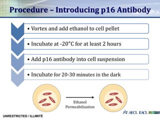
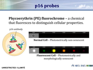
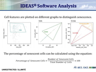
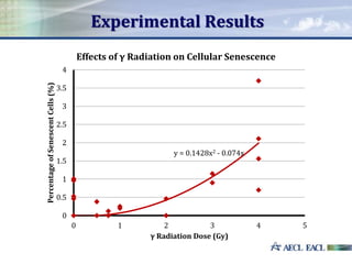
Investigating Cellular Senescence
Following Radiation Exposure
Stephen Liu, Mihai Dumbrava,
Adrienne Wan and Laura Paterson
UNRESTRICTED / ILLIMITÉ
Cellular Senescence
Cellular senescence refers to a process in which cell growth
and development are irreversibly halted.
No known factor can induce
senescent cells to re-enter
the cell cycle.
Although they no longer
divide, senescent cells remain
metabolically active.
UNRESTRICTED / ILLIMITÉ
Types of Senescence
Replicative
Senescence Stress Induced
Premature
Senescence (SIPS)
UNRESTRICTED / ILLIMITÉ
Gamma Radiation
Gamma (γ) radiation is a form
of indirectly ionizing radiation.
It can damage DNA through two
different processes:
1. Direct Action
2. Indirect Action
UNRESTRICTED / ILLIMITÉ
Lymphocytes
Lymphocytes are a type of white blood cell which
recognize and respond to particular pathogens.
They are one of the most radiosensitive and long-
lived cell types in the human body.
UNRESTRICTED / ILLIMITÉ
Gamma Radiation Doses
The Lethal Dose 50 (LD50) is between 4 to 7 Gy for humans
(1 Gy= 1 J/kg).
UNRESTRICTED / ILLIMITÉ
Absorbed Gamma
Radiation Dose (Gy)
Irradation Time (s) Dose Rate (Gy/s) Blood Volume (mL)
0 - - 2.5
0.5 3.4 0.0607 2.5
0.75 7.5 0.0607 2.5
1 11.6 0.0607 2.5
2 28 0.0611 2.5
3 44.4 0.0611 2.5
4 60.8 0.0611 2.5
SIPS Pathway
UNRESTRICTED / ILLIMITÉ
Metabolic
processes
Gamma
radiation
exposure
DNA damage
which can
lead to
senescence
Biomarkers of Senescent Cells
UNRESTRICTED / ILLIMITÉ
Purpose
This project sets out to determine a relationship
between the amount of absorbed gamma radiation
and the occurrence of cellular senescence.
Absorbed Gamma Radiation
Percentageof
Senescentcells
UNRESTRICTED / ILLIMITÉ
Hypothesized
Correlation
Procedure – Isolating Lymphocytes
UNRESTRICTED / ILLIMITÉ
• Irradiate human blood samples
• Dilute blood using PBS
• Carefully layer diluted blood samples on Ficoll
• Centrifuge to separate blood into different layers
• Wash lymphocytes, transfer into RPMI Media
• Incubate for 48 hours at 37°C, 5% CO2
Procedure – Introducing p16 Antibody
UNRESTRICTED / ILLIMITÉ
Ethanol
Permeabilization
• Vortex and add ethanol to cell pellet
• Incubate at -20°C for at least 2 hours
• Add p16 antibody into cell suspension
• Incubate for 20-30 minutes in the dark
Flow Cytometry
1. Cell sample is inserted into flow
cytometer through a small nozzle
2. Cells move in a single file past
lasers
3. p16 probes are excited by these
lasers to emit light in a specific
channel
4. Detectors measure the light emitted
5. The amount of cells, as well as their
morphological and photometric
features, is determined
UNRESTRICTED / ILLIMITÉ
p16 probes
UNRESTRICTED / ILLIMITÉ
Phycoerythrin (PE) fluorochrome – a chemical
that fluoresces to distinguish cellular properties.
p16 antibody
Normal Cell – Photometrically non-senescent
Fluorescent Cell – Photometrically and
morphologically senescent
IDEAS® Software Analysis
Cell features are plotted on different graphs to distinguish senescence.
The percentage of senescent cells can be calculated using the equation:
UNRESTRICTED / ILLIMITÉ
Experimental Results
y = 0.1428x2 - 0.074x
0
0.5
1
1.5
2
2.5
3
3.5
4
0 1 2 3 4 5
PercentageofSenescentCells(%)
γ Radiation Dose (Gy)
Effects of γ Radiation on Cellular Senescence
Discussion – Acute Radiation SyndromeSenescencein%
Error Analysis
Laboratory
Procedure
Software
Analysis
Blood
Donors
Limited
Sample
Size
• Sample
contamination
• Cell loss
• Incorrect
morphology
• Different
radiation
exposure
• Lifestyle
choices
• Insufficient
data to draw
conclusions
UNRESTRICTED / ILLIMITÉ
Application
THE BRIGHT SIDE
Tumor Suppression
Cellular senescence can
stop the uncontrolled
division of cancerous
cells
Aging
Senescent cells can disrupt cell
growth, differentiation, and
initiate or promote age-related
diseases
UNRESTRICTED / ILLIMITÉ
THE DARK SIDE
Future Work
Radiation Therapy – determining the amount of
radiation which will maximize the occurrence of cellular
senescence.
Cell
Repair
Cell
Misrepair
Cell
Death
Normal
Cell
Division
Determining Accidental Radiation Dose
Conclusion
There is a positive quadratic correlation between the
absorbed gamma radiation dose and the percentage of
senescent lymphocytes.
Ranging from radiation therapy to age-related diseases,
cellular senescence has a variety of applications and
drawbacks.
This research has created a foundation for diagnosing an
individual’s original radiation dose through the analysis of
senescent cells.
Acknowledgements
Works Cited
Abcam. "Introduction to Flow Cytometry." Introduction to Flow Cytometry. Abcam, 2014. Web. 30 July
2014.
AbD Serotec. "Fluorochromes and Light." Fluorochromes and Light. Bio-Rad Laboratories, 2014. Web.
01 Aug. 2014.
Hall, Eric J., and Amato J. Giaccia. Radiobiology for the Radiologist. Philadelphia: Lippincott Williams &
Wilkins, 2006. Print.
International Atomic Energy Agency. Radiation Biology: A Handbook for Teachers and Students.
Vienna: International Atomic Energy Agency, 2010. IAEA. International Atomic Energy Agency,
2010. Web. 1 Aug. 2014.
Jefferson Lab. "Radiation Biological Effects." Radiation Biological Effects. Jefferson Lab, n.d. Web. 10
July 2014.
Rahman, Misha. "Introduction to Flow Cytometry." Introduction to Flow Cytometry. Bio-Rad
Laboratories, 2014. Web. 24 July 2014.
Rodier, Francis, and Judith Campisi. "Four Faces of Cellular Senescence." Four Faces of Cellular
Senescence. The Rockefeller University Press, 14 Feb. 2011. Web. 24 July 2014.
Tough, D. F., and J. Sprent. "Lymphocyte life-span and memory." National Center for Biotechnology
Information. U.S. National Library of Medicine, May 1995. Web. 01 Aug. 2014.
The End
Thank you for listening!
Are there any questions?

Cellular Senescence Presentation

  • 1.
    Investigating Cellular Senescence FollowingRadiation Exposure Stephen Liu, Mihai Dumbrava, Adrienne Wan and Laura Paterson UNRESTRICTED / ILLIMITÉ
  • 2.
    Cellular Senescence Cellular senescencerefers to a process in which cell growth and development are irreversibly halted. No known factor can induce senescent cells to re-enter the cell cycle. Although they no longer divide, senescent cells remain metabolically active. UNRESTRICTED / ILLIMITÉ
  • 3.
    Types of Senescence Replicative SenescenceStress Induced Premature Senescence (SIPS) UNRESTRICTED / ILLIMITÉ
  • 4.
    Gamma Radiation Gamma (γ)radiation is a form of indirectly ionizing radiation. It can damage DNA through two different processes: 1. Direct Action 2. Indirect Action UNRESTRICTED / ILLIMITÉ
  • 5.
    Lymphocytes Lymphocytes are atype of white blood cell which recognize and respond to particular pathogens. They are one of the most radiosensitive and long- lived cell types in the human body. UNRESTRICTED / ILLIMITÉ
  • 6.
    Gamma Radiation Doses TheLethal Dose 50 (LD50) is between 4 to 7 Gy for humans (1 Gy= 1 J/kg). UNRESTRICTED / ILLIMITÉ Absorbed Gamma Radiation Dose (Gy) Irradation Time (s) Dose Rate (Gy/s) Blood Volume (mL) 0 - - 2.5 0.5 3.4 0.0607 2.5 0.75 7.5 0.0607 2.5 1 11.6 0.0607 2.5 2 28 0.0611 2.5 3 44.4 0.0611 2.5 4 60.8 0.0611 2.5
  • 7.
    SIPS Pathway UNRESTRICTED /ILLIMITÉ Metabolic processes Gamma radiation exposure DNA damage which can lead to senescence
  • 8.
    Biomarkers of SenescentCells UNRESTRICTED / ILLIMITÉ
  • 9.
    Purpose This project setsout to determine a relationship between the amount of absorbed gamma radiation and the occurrence of cellular senescence. Absorbed Gamma Radiation Percentageof Senescentcells UNRESTRICTED / ILLIMITÉ Hypothesized Correlation
  • 10.
    Procedure – IsolatingLymphocytes UNRESTRICTED / ILLIMITÉ • Irradiate human blood samples • Dilute blood using PBS • Carefully layer diluted blood samples on Ficoll • Centrifuge to separate blood into different layers • Wash lymphocytes, transfer into RPMI Media • Incubate for 48 hours at 37°C, 5% CO2
  • 11.
    Procedure – Introducingp16 Antibody UNRESTRICTED / ILLIMITÉ Ethanol Permeabilization • Vortex and add ethanol to cell pellet • Incubate at -20°C for at least 2 hours • Add p16 antibody into cell suspension • Incubate for 20-30 minutes in the dark
  • 12.
    Flow Cytometry 1. Cellsample is inserted into flow cytometer through a small nozzle 2. Cells move in a single file past lasers 3. p16 probes are excited by these lasers to emit light in a specific channel 4. Detectors measure the light emitted 5. The amount of cells, as well as their morphological and photometric features, is determined UNRESTRICTED / ILLIMITÉ
  • 13.
    p16 probes UNRESTRICTED /ILLIMITÉ Phycoerythrin (PE) fluorochrome – a chemical that fluoresces to distinguish cellular properties. p16 antibody Normal Cell – Photometrically non-senescent Fluorescent Cell – Photometrically and morphologically senescent
  • 14.
    IDEAS® Software Analysis Cellfeatures are plotted on different graphs to distinguish senescence. The percentage of senescent cells can be calculated using the equation: UNRESTRICTED / ILLIMITÉ
  • 15.
    Experimental Results y =0.1428x2 - 0.074x 0 0.5 1 1.5 2 2.5 3 3.5 4 0 1 2 3 4 5 PercentageofSenescentCells(%) γ Radiation Dose (Gy) Effects of γ Radiation on Cellular Senescence
  • 16.
    Discussion – AcuteRadiation SyndromeSenescencein%
  • 17.
    Error Analysis Laboratory Procedure Software Analysis Blood Donors Limited Sample Size • Sample contamination •Cell loss • Incorrect morphology • Different radiation exposure • Lifestyle choices • Insufficient data to draw conclusions UNRESTRICTED / ILLIMITÉ
  • 18.
    Application THE BRIGHT SIDE TumorSuppression Cellular senescence can stop the uncontrolled division of cancerous cells Aging Senescent cells can disrupt cell growth, differentiation, and initiate or promote age-related diseases UNRESTRICTED / ILLIMITÉ THE DARK SIDE
  • 19.
    Future Work Radiation Therapy– determining the amount of radiation which will maximize the occurrence of cellular senescence. Cell Repair Cell Misrepair Cell Death Normal Cell Division Determining Accidental Radiation Dose
  • 20.
    Conclusion There is apositive quadratic correlation between the absorbed gamma radiation dose and the percentage of senescent lymphocytes. Ranging from radiation therapy to age-related diseases, cellular senescence has a variety of applications and drawbacks. This research has created a foundation for diagnosing an individual’s original radiation dose through the analysis of senescent cells.
  • 21.
  • 22.
    Works Cited Abcam. "Introductionto Flow Cytometry." Introduction to Flow Cytometry. Abcam, 2014. Web. 30 July 2014. AbD Serotec. "Fluorochromes and Light." Fluorochromes and Light. Bio-Rad Laboratories, 2014. Web. 01 Aug. 2014. Hall, Eric J., and Amato J. Giaccia. Radiobiology for the Radiologist. Philadelphia: Lippincott Williams & Wilkins, 2006. Print. International Atomic Energy Agency. Radiation Biology: A Handbook for Teachers and Students. Vienna: International Atomic Energy Agency, 2010. IAEA. International Atomic Energy Agency, 2010. Web. 1 Aug. 2014. Jefferson Lab. "Radiation Biological Effects." Radiation Biological Effects. Jefferson Lab, n.d. Web. 10 July 2014. Rahman, Misha. "Introduction to Flow Cytometry." Introduction to Flow Cytometry. Bio-Rad Laboratories, 2014. Web. 24 July 2014. Rodier, Francis, and Judith Campisi. "Four Faces of Cellular Senescence." Four Faces of Cellular Senescence. The Rockefeller University Press, 14 Feb. 2011. Web. 24 July 2014. Tough, D. F., and J. Sprent. "Lymphocyte life-span and memory." National Center for Biotechnology Information. U.S. National Library of Medicine, May 1995. Web. 01 Aug. 2014.
  • 23.
    The End Thank youfor listening! Are there any questions?

Editor's Notes

  • #3 This differs from cell death in that senescent cells are still metabolically active, yet it differs from quiescence in that senescent cells cannot be re-induced to replicate further.
  • #4 Ste
  • #5 Ste
  • #6 Their long-living nature makes them useful subjects in investigating the effects of ionizing radiation on human cells since they, unlike shorter lived cells, are not taken out of circulation before the blood can be examined.  
  • #7 Ste
  • #8 Ste
  • #10 Purpose: Ste
  • #11 Ste
  • #15 characteristics
  • #20 stops the uncontrollable division in tumors without disturbing the surrounding cells.
  • #21 Following mortality rate, even higher doses of radiation (>4 Gy) may lead to more senescent cells up until cell death is certain.