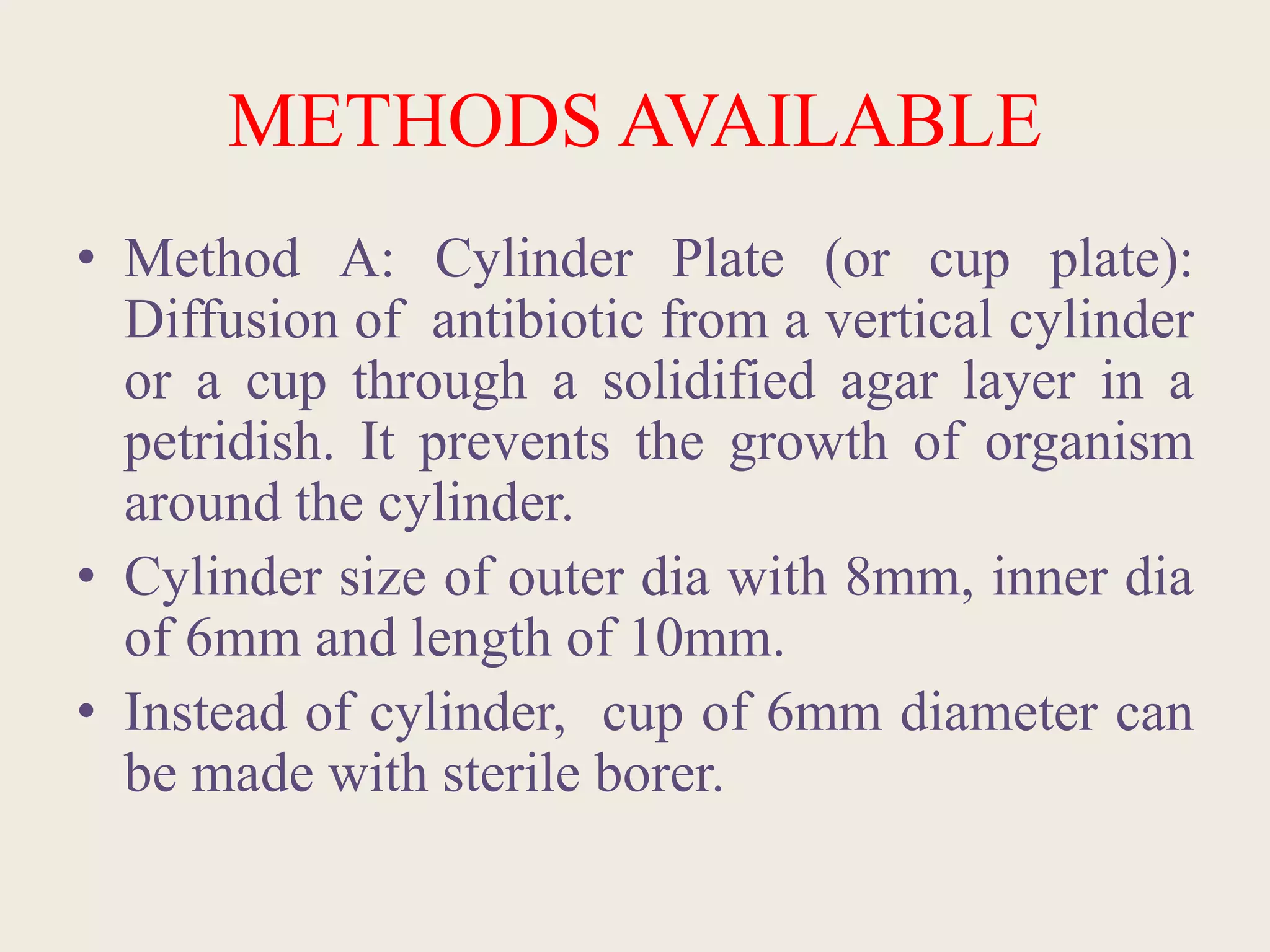
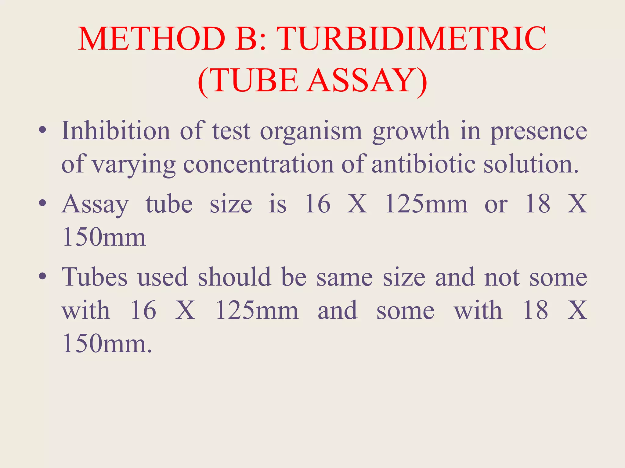
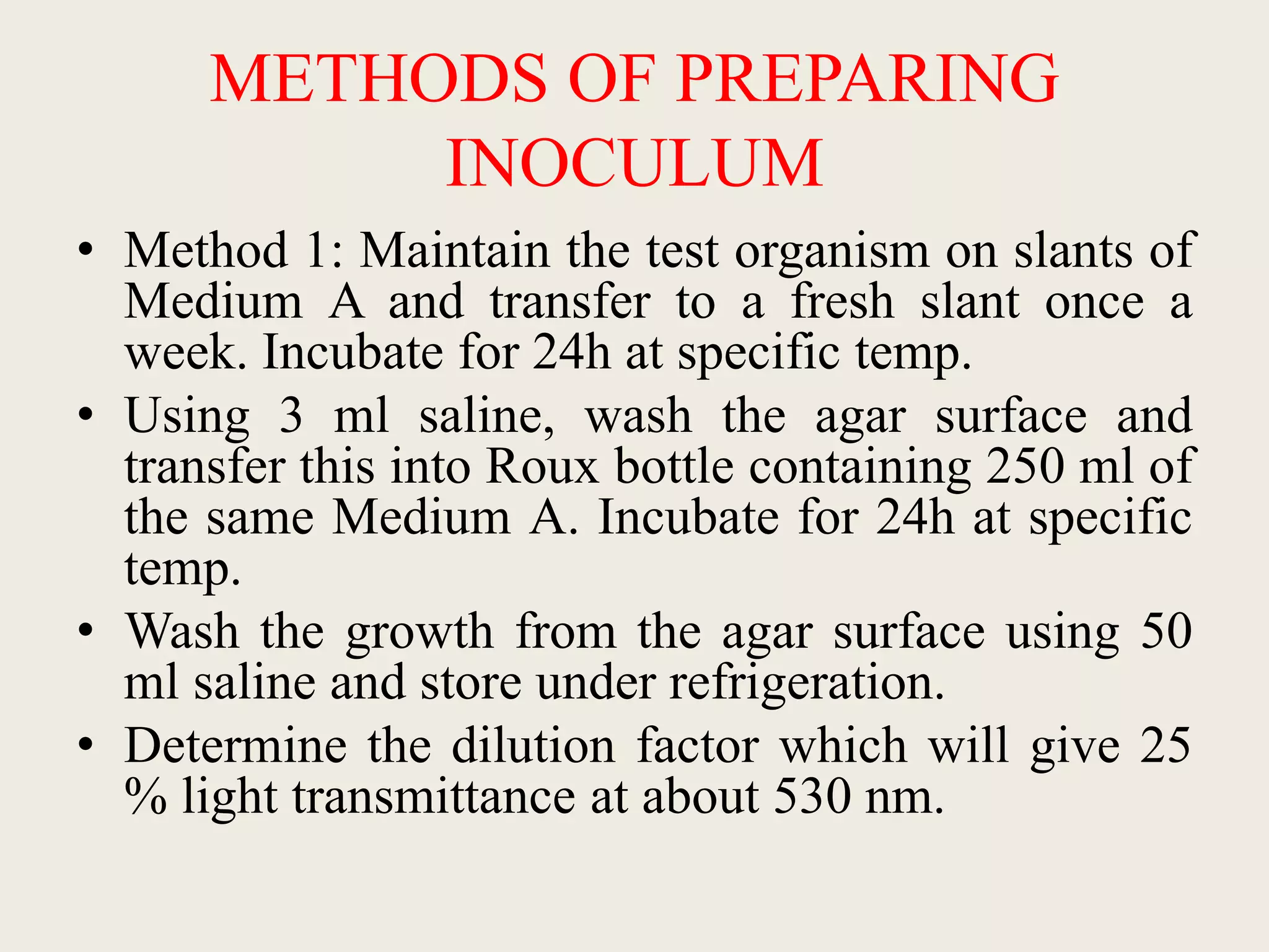
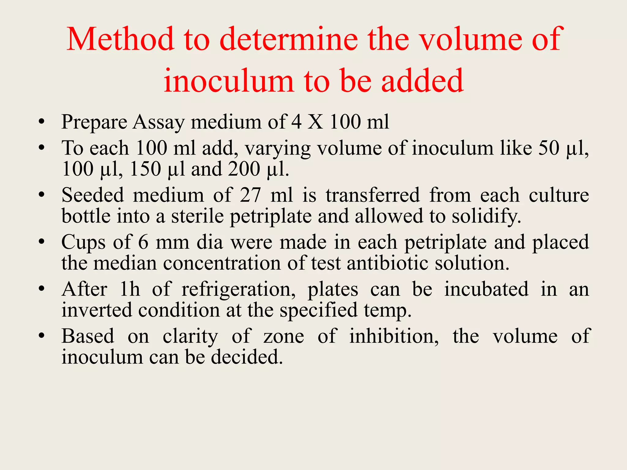
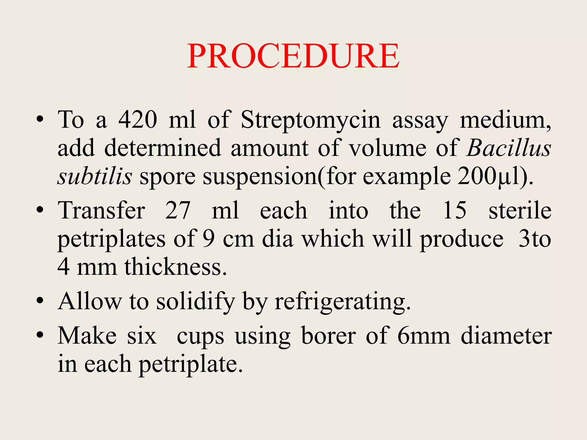
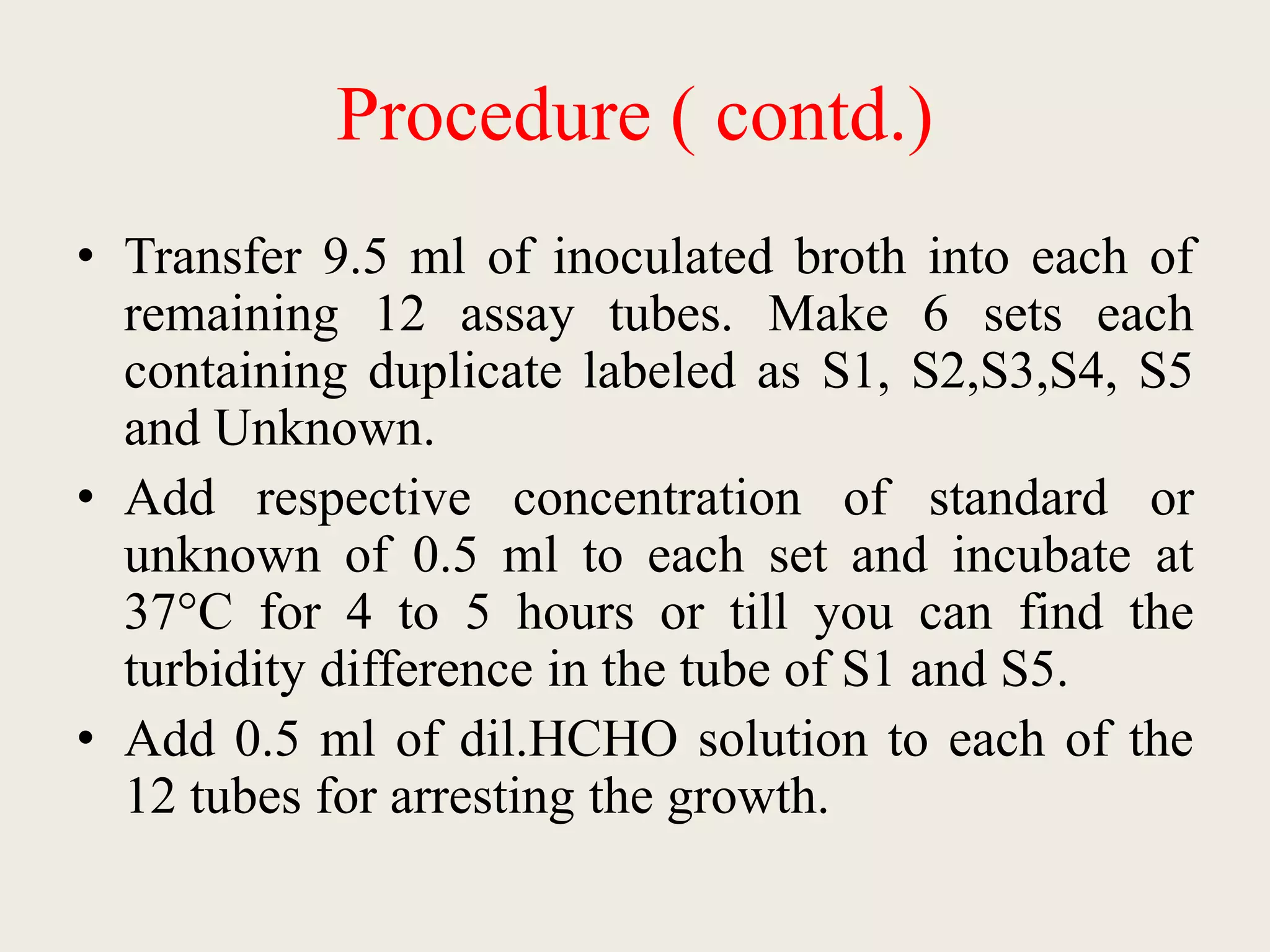
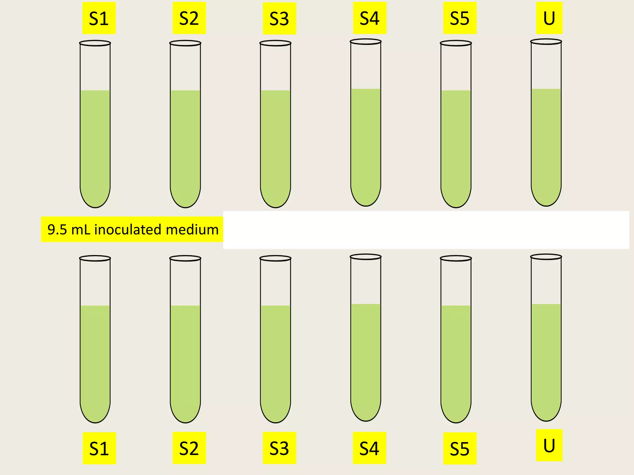
This document describes the microbiological assay method for quantifying antibiotics. It involves preparing standard solutions of known antibiotic concentrations and measuring their zones of inhibition against a test microbe. An unknown sample is also tested and its zone of inhibition is compared to the standard curve to determine its concentration. The key steps are:
1) Preparing standard solutions in increasing concentrations and an unknown at a median level.
2) Inoculating agar plates with a test microbe and creating wells for the solutions.
3) Measuring the zones of inhibition and calculating averages for the standards.
4) Comparing the unknown's zone of inhibition to the standard curve to determine its concentration.
Two general methods are used for microbiological assays
Method A: Cylinder plate method or cup plate method.
Method B: Tube assay method or titrimetric method.
This document provides information on microbiological assays for vitamins B2 and B12. It discusses the underlying principles, which involve measuring the growth response of test microorganisms to different concentrations of the vitamin being assayed. Two common methods are described: the cylinder-plate method and the turbidimetric tube assay method. Specific details are given on reagents, preparation of inoculum, procedures, and interpretation of results for assays of vitamins B2 and B12 using Lactobacillus species as the test microorganisms.
Methods for standardization of antibiotics NISHA MANDLOI
This document discusses methods for standardizing antibiotics. It notes that there are three important points for standardization: following FDA regulations, referring to FDA methods for individual antibiotics, and measuring inhibition of microbial growth. Two common assay methods are described: the cylinder-plate method which measures zones of inhibition, and the turbidimetric method which measures prevention of microbial growth. Procedures for preparing media, buffers, standards and samples are outlined. Streptomycin is used as an example antibiotic, noting it is bactericidal, derived from Streptomyces griseus, and tested using Bacillus subtilis.
Bacterial endotoxins tests are used to detect and quantify endotoxins from gram-negative bacteria. Endotoxins are heat-stable lipopolysaccharides in bacterial cell walls. The test uses amoebocyte lysate from horseshoe crabs to detect endotoxins. There are several methods to conduct the test including gel clot formation, turbidity measurement, and chromogenic assays. Proper controls and standard curves must be established to validate the test results. The test is used to ensure medical products and devices are free of endotoxins, which can cause adverse reactions in humans.
This document describes the process of microbiological assay, specifically as it relates to antibiotics and vitamins. There are two main methods described - the cylinder plate method and the turbidimetric tube assay method. For both methods, standard and test solutions are prepared along with appropriate culture media and test organisms. Zones of inhibition are measured for the cylinder plate method to determine potency, while growth is measured spectrometrically for the turbidimetric tube assay method. Requirements, procedures, and interpretation of results are provided for microbiological assay of both antibiotics and specific vitamins like vitamin B12.
The efficacy of antimicrobial preservation of a pharmaceutical preparation on its own or, if necessary, with the addition of a suitable preservative has to be ascertained during the development of the product.
The primary purpose of adding antimicrobial preservatives to dosage forms is to prevent adverse effects arising from contamination by micro-organisms that may be introduced inadvertently during or subsequent
to the manufacturing process.
However, antimicrobial agents should not be used solely to reduce the viable microbial count as a substitute for good manufacturing procedures.
There may be situations where a preservative system may have to be used to minimise proliferation of micro-organisms in preparations that are not required to be sterile.
Microbiological assays use microorganisms to determine the potency of drugs. There are two main methods - the cylinder-plate method which measures inhibition zone diameters, and the turbidimetric method which measures absorbance changes in liquid cultures. Standard curves are prepared using known concentrations of a reference standard. Test samples are run alongside at assumed concentrations and their potency determined by comparing results to the standard curve. Proper preparation of media, buffers, microorganism cultures and standards is required for accurate and reproducible assays.
The document discusses the microbiological assay of antibiotics, which quantitatively estimates antibiotics by measuring their ability to inhibit the growth of microorganisms. There are two main methods - the cylinder-plate method which measures zones of inhibition, and the turbidimetric method which compares the turbidity of bacterial solutions with different antibiotic concentrations. The assay is useful for demonstrating antibiotic efficacy, detecting subtle molecular changes, and resolving doubts about antibiotic potency changes.
Anti Microbiological Assay Test or Antibiotic Assay Test of Pharmaceutical Pr...ijtsrd
In this paper, we are going to discuss the anti microbiological assay of the antibiotics. Aim of this paper is to predict the potency of the antibiotic preparation in reference with the working standard of the antibiotic and using the mathematical model in order to obtain the potency of the preparation in regards to its claim. Faiz Hashmi "Anti-Microbiological Assay Test or Antibiotic Assay Test of Pharmaceutical Preparation Containing Antibiotics using Cylinder Plate Method'" Published in International Journal of Trend in Scientific Research and Development (ijtsrd), ISSN: 2456-6470, Volume-3 | Issue-5 , August 2019, URL: https://www.ijtsrd.com/papers/ijtsrd27940.pdfPaper URL: https://www.ijtsrd.com/pharmacy/analytical-chemistry/27940/anti-microbiological-assay-test-or-antibiotic-assay-test-of-pharmaceutical-preparation-containing-antibiotics-using-%E2%80%98cylinder-plate-method%E2%80%99/faiz-hashmi
This document describes microbiological assays for Vitamin B6 and Vitamin B12.
For the Vitamin B12 assay, it uses E. coli M200 and measures zone diameters to determine Vitamin B12 concentrations. Standard solutions of cyanocobalamin are prepared and tested alongside sample solutions.
The Vitamin B6 assay uses Kloeckera brevis yeast and measures zone diameters from standard pyridoxine solutions and processed sample solutions to generate a standard curve and determine Vitamin B6 content. Procedures for culture maintenance, inoculum preparation, sample digestion, and running the cup plate assay are provided.
Sterility testing is required for all products labeled as sterile to ensure they have been effectively sterilized. Tests are conducted using specific culture media and procedures to detect any viable bacteria, fungi, or yeasts. The USP outlines sterility testing methods for various pharmaceutical products and devices, including membrane filtration and direct inoculation. Interpretation of results involves incubating samples and checking for any microbial growth over time, with growth indicating test failure.
This document discusses assessing the value of new antibiotics through health technology assessment (HTA). It identifies four benefits typically included in HTA evaluations and six additional benefits not traditionally considered but relevant for antibiotics. These additional benefits include insurance value, diversity value, diagnostic value, uniqueness, enablement value, and spectrum value. The document notes that HTA methods may not fully capture the value of antibiotics in addressing antimicrobial resistance and that new payment mechanisms are needed given antibiotics must have limited use to delay resistance development.
Preservatives are added to pharmaceutical products to prevent microbial growth and extend shelf life. An ideal preservative kills microbes rapidly at low concentrations, is non-toxic, stable, and does not interact negatively with the product ingredients. A preservative efficacy test evaluates the ability of a preservative system to inhibit microbial growth when challenged with common test microbes like S. aureus and P. aeruginosa over 28 days. The test involves inoculating product samples, incubating them, and conducting plate counts to determine microbial survival rates over time. This ensures the preservative maintains effective antimicrobial activity as required by pharmacopeia standards.
Microbiological assay-Principles and methods of different microbiological assay.someshwar mankar
Principles and methods of different microbiological assay. Methods for standardization of
antibiotics, vitamins and amino acids. Assessment of a new antibiotic.
This document provides information on pyrogen and endotoxin testing according to specifications from the Indian Pharmacopoeia (IP), British Pharmacopoeia (BP), and United States Pharmacopoeia (USP). It describes testing methods including the rabbit pyrogen test and Limulus Amebocyte Lysate (LAL) test for endotoxins. Key steps of the tests are outlined, such as administration of samples to rabbits or incubation with horseshoe crab lysate, followed by interpretation of temperature changes or gel formation to determine passing or failing of the tests.
Endotoxin Testing is performed to ensure that injectable preparations and medical devices are free from pyrogens and safe for human use.
Pyrogens constitute a heterogeneous group of fever causing substances which comprise both microbial and non-microbial substances. The most potent and most widely known are the endotoxins or lipopolysaccharides (LPS), which are cell wall components of gram-negative bacteria. Gram-positive bacteria are also sources of pyrogens, in particular lipoteichoic acid (LTA), as are particles from yeasts and viruses. Non-microbial pyrogens often emanate from production environments. Small particles of packaging materials are a typical example.
The document describes procedures for the microbiological assay of antibiotics and vitamins. There are two main methods for antibiotic assay - the cylinder-plate method and the turbidimetric method. The cylinder-plate method involves measuring zones of inhibition around cylinders containing different concentrations of antibiotics in an agar plate inoculated with a test microorganism. The turbidimetric method involves measuring inhibition of microbial growth in liquid medium containing varying antibiotic concentrations. Standards curves are constructed to determine unknown concentrations. Vitamin assays similarly rely on microbial growth responses to different concentrations of vitamins in culture media. Procedures for assaying vitamins B12 and B2 using specific test microorganisms like Lactobacillus leichmannii are provided.
This document discusses microbiological assays for testing antibiotics. It describes biological assays using microorganisms and the two main types of microbiological assays: agar diffusion assays and turbidimetric assays. The agar diffusion assay uses discs containing antibiotics placed on an agar plate seeded with bacteria, and measures zones of inhibition of bacterial growth. The turbidimetric assay measures inhibition of bacterial growth in liquid culture containing known concentrations of antibiotics.
Sterility testing attempts to determine if viable microorganisms are present in pharmaceutical products by testing samples from a batch. It is done after sterilization but cannot guarantee sterility of the entire batch. Major factors that can affect the results are the testing environment, culture conditions, sampling procedure, and test method used. Common test methods include membrane filtration and direct inoculation into culture media like fluid thioglycollate and soya-bean casein digest media. Observation over the incubation period can indicate if the test and batch pass or fail sterility requirements. Guidelines stress the level of assurance depends on manufacturing conditions and batch homogeneity.
Assessment of microbial contamination and spoilage. PHARMACEUTICAL MICROBIOLO...Ms. Pooja Bhandare
PHARMACEUTICAL MICROBIOLOGY (BP303T)Unit-VPart-2
Assessment of microbial contamination and spoilage.
Assessment of microbial contamination and spoilage
1. Physical and chemical changes:
2. Assessment of viable microorganisms in non-sterile products:
3. Sterility test:
4. Estimation of pyrogens:
Microbial Limit Tests:
Total Aerobic Microbial Count:
Membrane Filtration.
Plate Count Methods.
Pour Plate Method.
Surface spread Method.
Most Probable Number(MPN)
The document discusses pyrogen testing techniques including the rabbit test and LAL (Limulus Amebocyte Lysate) test. It provides details on how to conduct the rabbit test, including temperature monitoring and criteria for a passing result. For the LAL test, it describes the mechanism, different methods (gel clot, turbidimetric, chromogenic), and procedures for confirming lysate sensitivity and determining endotoxin levels in samples. It notes that various pharmacopeias like IP, BP, and USP specify methods for the LAL test.
This document summarizes sterility testing procedures for pharmaceutical products. Sterility testing aims to detect any viable microorganisms that may be present. Samples are inoculated into fluid thioglycollate medium, alternative thioglycollate medium, or soybean-casein digest medium and incubated with test microbes like S. aureus, C. sporogenes, P. aeruginosa, B. subtilis, A. brasiliensis or C. albicans. Tests are done using either membrane filtration or direct inoculation methods depending on the product type and volume. After incubation, the results are observed and interpreted to determine if the product passes or fails sterility requirements.
Evaluation of Bactericidal and BacteriostaticRajsingh467604
What are disinfectants?
As per the definition given by WHO ( World health organization ) : a disinfectant is a chemical agent, which destroys or inhibits growth of pathogenic microorganisms in the non-sporing or vegetative state.
Why Evaluation?
Evaluation of disinfectants is used to check the ability or efficacy of any disinfectant against specific microorganisms to establish its effectiveness.
Evaluation tests of bactericide.
1. RIDEAL WALKER TEST
This test is also known as the phenol coefficient test,in which any chemical is compared with phenol for its antimicrobial activity.
The result is shown in the form of phenol coefficient.
▪ If a phenol coefficient of a given test disinfectant is less than 1, it means that disinfectant is less effective than phenol.
▪ If a phenol coefficient of a given test disinfectant is more than 1, it means that disinfectant is more effective than phenol.
Procedure
1.1 Different dilutions of the test disinfectant and phenol are prepared and 5 ml of each dilution is inoculated with 0.5ml of the 24 hour growth culture of the organisms.
1.2 All tubes(Disinfectant + organisms & phenol + organisms) are placed in a water bath ( at 17.5° C)
1.3 Subcultures of each reaction mixture are taken and transferred to 5ml sterile broth at an interval of 2.5 minutes from zero to 10 mintues.
1.4 Broth tubes are incubated at 37° C for 2 to 3 days & examined for the presence or absence of the growth.
1.5 Then the Rideal Walker coefficient is calculated :
2. CHICK MARTIN TEST.
CHICK MARTIN test is performed in the much similar way as the RIDEAL Walker test but with a little variation.
Principle : This test is carried out in the presence of organic matter like 3% human feces or dried yeast.
Procedure
2.1 Serial dilutions of test solution and phenol is prepared in distilled water.
2.2 To this 3% yeast suspension is also added.
2.3 To this solution the S. typhi is added
2.4 After contact time of 30 mins the above mixture is transferred to the freshly prepared 10 ml of broth.
2.5 The test tubes are incubated at 37°C for 48 hours.
2.6 Presence or absence of the growth is calculated.
Evaluation tests of Bacteriostatic.
1. Tube dilution & Agar plate Method
1.1 The chemical agent is incorporated into nutrient broth or agar medium and inoculated with test micro-organisms.
1.2 These tubes are incubated at 30° TO 35°C for 2 to 3 days and then the results in the form of turbidity or colonies are observed.
1.3 The results are recorded and the activity of the given disinfectant is compared.
2. Cup plate method
2.1 Agar is melted and cooled at 45° Celsius.
2.2 Then inoculated with test micro-organisms and poured into a sterile petri plate.
2.3 In the cup plate method, when the inoculated agar has solidified, holes around 8mm in diameter are cut in the medium with a steel cork borer.
2.4 Now the antimicrobial agents are directly placed in the holes.
The document discusses three main methods for the bacterial endotoxin test - gel clot, turbidimetric, and chromogenic. The gel clot method is the simplest but least quantitative, while turbidimetric and chromogenic methods allow for more automation and precision using spectrophotometry. All three methods use Limulus amebocyte lysate and detect endotoxins through coagulation reactions. The choice of method depends on factors like testing volumes, sample properties, required sensitivity, and compliance needs. Photometric methods have advantages of automation and precision but higher costs, while gel clot is inexpensive but less quantitative.
Sterility indicators are used to check the quality and monitoring of sterilization processes. They can indicate whether microbial growth occurs or sterilization was effective. There are several types of sterility indicators for different sterilization methods including dry heat, moist heat, gaseous, radiation, and filtration sterilization. Physical indicators monitor parameters like temperature and pressure. Chemical indicators involve temperature sensitive solutions or chemicals that change color. Biological indicators use microbes like Bacillus species to determine sterilization effectiveness by observing growth or no growth.
Prof.Mr.Kiran K. Shinde (M.Pharm), Assistant professor (VNIPRC)
Pharmaceutical microbiology (Second year b.pharm) (3rd semester)
Introduction
Methods Of Different microbiological assays
Principles of Assays with Procedure
Methods For Standardization of
1. Antibiotics
2. Vitamins
3. Amino Acids
Assessment of new Antibiotic
The document describes an experiment to determine the potency of amoxycillin 3H2O using a bioassay. The experiment uses Bacillus subtilis and involves preparing standard and sample dilutions of amoxycillin to test using cylinder plate assays. Zones of inhibition are measured after incubating the plates and potency is calculated using a formula comparing the sample and standard results.
Microbial Assay of Antibiotics
STANDARD PREPARATION AND UNITS OF ACTIVITY
Preparation of media
Buffer solutions
Standard solution
Sample solution
Test organisms
Preparation of inoculum Method -1
Method 2
Method 3
Method 4
Determination of Inoculum
Apparatus
Assay design
Assay method
cylinder plate method
One level assay with standard curve
Estimation of potency
Turbidimetric method
This document provides information on techniques for serological tests. It discusses the materials needed, including glassware, incubators, and rotating machines. It describes how to collect, prepare, and preserve serological specimens like serum, plasma and cerebrospinal fluid. Proper shipment of specimens is also covered. The document explains complement inactivation through heating serum to prevent interference. Dilution techniques like serial dilution are outlined, along with calculating titers from dilution endpoints.
Principles and methods of different microbiological assay, methods for standa...Ms. Pooja Bhandare
PHARMACEUTICAL MICROBIOLOGY (BP303T)Unit-IV Part-2 Principles and methods of different microbiological assay, methods for standardization of antibiotics.
Introduction: Principles Advantages of Microbial Assay: Disadvantages of Microbial Assay: MICROBIOLOGICAL ASSAY OF ANIBIOTICS PRINCIPLE Media used for antibiotics assay Standard Preparation. Buffer Solutions Preparation of the Sample Solution: Test Organisms Preparation of inoculum: Methods of preparation of test organism suspension: Assay Methods: Method A: Cup-plate or Cylinder Plate Method.
Method B: Turbidimetric or Tube assay Method
Validation protocol for hold time study of collected water samplescubi333
This document outlines a validation protocol to establish a 72-hour hold time for various water quality parameters in collected water samples. The protocol involves testing samples initially and after 24, 48, and 72 hours of storage at 2-8°C for parameters like pH, conductivity, heavy metals, nitrates, TOC, microbial counts, and bacterial endotoxins. Acceptance criteria are based on meeting specifications for each water type. A final report will determine the validated hold time based on conclusion from the study results.
Microbial assays or microbiological assays could be a sort of bioassays designed to analyse the compounds or substances that have impact on micro-organisms. They help to estimate concentration and efficiency of antibiotics. Also facilitate in determination of the simplest anti-biotic appropriate for patient recovery.
This document provides information on a human orosomucoid 2 (ORM2) ELISA kit that allows for the quantitative determination of ORM2 concentrations in biological samples like serum, plasma, tissue homogenates, and cell culture supernatants. It describes the intended use, test principle, materials included in the kit, sample collection and storage recommendations, limitations of the procedure, reagent preparation instructions, and the assay procedure.
This document summarizes a seminar on bioassay of official drugs. It defines bioassay, describes the principles and importance of bioassay, and outlines common types including heparin sodium, oxytocin, streptokinase, and vitamin D. Limitations of bioassay are also noted. Methods for each drug are provided, including preparation of standards and testing solutions, procedures, and statistical analysis of results.
Preparation of large volume and small volume parenteralsangram maskar
Large volume parenterals are sterile aqueous drug products packaged in single-dose containers holding 100 mL or more. Small volume parenterals are packaged in containers holding 100 mL or less. Both are administered via intravenous, intramuscular, or subcutaneous routes. Key differences include that small volume parenterals may use preservatives and not require isotonicity, while large volume parenterals must be isotonic. Both undergo processes like cleaning, preparation, sterilization, filling, and packaging to ensure sterility. They are tested using methods like sterility testing, particulate testing, and pyrogen testing to ensure quality and safety.
This document provides information on techniques and procedures for serological tests. It discusses the necessary materials, including glassware, incubators, and rotating machines. It describes how to collect, prepare, and preserve serum, plasma, and cerebrospinal fluid specimens. Proper collection involves centrifugation to separate serum or plasma from whole blood. Specimens should be shipped promptly if not tested immediately. The document also covers complement inactivation through heating serum to prevent interference, and dilution techniques including serial dilution used to determine antibody titers.
Oxidation of a Glycosylated Therapeutic ProteinKBI Biopharma
A presentation by Jimmy Smedley, Ph.D., (KBI Group Leader, Biopharmaceutical Development) from the Formulation Strategies for Protein Therapeutics Conference.
This document outlines procedures for performing microbial limit tests on pharmaceutical products. The tests are designed to qualitatively or quantitatively estimate the number of viable aerobic microorganisms present or detect designated microbial species. Several methods are described, including membrane filtration, pour plate, spread plate, and multiple tube dilution. Specific procedures are provided for testing for total aerobic count, E. coli, and Salmonella. Controls and interpretation of results are also described to validate the testing methods.
Microbiological Assay of Vitamin & Amino acid Assessment of a New Antibiotic...Ms. Pooja Bhandare
The document discusses methods for assessing new antibiotics through microbiological assays. It describes how the minimum inhibitory concentration (MIC) can be determined using either liquid or solid dilution methods. The liquid dilution method involves setting up a series of test tubes with doubling dilutions of the antibiotic being tested and incubating with a test microorganism. The solid dilution method incorporates the antibiotic dilutions into an agar plate which is then inoculated. After incubation, the MIC is the lowest concentration that inhibits microbial growth. These assays help establish the efficacy and potency of new antibiotics.
This document describes a method for extracting glycogen from chicken liver. Glycogen is a stored form of glucose found primarily in the liver and muscle. The method involves mincing chicken liver, homogenizing it, boiling and centrifuging to separate glycogen. Glycogen is then precipitated using ethanol, dissolved in water, and hydrolyzed using acid. A positive result in Molisch's and iodine tests confirms the presence of glycogen. The purpose is to isolate glycogen from chicken liver through a series of extraction and precipitation steps.
This document describes the process of extracting glycogen from chicken liver. Glycogen is the major glucose storage polymer found in animals like chicken liver. The method involves mincing chicken liver, homogenizing it, centrifuging to collect the supernatant, precipitating glycogen using ethanol, and performing tests like Molisch's test and iodine test to identify glycogen. The Molisch test on the extracted glycogen sample produced a purple ring, confirming the presence of glycogen.
Optimization of ABE Fermentation from Rice Husk Medium using Clostridium acet...pranavdadhich
A chemically defined medium was optimised for the biomass production of Clostridium acetobutylicum in the fermentor using rice husk as the carbon source.
The researchers developed a micro-formulation method to formulate new chemical entities for intravenous injection using only 2-3 mg of the compound. They tested solubility of compounds in various formulation bases using a 50 microliter scale, requiring 100-150 micrograms of compound per test. Results were generally comparable to conventional formulation methods using larger quantities. The micro-formulation approach allows rapid early stage animal screening with minimal compound quantities.
This document provides guidelines for developing specifications for new drug substances and products according to ICH Q6A. It discusses universal and specific tests/criteria that should be included for drug substances and products, such as identification, description, assay, impurities, dissolution, disintegration, content uniformity, and microbial limits. The document gives acceptance criteria and justification for key tests like dissolution, discussing how to set Q values and limits based on biobatch results and BCS classification. It also provides guidance on other tests for oral liquids, parenterals and solid dosage forms.
Each chromosome in the somatic-cell complement can be uniquely identified by following a number of different banding procedures.
The banding patterns are highly characteristic. The International System for Cytogenetic Nomenclature (ISCN) provides schematic representations, or Ideograms, of human chromosomes corresponding to approx. 400, 550, and 850 bands per haploid set (I).
Although under constant revision, its principles rest on a numbering system based on major bands as they appear from the centromere outward along each chromosome arm.
To the cytogeneticist, the appearance of well-prepared, clearly banded chromosomes has an aesthetic appeal that is often difficult for the non-cytogeneticist to comprehend.
Similar to Micro assay dr. saisivam (04-03-2016) (20)
Current Ms word generated power point presentation covers major details about the micronuclei test. It's significance and assays to conduct it. It is used to detect the micronuclei formation inside the cells of nearly every multicellular organism. It's formation takes place during chromosomal sepration at metaphase.
The binding of cosmological structures by massless topological defectsSérgio Sacani
Assuming spherical symmetry and weak field, it is shown that if one solves the Poisson equation or the Einstein field
equations sourced by a topological defect, i.e. a singularity of a very specific form, the result is a localized gravitational
field capable of driving flat rotation (i.e. Keplerian circular orbits at a constant speed for all radii) of test masses on a thin
spherical shell without any underlying mass. Moreover, a large-scale structure which exploits this solution by assembling
concentrically a number of such topological defects can establish a flat stellar or galactic rotation curve, and can also deflect
light in the same manner as an equipotential (isothermal) sphere. Thus, the need for dark matter or modified gravity theory is
mitigated, at least in part.
ESR spectroscopy in liquid food and beverages.pptxPRIYANKA PATEL
With increasing population, people need to rely on packaged food stuffs. Packaging of food materials requires the preservation of food. There are various methods for the treatment of food to preserve them and irradiation treatment of food is one of them. It is the most common and the most harmless method for the food preservation as it does not alter the necessary micronutrients of food materials. Although irradiated food doesn’t cause any harm to the human health but still the quality assessment of food is required to provide consumers with necessary information about the food. ESR spectroscopy is the most sophisticated way to investigate the quality of the food and the free radicals induced during the processing of the food. ESR spin trapping technique is useful for the detection of highly unstable radicals in the food. The antioxidant capability of liquid food and beverages in mainly performed by spin trapping technique.
Immersive Learning That Works: Research Grounding and Paths ForwardLeonel Morgado
We will metaverse into the essence of immersive learning, into its three dimensions and conceptual models. This approach encompasses elements from teaching methodologies to social involvement, through organizational concerns and technologies. Challenging the perception of learning as knowledge transfer, we introduce a 'Uses, Practices & Strategies' model operationalized by the 'Immersive Learning Brain' and ‘Immersion Cube’ frameworks. This approach offers a comprehensive guide through the intricacies of immersive educational experiences and spotlighting research frontiers, along the immersion dimensions of system, narrative, and agency. Our discourse extends to stakeholders beyond the academic sphere, addressing the interests of technologists, instructional designers, and policymakers. We span various contexts, from formal education to organizational transformation to the new horizon of an AI-pervasive society. This keynote aims to unite the iLRN community in a collaborative journey towards a future where immersive learning research and practice coalesce, paving the way for innovative educational research and practice landscapes.
When I was asked to give a companion lecture in support of ‘The Philosophy of Science’ (https://shorturl.at/4pUXz) I decided not to walk through the detail of the many methodologies in order of use. Instead, I chose to employ a long standing, and ongoing, scientific development as an exemplar. And so, I chose the ever evolving story of Thermodynamics as a scientific investigation at its best.
Conducted over a period of >200 years, Thermodynamics R&D, and application, benefitted from the highest levels of professionalism, collaboration, and technical thoroughness. New layers of application, methodology, and practice were made possible by the progressive advance of technology. In turn, this has seen measurement and modelling accuracy continually improved at a micro and macro level.
Perhaps most importantly, Thermodynamics rapidly became a primary tool in the advance of applied science/engineering/technology, spanning micro-tech, to aerospace and cosmology. I can think of no better a story to illustrate the breadth of scientific methodologies and applications at their best.
The cost of acquiring information by natural selectionCarl Bergstrom
This is a short talk that I gave at the Banff International Research Station workshop on Modeling and Theory in Population Biology. The idea is to try to understand how the burden of natural selection relates to the amount of information that selection puts into the genome.
It's based on the first part of this research paper:
The cost of information acquisition by natural selection
Ryan Seamus McGee, Olivia Kosterlitz, Artem Kaznatcheev, Benjamin Kerr, Carl T. Bergstrom
bioRxiv 2022.07.02.498577; doi: https://doi.org/10.1101/2022.07.02.498577
Mending Clothing to Support Sustainable Fashion_CIMaR 2024.pdfSelcen Ozturkcan
Ozturkcan, S., Berndt, A., & Angelakis, A. (2024). Mending clothing to support sustainable fashion. Presented at the 31st Annual Conference by the Consortium for International Marketing Research (CIMaR), 10-13 Jun 2024, University of Gävle, Sweden.
Travis Hills of MN is Making Clean Water Accessible to All Through High Flux ...Travis Hills MN
By harnessing the power of High Flux Vacuum Membrane Distillation, Travis Hills from MN envisions a future where clean and safe drinking water is accessible to all, regardless of geographical location or economic status.
Describing and Interpreting an Immersive Learning Case with the Immersion Cub...Leonel Morgado
Current descriptions of immersive learning cases are often difficult or impossible to compare. This is due to a myriad of different options on what details to include, which aspects are relevant, and on the descriptive approaches employed. Also, these aspects often combine very specific details with more general guidelines or indicate intents and rationales without clarifying their implementation. In this paper we provide a method to describe immersive learning cases that is structured to enable comparisons, yet flexible enough to allow researchers and practitioners to decide which aspects to include. This method leverages a taxonomy that classifies educational aspects at three levels (uses, practices, and strategies) and then utilizes two frameworks, the Immersive Learning Brain and the Immersion Cube, to enable a structured description and interpretation of immersive learning cases. The method is then demonstrated on a published immersive learning case on training for wind turbine maintenance using virtual reality. Applying the method results in a structured artifact, the Immersive Learning Case Sheet, that tags the case with its proximal uses, practices, and strategies, and refines the free text case description to ensure that matching details are included. This contribution is thus a case description method in support of future comparative research of immersive learning cases. We then discuss how the resulting description and interpretation can be leveraged to change immersion learning cases, by enriching them (considering low-effort changes or additions) or innovating (exploring more challenging avenues of transformation). The method holds significant promise to support better-grounded research in immersive learning.
EWOCS-I: The catalog of X-ray sources in Westerlund 1 from the Extended Weste...Sérgio Sacani
Context. With a mass exceeding several 104 M⊙ and a rich and dense population of massive stars, supermassive young star clusters
represent the most massive star-forming environment that is dominated by the feedback from massive stars and gravitational interactions
among stars.
Aims. In this paper we present the Extended Westerlund 1 and 2 Open Clusters Survey (EWOCS) project, which aims to investigate
the influence of the starburst environment on the formation of stars and planets, and on the evolution of both low and high mass stars.
The primary targets of this project are Westerlund 1 and 2, the closest supermassive star clusters to the Sun.
Methods. The project is based primarily on recent observations conducted with the Chandra and JWST observatories. Specifically,
the Chandra survey of Westerlund 1 consists of 36 new ACIS-I observations, nearly co-pointed, for a total exposure time of 1 Msec.
Additionally, we included 8 archival Chandra/ACIS-S observations. This paper presents the resulting catalog of X-ray sources within
and around Westerlund 1. Sources were detected by combining various existing methods, and photon extraction and source validation
were carried out using the ACIS-Extract software.
Results. The EWOCS X-ray catalog comprises 5963 validated sources out of the 9420 initially provided to ACIS-Extract, reaching a
photon flux threshold of approximately 2 × 10⁻⁸ photons cm−2
s
−1
. The X-ray sources exhibit a highly concentrated spatial distribution,
with 1075 sources located within the central 1 arcmin. We have successfully detected X-ray emissions from 126 out of the 166 known
massive stars of the cluster, and we have collected over 71 000 photons from the magnetar CXO J164710.20-455217.
2. DEFINITION
• Therapeutic efficacy of Antibiotics can be
exhibited by inhibiting the microbial growth.
• Any small change in the structure of antibiotic
can be easily detected by this method. Even
chemical methods may not reveal this.
3. PRINCIPLE
• Standard graph: known concentration Vs. ZOI.
• Comparing the zone of inhibition produced by
standard with that of unknown
4. METHODS AVAILABLE
• Method A: Cylinder Plate (or cup plate):
Diffusion of antibiotic from a vertical cylinder
or a cup through a solidified agar layer in a
petridish. It prevents the growth of organism
around the cylinder.
• Cylinder size of outer dia with 8mm, inner dia
of 6mm and length of 10mm.
• Instead of cylinder, cup of 6mm diameter can
be made with sterile borer.
5. METHOD B: TURBIDIMETRIC
(TUBE ASSAY)
• Inhibition of test organism growth in presence
of varying concentration of antibiotic solution.
• Assay tube size is 16 X 125mm or 18 X
150mm
• Tubes used should be same size and not some
with 16 X 125mm and some with 18 X
150mm.
6.
7. STANDARD PREPARATION AND
ITS UNITS OF ACTIVITY
• Standard preparation is an authentic sample.
• The potency expressed in International Units
or in µg or mg of the pure antibiotic.
• Maintained at the Central Drug Laboratory,
Calcutta.
• A standard preparation may be replaced by a
working standard prepared by any laboratory
which should be compared at definite intervals
under varying conditions with the standard.
8. MEDIA
• The media required for the preparation of test
organism inocula is to be referred from Indian
Pharmacopoeia. They are termed as Media A
to J. The pH of the medium after sterilization
can be adjusted using sterile 1M NaOH or 1M
HCl.
10. PREPARATION OF STANDARD
• Dissolve the weighed quantity of the standard
(if stated, previously dried) in the specified
solvent to get the required concentration.
• Store in a refrigerator and use within the
specified period.
• On the day of assay from the stock solution,
test solutions of 5 or more are prepared in
increasing concentration at the ratio of 1:1.25
using final diluents specified.
11. PREPARATION OF SAMPLE
• By assuming the potency labeled as correct,
the sample can also be similarly prepared as
like standard preparation both in case of stock
and in case of test solution.
• The assay with 5 levels of the standard
requires only one level of the sample or
unknown at a concentration assumed equal to
the median level of the standard.
12. FINAL DILUENT FOR THE TEST
SOLUTION
• It can be simply water or various buffer
solutions coded as B1 to B6.
14. Table 3 details of the Indian
Pharmacopoeia (2010)
• It furnishes following details:
• Name of the antibiotic to be assayed
• Assay method to be followed (method A or B)
• Whether to be dried before making the
solution or not. For example,
Bleomycin = dry at 25°C for 4h
Novobiocin = dry at 100 °C for 4h
Gentamycin = Dry at 110°C for 3h
15. Table 3 details (contd.)
• What is the initial solvent for preparing the standard
stock solution?
• Solvents = Water, DMF, 0.01M HCl, B1, B6,
Methanol, B2, Ethanol
• What is the final stock concentration per ml?
• Within how many days you have to use the stock?
• What is the final diluent for making the test
solutions?
• What is the median dose in µg or units per ml to be
selected?
• What is the incubation temperature?
17. METHODS OF PREPARING
INOCULUM
• Method 1: Maintain the test organism on slants of
Medium A and transfer to a fresh slant once a
week. Incubate for 24h at specific temp.
• Using 3 ml saline, wash the agar surface and
transfer this into Roux bottle containing 250 ml of
the same Medium A. Incubate for 24h at specific
temp.
• Wash the growth from the agar surface using 50
ml saline and store under refrigeration.
• Determine the dilution factor which will give 25
% light transmittance at about 530 nm.
18. Method 2
• Proceed as per method 1 but incubate the Roux bottle
for 5 days.
• Centrifuge and decant the supernatant.
• Resuspend the sediment with 50 to 70 ml saline and
heat the suspension for 30 minutes at 70°C.
• Wash the spore suspension 3 times with 50 to 70 ml
saline. Resuspend in 50 to 70 ml saline and heat shock
again for 30 minutes.
• Determine the dilution factor to get 25% light
transmittance and dilute using sterile saline
• Store the spore suspension under refrigeration.
19. Method 3
• Maintain on 10 ml agar slants of Medium G.
• Incubate at 32°C to 35 °C for 24h. Rest is
similar to Method 1.
20. Method to determine the volume of
inoculum to be added
• Prepare Assay medium of 4 X 100 ml
• To each 100 ml add, varying volume of inoculum like 50 µl,
100 µl, 150 µl and 200 µl.
• Seeded medium of 27 ml is transferred from each culture
bottle into a sterile petriplate and allowed to solidify.
• Cups of 6 mm dia were made in each petriplate and placed
the median concentration of test antibiotic solution.
• After 1h of refrigeration, plates can be incubated in an
inverted condition at the specified temp.
• Based on clarity of zone of inhibition, the volume of
inoculum can be decided.
21. TABLE 5 DETAILS OF INDIAN
PHARMACOPOEIA
• Which method for preparing the inoculum?
• Which medium for inoculum prepartion?
• The incubation temperature and time
• Suggested dilution factor for the inoculum
• Assay medium to be used
• Amount of inoculum to be added
• What are the antibiotics can be assayed using
the specific organism?
22. ASSAY DESIGNS
• One level factorial assay: Prepare 5 test
dilutions of the standard and a solution of a
single median test level of the unknown.
• Two or three level factorial assay: Prepare
solutions of 3 or 2 corresponding test dilutions
for both the standard and the unknown.
23. ONE LEVEL FACTORIAL ASSAY
• Means 5 test dilutions of the standard and one
unknown concentration of the sample.
• Labelled as S1,S2,S3, S4 and S5 for test
dilutions of the standard and UK for the
sample.
24. TO ASSAY THE STREPTOMYCIN
• Pharmacopoeial suggested method: Both A and B.
• Test organism suggested: Bacillus subtilis
ATCC6633(MTCC 619).
• Dry the standard and sample of 100mg each at
60ºC for 3h.
• Initial solvent : Water sterile
• Stock concentration: 1 mg/ml
• Use the stock within 30 days
• Final diluent for test dilution : Sterile water
25. PROCEDURE
• To a 420 ml of Streptomycin assay medium,
add determined amount of volume of Bacillus
subtilis spore suspension(for example 200µl).
• Transfer 27 ml each into the 15 sterile
petriplates of 9 cm dia which will produce 3to
4 mm thickness.
• Allow to solidify by refrigerating.
• Make six cups using borer of 6mm diameter
in each petriplate.
26. PROCEDURE(Contd.)
• Each standard test dilution is to be tested in
triplicate except S3.
• Similarly prepare 3 plates for Unknown.
• Alternative cups are to be filled with 50 µl of S3
solutions in all the petriplates of standard test
dilution and unknown.
• Refrigerate for half an hour to allow the diffusion
of antibiotic solution and also to minimize the
growth of test organism during diffusion.
29. Diameter of zone of inhibition
(cm)
Diameter of zone of
inhibition(cm)
1 3 5 Avg. 2 4 6 Avg.
S1:S3
S1:S3
S1:S3
S2:S3
S2:S3
S2:S3
S4:S3
S4:S3
S4:S3
S5:S3
S5:S3
S5:S3
UK:S3
UK:S3
UK:S3
OBSERVATIONS
30. Concn. of
std.antibiotic
solutions
Log
Concentration
Actual zone of
inhibition
Corrected Zone
of inhibition(cm)
S1: 20 units/ml 1.3010 Average of 9
readings =
S2: 25 units/ml
1.3979 Average of 9
readings =
S3: 30 units/ml 1.477 Average of 36
readings=
S4: 35 units/ml 1.544 Average of 9
readings =
S5: 40 units/ml 1.6020 Average of 9
readings =
Unknown(UK) Average of 9
readings =
32. Concn. of
std.antibiotic
solutions
Log
Concentration
Actual zone of
inhibition
Corrected Zone of
inhibition(cm)
S S3
S1: 20 units/ml 1.3010 1.25 1.36 1.38 -1.36 = 0.02 is less in
S3 avg and hence, S1 will be
corrected as 1.25 + 0.02 =
1.27
S2: 25 units/ml 1.3979 1.36 1.38 1.36
S3: 30 units/ml 1.477 1.38(Avg. of
36 readings)
1.38
S4: 35 units/ml 1.544 1.51 1.42 1.42-1.38 =0.04 in excess
and hence S4 will be
corrected as 1.51 – 0.04
=1.47
S5: 40 units/ml 1.6020 1.51 1.38 Average of 9 readings =1.51
Unknown(UK) May be 1.58
equals to 37.5
units
1.5 1.38 Average of 9 readings =1.5
34. FORMULA FOR ESTIMATION OF
POTENCY
• If we are not able to plot a graph as explained
before, then following formula can be used.
3a+ 2b+c-e 3e+ 2d+c –a
L = ------------- H = --------------
5 5
Where L= calculated zone diameter for the lowest
concentration of standard response line
H= Calculated zone diameter for the highest
concentration of standard response line
35. Contd.
• C= average zone dia of 36 readings of S3
• a,b,d,e = corrected average values for each
concentration of standard response line
• A plot can be drawn only by using L and H
value on Y axis and concentration on X axis.
36. TURBIDIMETRIC ASSAY OF
STREPTOMYCIN
• Test organism : 18 to 24 h culture
• Medium: 150 ml assay broth
• 15 sterile cotton plugged tubes of 16 X 125mm
or 18 X 150 mm, out of which 12 tubes for
assay and 3 for control
• Dilute Formaldehyde solution( 17 ml of
formalin + 33 ml of sterile water)
• Sterile pipettes of 10 ml, 5 ml capacity
37. PROCEDURE
• Transfer 10 ml of assay broth using sterile pipette
into the assay tube and label as negative control.
• Add 1 ml of 18 to 24h culture into the remaining
140 ml broth.
• Transfer 10 ml into the assay tube and label as
positive control.
• Transfer 10 ml into another assay tube and add
0.5ml of dilute formaldehyde solution and label as
blank.
38. Procedure ( contd.)
• Transfer 9.5 ml of inoculated broth into each of
remaining 12 assay tubes. Make 6 sets each
containing duplicate labeled as S1, S2,S3,S4, S5
and Unknown.
• Add respective concentration of standard or
unknown of 0.5 ml to each set and incubate at
37°C for 4 to 5 hours or till you can find the
turbidity difference in the tube of S1 and S5.
• Add 0.5 ml of dil.HCHO solution to each of the
12 tubes for arresting the growth.
39. Procedure ( contd.)
• Set the turbidometer at the λmax of 550 nm.
• Set zero with blank prepared previously.
• Measure the turbidities by starting from low to
high (means from S5 to S1) and then measure the
turbidity of Unknown.
• Plot a graph and find out the concentration of
unknown. If graphical points are not connectable,
then use the formula of L and H for drawing the
graph to find out the unknown concentration.
40. -ve control +ve control Blank
150 mL
10 mL 10 mL
10 mL
0.5 mL
41. 9.5 mL inoculated medium + 0.5 mL different conc. of Antibiotic + 0.5 mL Formaldehyde
S1
S1
S2
S2
S3
S3
S4
S4
S5
S5
U
U
42. Remember that Research is a
matter of honesty. Even result
opposite to that of your
expectation is also a research.