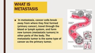
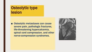
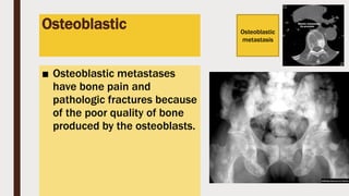
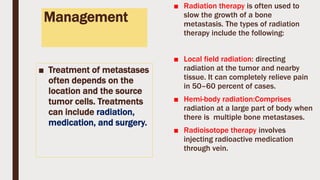
METASTATIC BONY
LESION
DR.ABU BAKAR SIDDIQUE
MS(ORTHO),WOC-SICOT FELLOW,SPINE MICROSURGERY
FELLOW,OBSERVERSHIP ON SPINE DEFORMITY CORRECTION
ASSOCIATE PROFESSOR
Introduction
■ Bone metastases are tumors that occur when cancer
cells break away from the place where they started
growing and move into bone tissue.
■ Bone metastases are considered a form of advanced
cancer. These secondary cancers within the bone are
difficult to cure, but treatments are available to lessen
the symptoms and lengthen life.
■ The skeleton is the third (after the liver and lungs)
most common organ to be affected by metastatic
cancer
WHAT IS
METASTASIS
■ In metastasis, cancer cells break
away from where they first formed
(primary cancer), travel through the
blood or lymph system, and form
new tumors (metastatic tumors) in
other parts of the body. The
metastatic tumor is the same type of
cancer as the primary tumor.
Incidence
■ Metastases are a frequent complication of cancer
■ Secondary malignant bone tumors are estimated to be 50 to 100 times as
common as primary bone cancers.
■ Up to 70 percent of patients with advanced breast or prostate cancer and
approximately 15 to 30 percent of patients with carcinoma of the lung,
colon, stomach, bladder, uterus, rectum, thyroid, or kidney.
■ 350,000 people die with bone metastases annually in the United States
■ once tumors metastasize to bone, they are usually incurable:
Types
■ Osteoblastic
■ Osteolytic
■ Mixed
Osteolytic type
lesion
■ Osteolytic metastases can cause
severe pain, pathologic fractures,
life-threatening hypercalcemia,
spinal cord compression, and other
nerve-compression syndromes.
■ Metastatic lesion rt femur with primary lesion in rt lung revealed in bone
scintigraphy,CT& MRI.( for full description please see next slide)
■ A case of bone metastasis located in the middle of femur (patient
#1). Fifty-year-old male patient with lung cancer. At first, the lesion
was suspected to be a primary femoral bone tumor. Then the
diagnosis was changed to metastatic tumor based on the result of
biopsy, which showed a poorly- differentiated carcinoma. Plain X-ray
and MRI (T1WI) showed an excentric bone tumor in the right femoral
diaphysis (a, b). BS showed high accumulation of 99m Tc-HMDP in
the corresponding area (c). FDG-PET was performed to detect the
primary tumor, and therefore, FOV of PET scan did not include full-
length of the femur. Whole-body scan from inguinal area to face
showed high accumulation of FDG in the right upper lobe which was
diagnosed as lung cancer later
Poorly differentiated lung cancer with
metastasis.
A case of osteolytic-type bone metastasis (patient #2). Eighty-year-old male
patient with a chest wall tumor. BS showed heterogeneous uptake in bilateral
ribs (a). BS diagnosis without clinical information could not detect osteolytic
lesion in the left upper rib, whereas BS diagnosis with clinical information was
able to detect the osteolysis. Chest X-ray revealed chest wall tumor, but no
osteolytic lesion (b). FDG-PET showed high accumulation of FDG in the left upper
chest wall (c). MRI: Gd enhanced 3D-VIBE (fat-suppressed 3D gradient-echo
technique with a volumetric interpolated breath- hold examination). Gd enhanced
MRI showed poorly enhanced left rib tumor surrounded by well enhanced
capsule (d). Further evaluation including biopsy and immunohistochemical
analysis sug- gested that a primary bone tumor was negative and metastasis
from a poorly differentiated lung cancer was highly suspected. This patient was
treated by localized radiotherapy against the chest wall tumor; however, he died
of multiple mediastinal lymph node, bone and liver metastases. According to the
clinical course, the final diagnosis of this patient was established as lung cancer
Osteoblastic
■ Osteoblastic metastases
have bone pain and
pathologic fractures because
of the poor quality of bone
produced by the osteoblasts.
Osteoblastic
metastasis
■ Most patients with breast
cancer have predominantly
osteolytic lesions, although
at least 15 to 20 percent of
them have predominantly
osteoblastic lesions.
■ Only in multiple myeloma do
purely lytic bone lesions
■ The lesions in prostate
cancer are predominantly
osteoblastic,
■ And bone metastases tend to
be the only site of metastasis
in prostatic cancer.
■ Agents that block bone
resorption can decrease bone
pain and the risk of
pathologic fractures in
prostatic carcinoma.
Why metastasis
happen in bone?
■ Several factors account for
the frequency of bone
metastasis.
■ Blood flow is high in areas
of red marrow,
■ tumor cells produce
adhesive molecules that
bind them to marrow
stromal cells and bone
matrix.
■ Varieties of growth factors, which
are released and activated during
bone resorption, provide fertile
ground in which tumor cells can
grow..
■ GF including transforming growth
factor β, insulin-like growth factors
I and II, fibroblast growth factors,
platelet-derived growth factors,
bone morphogenetic proteins, and
calcium
COMMOM PRIMARY
SITES
■ THYROID
■ BREAST
■ LUNG
■ GIT
■ KIDNEY
■ PROSTATE
■ BLADDER
Clinical presentations
■ Bone pain is the most common complication of metastatic bone disease,
resulting from structural damage, periosteal irritation, and nerve entrapment
■ Hypercalcemia occurs in 5-10% of all patients with advanced cancer but is most
common in patients with breast carcinoma, multiple myeloma, and squamous
carcinomas of the lung
■ Pathologic fractures are a relatively late complication of bone involvement
■ General symptoms and Signs
■ Sighs and symptoms due to primary lesion
Diagnosis
■ Modern imaging technique
-Bone Scintigraphy (isotope bone scan)
- Positron emission tomography
-Whole body MRI.
■ Complimentary imaging technique
-X-Ray,CT scan,MRI to determine the extent of metastasis
and its characteristics.
DIAGNOSIS-CONT.
■ Hyperkalamia: Impotant but non
specific serum marker
■ Biopsy : Is the final stage in
diagnostic process to confirm bony
metastasis
■ Guided biopsy: CT,MRI
■ Lequid Biopsy seems to be the
most promising diagnostic method for
detection of bone metastases based
on tumor specific DNA mutation.
Bone marker in metastatic diseases
■ Levels of bone-specific
alkaline phosphatase,
osteocalcin, and type I
procollagen C-propeptide in
serum are indicators of
osteoblast activity,
Management
■ Treatment of metastases
often depends on the
location and the source
tumor cells. Treatments
can include radiation,
medication, and surgery.
■ Radiation therapy is often used to
slow the growth of a bone
metastasis. The types of radiation
therapy include the following:
■ Local field radiation: directing
radiation at the tumor and nearby
tissue. It can completely relieve pain
in 50–60 percent of cases.
■ Hemi-body radiation:Comprises
radiation at a large part of body when
there is multiple bone metastases.
■ Radioisotope therapy involves
injecting radioactive medication
through vein.
MEDICATION
■ bone-building medications, such as
bisphosphonates, to help reduce
bone damage
■ chemotherapy to kill tumor cells
and reduce tumor size
■ hormone therapy to slow certain
hormones for cancers like breast
cancer and prostate cancer
■ pain medications
Surgery
■ Surgical excision of tumor
■ Surgical excision after neo adjuvant
therapy
■ Radical excision/Amputation
■ Limb spearing surgery with custom
made device reinforcement.
■ Bone cement reinforcement
■ Radio frequency ablation
■ Cryoablation.
■ Bone fixation for pathological
fracture when required.

Metastatic bony lesion

  • 1.
    METASTATIC BONY LESION DR.ABU BAKARSIDDIQUE MS(ORTHO),WOC-SICOT FELLOW,SPINE MICROSURGERY FELLOW,OBSERVERSHIP ON SPINE DEFORMITY CORRECTION ASSOCIATE PROFESSOR
  • 2.
    Introduction ■ Bone metastasesare tumors that occur when cancer cells break away from the place where they started growing and move into bone tissue. ■ Bone metastases are considered a form of advanced cancer. These secondary cancers within the bone are difficult to cure, but treatments are available to lessen the symptoms and lengthen life. ■ The skeleton is the third (after the liver and lungs) most common organ to be affected by metastatic cancer
  • 3.
    WHAT IS METASTASIS ■ Inmetastasis, cancer cells break away from where they first formed (primary cancer), travel through the blood or lymph system, and form new tumors (metastatic tumors) in other parts of the body. The metastatic tumor is the same type of cancer as the primary tumor.
  • 4.
    Incidence ■ Metastases area frequent complication of cancer ■ Secondary malignant bone tumors are estimated to be 50 to 100 times as common as primary bone cancers. ■ Up to 70 percent of patients with advanced breast or prostate cancer and approximately 15 to 30 percent of patients with carcinoma of the lung, colon, stomach, bladder, uterus, rectum, thyroid, or kidney. ■ 350,000 people die with bone metastases annually in the United States ■ once tumors metastasize to bone, they are usually incurable:
  • 5.
  • 6.
    Osteolytic type lesion ■ Osteolyticmetastases can cause severe pain, pathologic fractures, life-threatening hypercalcemia, spinal cord compression, and other nerve-compression syndromes.
  • 7.
    ■ Metastatic lesionrt femur with primary lesion in rt lung revealed in bone scintigraphy,CT& MRI.( for full description please see next slide)
  • 8.
    ■ A caseof bone metastasis located in the middle of femur (patient #1). Fifty-year-old male patient with lung cancer. At first, the lesion was suspected to be a primary femoral bone tumor. Then the diagnosis was changed to metastatic tumor based on the result of biopsy, which showed a poorly- differentiated carcinoma. Plain X-ray and MRI (T1WI) showed an excentric bone tumor in the right femoral diaphysis (a, b). BS showed high accumulation of 99m Tc-HMDP in the corresponding area (c). FDG-PET was performed to detect the primary tumor, and therefore, FOV of PET scan did not include full- length of the femur. Whole-body scan from inguinal area to face showed high accumulation of FDG in the right upper lobe which was diagnosed as lung cancer later
  • 10.
    Poorly differentiated lungcancer with metastasis.
  • 11.
    A case ofosteolytic-type bone metastasis (patient #2). Eighty-year-old male patient with a chest wall tumor. BS showed heterogeneous uptake in bilateral ribs (a). BS diagnosis without clinical information could not detect osteolytic lesion in the left upper rib, whereas BS diagnosis with clinical information was able to detect the osteolysis. Chest X-ray revealed chest wall tumor, but no osteolytic lesion (b). FDG-PET showed high accumulation of FDG in the left upper chest wall (c). MRI: Gd enhanced 3D-VIBE (fat-suppressed 3D gradient-echo technique with a volumetric interpolated breath- hold examination). Gd enhanced MRI showed poorly enhanced left rib tumor surrounded by well enhanced capsule (d). Further evaluation including biopsy and immunohistochemical analysis sug- gested that a primary bone tumor was negative and metastasis from a poorly differentiated lung cancer was highly suspected. This patient was treated by localized radiotherapy against the chest wall tumor; however, he died of multiple mediastinal lymph node, bone and liver metastases. According to the clinical course, the final diagnosis of this patient was established as lung cancer
  • 12.
    Osteoblastic ■ Osteoblastic metastases havebone pain and pathologic fractures because of the poor quality of bone produced by the osteoblasts. Osteoblastic metastasis
  • 13.
    ■ Most patientswith breast cancer have predominantly osteolytic lesions, although at least 15 to 20 percent of them have predominantly osteoblastic lesions. ■ Only in multiple myeloma do purely lytic bone lesions ■ The lesions in prostate cancer are predominantly osteoblastic, ■ And bone metastases tend to be the only site of metastasis in prostatic cancer. ■ Agents that block bone resorption can decrease bone pain and the risk of pathologic fractures in prostatic carcinoma.
  • 14.
    Why metastasis happen inbone? ■ Several factors account for the frequency of bone metastasis. ■ Blood flow is high in areas of red marrow, ■ tumor cells produce adhesive molecules that bind them to marrow stromal cells and bone matrix. ■ Varieties of growth factors, which are released and activated during bone resorption, provide fertile ground in which tumor cells can grow.. ■ GF including transforming growth factor β, insulin-like growth factors I and II, fibroblast growth factors, platelet-derived growth factors, bone morphogenetic proteins, and calcium
  • 15.
    COMMOM PRIMARY SITES ■ THYROID ■BREAST ■ LUNG ■ GIT ■ KIDNEY ■ PROSTATE ■ BLADDER
  • 16.
    Clinical presentations ■ Bonepain is the most common complication of metastatic bone disease, resulting from structural damage, periosteal irritation, and nerve entrapment ■ Hypercalcemia occurs in 5-10% of all patients with advanced cancer but is most common in patients with breast carcinoma, multiple myeloma, and squamous carcinomas of the lung ■ Pathologic fractures are a relatively late complication of bone involvement ■ General symptoms and Signs ■ Sighs and symptoms due to primary lesion
  • 17.
    Diagnosis ■ Modern imagingtechnique -Bone Scintigraphy (isotope bone scan) - Positron emission tomography -Whole body MRI. ■ Complimentary imaging technique -X-Ray,CT scan,MRI to determine the extent of metastasis and its characteristics.
  • 18.
    DIAGNOSIS-CONT. ■ Hyperkalamia: Impotantbut non specific serum marker ■ Biopsy : Is the final stage in diagnostic process to confirm bony metastasis ■ Guided biopsy: CT,MRI ■ Lequid Biopsy seems to be the most promising diagnostic method for detection of bone metastases based on tumor specific DNA mutation.
  • 19.
    Bone marker inmetastatic diseases ■ Levels of bone-specific alkaline phosphatase, osteocalcin, and type I procollagen C-propeptide in serum are indicators of osteoblast activity,
  • 20.
    Management ■ Treatment ofmetastases often depends on the location and the source tumor cells. Treatments can include radiation, medication, and surgery. ■ Radiation therapy is often used to slow the growth of a bone metastasis. The types of radiation therapy include the following: ■ Local field radiation: directing radiation at the tumor and nearby tissue. It can completely relieve pain in 50–60 percent of cases. ■ Hemi-body radiation:Comprises radiation at a large part of body when there is multiple bone metastases. ■ Radioisotope therapy involves injecting radioactive medication through vein.
  • 21.
    MEDICATION ■ bone-building medications,such as bisphosphonates, to help reduce bone damage ■ chemotherapy to kill tumor cells and reduce tumor size ■ hormone therapy to slow certain hormones for cancers like breast cancer and prostate cancer ■ pain medications
  • 22.
    Surgery ■ Surgical excisionof tumor ■ Surgical excision after neo adjuvant therapy ■ Radical excision/Amputation ■ Limb spearing surgery with custom made device reinforcement. ■ Bone cement reinforcement ■ Radio frequency ablation ■ Cryoablation. ■ Bone fixation for pathological fracture when required.