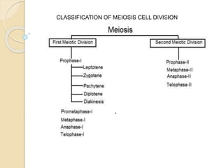
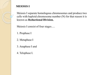
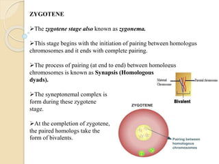
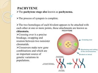
SHRI SHIVAJI SCIENCE COLLEGE,
AMRAVATI
CLASS : XI ( 2020-2021)
SUBJECT : BIOLOGY
( CELL DIVISION)
BY PROF. S. G. MANKAR
CONTENT :
AMITOSIS CELL DIVISION
MEIOSIS CELL DIVISION
AMITOSIS ( DIRECT CELL DIVISION):
 Amitosis is a mode of division which nucleus elongates,
constricts in the middle and divides directly into two daughter
nuclei.
Remak (1955) discovered amitosis in RBCs of chick embryo, but
the term was coined by Flemming (1882).
Chromatin does not condense into definite chromosomes.
A spindle is not formed.
Chromatin distribution occurs unequally which causes
abnormalities in metabolism and reproduction.
Cytokinesis may or may not follow karyokinesis.
Amitosis occurs in mega-nucleus of paramecium, nuclei of
internodal cells of Chara, endosperm cells of seeds, cartilage cells
and diseased cells.
MEIOSIS ( REDUCTIONAL CELL DIVISON)
The meiotic cell division first time described by Van Beneden
in 1883
Meiotic cell division occurs in germ cells of all living organism.
During meiosis, the genetic material of a diploid germ cell
undergoes two nuclear divisions and resulting in to four haploid
daughter cells.
Each daughter cells has one half of the number of chromosomes
as the parent cell.
There are two successive nuclear divisions in meiosis as
compared to the one division found in mitosis.
The two stages of meiosis are…
1. Meiosis I
2. Meiosis II
 Meiosis I also called as Reductional Division
Meiosis II also called as Equational Division
Before a dividing cell enters meiosis, it undergoes a period of
growth called Interphase.
CLASSIFICATION OF MEIOSIS CELL DIVISION
Meiosis I separate homologous chromosomes and produce two
cells with haploid chromosome number (N) for that reason it is
known as Reductional Division.
Meiosis I consist of four stages….
1. Prophase I
2. Metaphase I
3. Anaphase I and
4. Telophase I.
MEIOSIS I
PROPHASE I
Prophase I is the longest in duration compared to
Prophase in mitosis.
It takes about 85 - 95 percent of the total time for
meiosis and also much more complex.
The Prophase I divided into 5 stages (Le Za Pa Di
Dia).
1. Leptotene
2. Zygotene
3. Pachytene
4. Diplotene and
5. Diakinesis
LEPTOTENE
The first stage of Prophase I is called Leptotene or leptonema.
All the chromosomes begin to condense, so, they become visible as
fine thread.
There is marked increase in the nuclear volume.
A process of 'homology search' which is essential to the initial
pairing of homologs, begins during leptonema.
ZYGOTENE
The zygotene stage also known as zygonema.
This stage begins with the initiation of pairing between homologus
chromosomes and it ends with complete pairing.
The process of pairing (at end to end) between homologus
chromosomes is known as Synapsis (Homologous
dyads).
The syneptonemal complex is
form during these zygotene
stage.
At the completion of zygotene,
the paired homologs take the
form of bivalents.
PACHYTENE
The pachytene stage also known as pachynema.
The process of synapsis is complete.
The two homologus of each bivalent appears to be attached with
each other at one or more points, these attachments are known as
chiasmata.
Crossing over is a precise
breakage, swapping and
reunion between two nonsister
chromatids.
Crossovers make new gene
combinations and which are
an important source of
genetic variations in
populations.
DIPLOTENE
The diplotene stage also known as diplonema.
DNA recombination is complete.
The chromatids continue to shorten and thicken and the four sister
chromatids in a group is called a tetrad.
The synaptonemal complex begins to break down.
The paired chromatids begin to pull apart,
causing the strands to separate longitudinally.
The chiasmata tend to become terminalised
as the meiotic prophase continues.
DIAKINESIS
The chromomes become shorter and thicker due to condensation.
Nucleolus and nuclear envelope disappear towards the end of
diakinesis.
The spindle apparatus becomes organized.
The centrioles migrate away from one another.
METAPHASE- I
All the bivalents migrate within a cell migrate to metaphase plate.
One homologue is pulled above the metaphase plate, the other below.
The centromeres of homologous chromosomes of each bivalent stretch out
on either side.
The centrioles are at opposite poles of the cell.
Spindle fibers from one
pole of the cell attach to
one chromosome and
spindle fibers from the
opposite pole attach to
the homologous
chromosome.
ANAPHASE- I
Chromosomes move to the opposite poles.
The microtubules and the kinetochore fibers interact, which cause the
movement.
A difference between mitosis and meiosis is that sister chromatids remain
joined after metaphase in meiosis I, whereas in mitosis they separate.
During Anaphase I original chromosomes separate, so reduction in the
number of chromosomes from 2N to N number, yet the sister chromatids
remain together.
TELOPHASE- I
The homologous chromosome complete their migration to the two poles b/c
shortning of spindles.
The nuclear envelope organized around two groups of chromosomes.
The nucleolus also reappears
CYTOKINESIS-I
Cytokinesis involves the formation of a cleavage furrow, resulting in the
pocketing of the cell into two cells.
At the end of Telophase I and Cytokinesis, two daughter cells are produced,
each with one half of the number of chromosomes (haploid set of
replicated chromosomes) of the original parent cell.
MEIOSIS II
Meiosis II is the second part of the meiotic process.
The Meiosis II consists
Prophase II
Each dyad is composed of a pair of sister chromatids attached by a
common centromere.
Metaphase II
Centromeres are positioned at the equatorial plane.
Anaphase II
Centromeres divide and the sister chromatids of each dyad are
pulled to opposite poles
Telophase II & Cytokinesis
One member of each pair of homologous chromosome present in each
pole.
Each chromosome is referred as monad (a combination of maternal
and paternal genetic information).
Nuclei reform around chromosomes at the poles.
Following cytokinesis and finally four haploid gametes result from
a single meiotic event
THANK YOU

Meosis cell division

  • 1.
    SHRI SHIVAJI SCIENCECOLLEGE, AMRAVATI CLASS : XI ( 2020-2021) SUBJECT : BIOLOGY ( CELL DIVISION) BY PROF. S. G. MANKAR
  • 2.
    CONTENT : AMITOSIS CELLDIVISION MEIOSIS CELL DIVISION
  • 3.
    AMITOSIS ( DIRECTCELL DIVISION):  Amitosis is a mode of division which nucleus elongates, constricts in the middle and divides directly into two daughter nuclei. Remak (1955) discovered amitosis in RBCs of chick embryo, but the term was coined by Flemming (1882). Chromatin does not condense into definite chromosomes. A spindle is not formed.
  • 4.
    Chromatin distribution occursunequally which causes abnormalities in metabolism and reproduction. Cytokinesis may or may not follow karyokinesis. Amitosis occurs in mega-nucleus of paramecium, nuclei of internodal cells of Chara, endosperm cells of seeds, cartilage cells and diseased cells.
  • 5.
    MEIOSIS ( REDUCTIONALCELL DIVISON) The meiotic cell division first time described by Van Beneden in 1883 Meiotic cell division occurs in germ cells of all living organism. During meiosis, the genetic material of a diploid germ cell undergoes two nuclear divisions and resulting in to four haploid daughter cells. Each daughter cells has one half of the number of chromosomes as the parent cell. There are two successive nuclear divisions in meiosis as compared to the one division found in mitosis.
  • 6.
    The two stagesof meiosis are… 1. Meiosis I 2. Meiosis II  Meiosis I also called as Reductional Division Meiosis II also called as Equational Division Before a dividing cell enters meiosis, it undergoes a period of growth called Interphase.
  • 7.
  • 8.
    Meiosis I separatehomologous chromosomes and produce two cells with haploid chromosome number (N) for that reason it is known as Reductional Division. Meiosis I consist of four stages…. 1. Prophase I 2. Metaphase I 3. Anaphase I and 4. Telophase I. MEIOSIS I
  • 9.
    PROPHASE I Prophase Iis the longest in duration compared to Prophase in mitosis. It takes about 85 - 95 percent of the total time for meiosis and also much more complex. The Prophase I divided into 5 stages (Le Za Pa Di Dia). 1. Leptotene 2. Zygotene 3. Pachytene 4. Diplotene and 5. Diakinesis
  • 10.
    LEPTOTENE The first stageof Prophase I is called Leptotene or leptonema. All the chromosomes begin to condense, so, they become visible as fine thread. There is marked increase in the nuclear volume. A process of 'homology search' which is essential to the initial pairing of homologs, begins during leptonema.
  • 11.
    ZYGOTENE The zygotene stagealso known as zygonema. This stage begins with the initiation of pairing between homologus chromosomes and it ends with complete pairing. The process of pairing (at end to end) between homologus chromosomes is known as Synapsis (Homologous dyads). The syneptonemal complex is form during these zygotene stage. At the completion of zygotene, the paired homologs take the form of bivalents.
  • 12.
    PACHYTENE The pachytene stagealso known as pachynema. The process of synapsis is complete. The two homologus of each bivalent appears to be attached with each other at one or more points, these attachments are known as chiasmata. Crossing over is a precise breakage, swapping and reunion between two nonsister chromatids. Crossovers make new gene combinations and which are an important source of genetic variations in populations.
  • 13.
    DIPLOTENE The diplotene stagealso known as diplonema. DNA recombination is complete. The chromatids continue to shorten and thicken and the four sister chromatids in a group is called a tetrad. The synaptonemal complex begins to break down. The paired chromatids begin to pull apart, causing the strands to separate longitudinally. The chiasmata tend to become terminalised as the meiotic prophase continues.
  • 14.
    DIAKINESIS The chromomes becomeshorter and thicker due to condensation. Nucleolus and nuclear envelope disappear towards the end of diakinesis. The spindle apparatus becomes organized. The centrioles migrate away from one another.
  • 15.
    METAPHASE- I All thebivalents migrate within a cell migrate to metaphase plate. One homologue is pulled above the metaphase plate, the other below. The centromeres of homologous chromosomes of each bivalent stretch out on either side. The centrioles are at opposite poles of the cell. Spindle fibers from one pole of the cell attach to one chromosome and spindle fibers from the opposite pole attach to the homologous chromosome.
  • 16.
    ANAPHASE- I Chromosomes moveto the opposite poles. The microtubules and the kinetochore fibers interact, which cause the movement. A difference between mitosis and meiosis is that sister chromatids remain joined after metaphase in meiosis I, whereas in mitosis they separate. During Anaphase I original chromosomes separate, so reduction in the number of chromosomes from 2N to N number, yet the sister chromatids remain together.
  • 17.
    TELOPHASE- I The homologouschromosome complete their migration to the two poles b/c shortning of spindles. The nuclear envelope organized around two groups of chromosomes. The nucleolus also reappears
  • 18.
    CYTOKINESIS-I Cytokinesis involves theformation of a cleavage furrow, resulting in the pocketing of the cell into two cells. At the end of Telophase I and Cytokinesis, two daughter cells are produced, each with one half of the number of chromosomes (haploid set of replicated chromosomes) of the original parent cell.
  • 19.
    MEIOSIS II Meiosis IIis the second part of the meiotic process. The Meiosis II consists Prophase II Each dyad is composed of a pair of sister chromatids attached by a common centromere. Metaphase II Centromeres are positioned at the equatorial plane. Anaphase II Centromeres divide and the sister chromatids of each dyad are pulled to opposite poles
  • 20.
    Telophase II &Cytokinesis One member of each pair of homologous chromosome present in each pole. Each chromosome is referred as monad (a combination of maternal and paternal genetic information). Nuclei reform around chromosomes at the poles. Following cytokinesis and finally four haploid gametes result from a single meiotic event
  • 22.